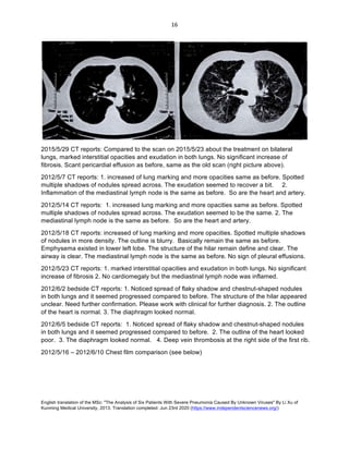
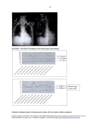
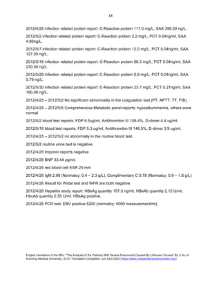
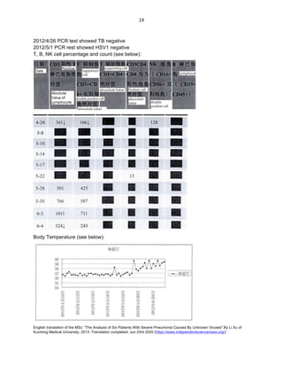
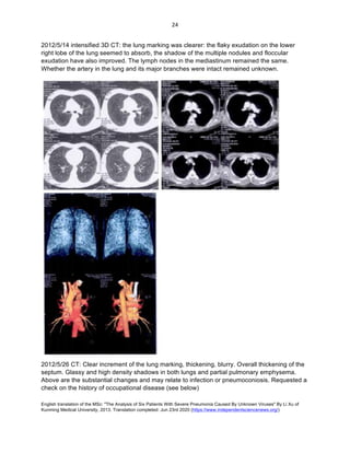
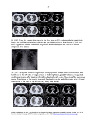
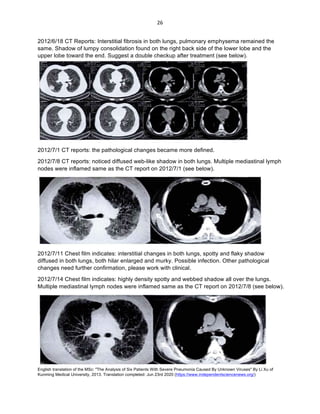
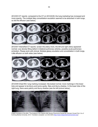
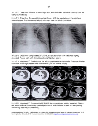
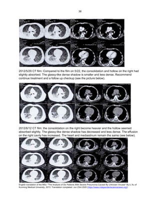
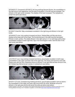
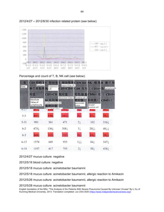
SlideShare a Scribd company logo
1 of 66
Download to read offline
English translation of the MSc: "The Analysis of Six Patients With Severe Pneumonia Caused By Unknown Viruses" By Li Xu of
Kunming Medical University, 2013. Translation completed: Jun 23rd 2020 (https://www.independentsciencenews.org/)
	
	
1	
Master's Thesis
"The Analysis of Six Patients With Severe Pneumonia Caused By
Unknown Viruses"
School/ College: No. 1 School of Clinical Medicine, Kun Ming Medical University
Student’s Name: Li Xu
Study field: Clinical Medicine and Emergency Medicine
May, 2013
Translation note: This translation was commissioned by Independent Science News
(https://www.independentsciencenews.org/). Where the author (Li Xu) provided it, we have
preserved the original English text (e.g. The Title and the Abstract). Page numbers match the
original pdf file. Thesis accessed June 10th 2020.
专 业 学 位 硕 士 学 位 论 文
单 位 代 码 : 分 类 号 :
学 号 : 密 级 :
尾 蚪 大
硕 士 学 位 论 文
未 知 病 毒 引 起 重 症 肺 炎 例 分 析
院 系 、 所
研 究 生 姓名
学 科 、 专 业
学 位 类 型
指 导 教 师
昆 明 医科大学第一临床 医学院
李 旭
临 床 医 学 、 急 诊 医 学
专 业 学 位
钱 传 云 教 授
二 一 三 年 五 月
English translation of the MSc: "The Analysis of Six Patients With Severe Pneumonia Caused By Unknown Viruses" By Li Xu of
Kunming Medical University, 2013. Translation completed: Jun 23rd 2020 (https://www.independentsciencenews.org/)
	
	
2	
尾 略 蚪
硕 士 学位论 文
未知病毒 引起 重症肺炎 例分析
申 请 人 姓 名 李 旭
学 科 、 专 、 丨 临床 医 学 、 急 谂 医学
申 请学位类型 专业学位
指 导 教 师 钱传 云 教授
完 成 日 期 二 〇 一三年五 月
English translation of the MSc: "The Analysis of Six Patients With Severe Pneumonia Caused By Unknown Viruses" By Li Xu of
Kunming Medical University, 2013. Translation completed: Jun 23rd 2020 (https://www.independentsciencenews.org/)
	
	
3	
Kun Ming Medical University
Master's Research Statement
I stated that this master's thesis was written by me and under the supervision of my professors.
It is based on the result of my independent study. Besides the citation and the
acknowledgement in the end, there are no other organizations have published such work. In this
paper, I have pointed out those who made contribution to this work and showed my appreciation.
If there is any part in this paper that is fraud, I would take full responsibility.
Author of the paper:
2013/5/28
Kun Ming Medical University
Authorization of the right of master research
This author understands that Kun Ming Medical University has the right to preserve and use this
master research. The author agrees on the reservation of the school and sending a digital copy
to related offices and department of the nation. The author permits the consultation and
borrowing. The school can decide to publish all or partial content (The confidential part is
marked out. After decrypting, it will also follow such rule); the school can keep this thesis in print
or by any other means.
Signature of the author: Signature of the Advisor:
2013/5/28 2013/5/28
My advisor and I agree to submit this paper to “Digital Chinese research periodical” of Chin Hua
University for digital publication and incorporation into CNKI series of data base. We will follow
the rules of Chinese Selected doctoral dissertations and master’s theses full-text database and
enjoy the related rights.
Signature of the author: Signature of the Advisor:
2013/5/28 2013/5/28
English translation of the MSc: "The Analysis of Six Patients With Severe Pneumonia Caused By Unknown Viruses" By Li Xu of
Kunming Medical University, 2013. Translation completed: Jun 23rd 2020 (https://www.independentsciencenews.org/)
	
	
4	
英文縮略词表
英文缩略语 英文 全称 中 文全称
体温
脉率
呼 吸频率
血压
心率
计算 机断 层扫 描
反应蛋 白
降钙 素原
血清 淀粉样蛋 白
二聚 体
纤维 蛋 白 降解产物
二氧化 碳分压
〇 血氧 分压
氢离子 浓度指数
〗 氧合指 数
机体反应水 平 分 级
型尿 钠肽
聚合酶链式 反应
人类 免疫缺 陷病 毒
国 际标准化 比值
心 电 图
脱氧核 糖核 酸
English translation of the MSc: "The Analysis of Six Patients With Severe Pneumonia Caused By Unknown Viruses" By Li Xu of
Kunming Medical University, 2013. Translation completed: Jun 23rd 2020 (https://www.independentsciencenews.org/)
	
	
5	
R N A


r
i b o n u c l e i c a c i d


核糖核酸
国 际 病毒分类委员 会
急 性呼吸 窘迫综合征
严重急 性呼 吸综合征
冠 状病毒
样 冠状病毒
重症加 强护理病房
English translation of the MSc: "The Analysis of Six Patients With Severe Pneumonia Caused By Unknown Viruses" By Li Xu of
Kunming Medical University, 2013. Translation completed: Jun 23rd 2020 (https://www.independentsciencenews.org/)
	
	
6	
Table of Contents
Chinese Abstract………………….………………………………………………………………….1
English Abstract………………….…………………………………………………………………...2
Medical Cases…………………...…………………………………………………………………...3
Comprehensive Analysis…………………………………………………………….………………52
Work Cited…………………………………………………………………………….………………58
Author’s publication…………………………………………………………………………………..59
Acknowledgements……..…………………………………………………………………………….60
English translation of the MSc: "The Analysis of Six Patients With Severe Pneumonia Caused By Unknown Viruses" By Li Xu of
Kunming Medical University, 2013. Translation completed: Jun 23rd 2020 (https://www.independentsciencenews.org/)
	
	
7	
昆 明 医科 大 学硕 士研 究 生学位 论文
未知 病毒引 起重症肺炎 例 分析
研究 生: 李旭
导 师 : 钱传 云教授
昆 明 医 科大学第 一 附属 医 院急诊 医学科 、
摘要
年 月 、 月 我院先后 收住 例 未知病毒 引 起相 关重症肺 炎 患 者 。 此
位患 者均为 同 一 矿洞 工人 工作环境 中 接触大量蝙蝠及蝙蝠粪便。 最终结 局
位 患者死亡 位患 者存活 。 据 中 国 科学 院 昆 明 动物研究所鉴定 此 位 患者工
作矿洞 内 蝙蝠正 为 中 华菊 头 蝠 然而我 国 科学家 在寻 找 病 原 的 过程 中 在
中 华菊头 蝠体 内 提取 出 了 样 冠状病 毒 。 本文针 对 例 患
者所感染 未知 病毒相关 重症 肺炎 的 诊治过 程及可 能 引 起 的 病 因 、 病原学进行推 断
与 分析 。
关键词 : 重症肺 炎 、 蝙 蝠 、 样 冠状病 毒
English translation of the MSc: "The Analysis of Six Patients With Severe Pneumonia Caused By Unknown Viruses" By Li Xu of
Kunming Medical University, 2013. Translation completed: Jun 23rd 2020 (https://www.independentsciencenews.org/)
	
	
8	
昆 明 医 科 大 学 硕 士 研究 生 学位 论文
:
:
: 、 、
English translation of the MSc: "The Analysis of Six Patients With Severe Pneumonia Caused By Unknown Viruses" By Li Xu of
Kunming Medical University, 2013. Translation completed: Jun 23rd 2020 (https://www.independentsciencenews.org/)
	
	
9	
Case 1
Patient Zhou, male, age 63, was admitted to the hospital on April 26, 2012. He had signs of
fever, coughing, difficulty in breathing, chest pain, and hiccups for more than ten days. 24 days
prior to the hospitalization, he was working in the mining well for half of a month. He worked 7
hours a day. After exposing to the mining well where there were many bats and bats’ feces, he
started to show signs of coughing and fever and had a 38 Celsius body temperature. He
immediately went to the local hospital. His fever went on and off in the next five consecutive
days. The actual treatment remained unknown. The highest body temperature was 40 Celsius
and the lower is 37 Celsius. He also experienced headache, dizziness, ear congestion and dry
cough. There was no pattern of his illness in daytime or night time, along with chest pain.
Difficulty in breathing was getting worse. Occasionally, having hiccups. No sign of nausea,
vomiting, or diarrhea. To pursue more treatment, the patient was admitted to my department.
Since the onset of the disease, the patient had felt lethargic. He has insomnia and loss of
appetite, but regular bowel movement and urination. Self-reported that he did not have a history
of high blood pressure, diabetes and heart disease or other chronic diseases, nor did he have
hepatitis, typhoid or any other contagious disease. He did not have surgical operation, trauma,
and blood transfusion in the past. He was not allergic to any medication or food. His vaccination
record remained unknown. Physical checkup: body temperature is 37.8 Celsius, pulse rate: 74
times/minute, respiratory rate: 20 times/ minute, blood pressure: 110/63 mmhg. The patient
stays alert and could answer all the questions. No sign of malnutrition or obesity. He was sent to
the room by stretcher. Skin and mucous membranes remained normal, and so were the pupils.
They were 3mm in diameter. The pupils remained sensitive to light. The chest and respiratory
movements were symmetrical. The breathing sounds were rough. Dry crackles were heard on
both bases of the lung. His heart rate is 74 beats/minute, regular heart rate and no heart
murmur from any of the heart valves. Softness of the abdomen, no pain when pressured, no
rebound tenderness, or guarding. Normal bowel sound: 5 times/minute. No inflammation at the
lower part of the legs. Regular body reflex. No pathological reflexes. The blood report from
04/25/2012: WBC12.10X109
/L, N%89.3, Hemoglobin: 178g/L; Comprehensive Metabolic Panel
was CRP 20.3 mg/L, blood ammonia: 43 umol/L; Normal result on the coagulation report. As the
CT scan showed, there was extensive and patchy consolidated exudate bilaterally, elevated
bronchovascular shadows and lung markings, some nodules in different sizes, parts were
calcified. Mediastinal lymph node enlargement, partially calcified.
Initial Diagnosis: 1. Fever, coughing, dyspnea, hiccup 2. Hyponatremia; 3. Malfunctioning in liver
and bladder
Method
The examination after hospitalization:
2012/4/25 Computed tomography report: extensive and patchy consolidated exudate over
bilateral lung, increased Broncho vascular shadows and lung markings, some nodules in
different sizes, parts were calcified. Mediastinal lymph node enlargement, partial were calcified.
2012/4/30 CT report: 1. No noticeable changes in the lung, little pleural effusion in both lungs..
English translation of the MSc: "The Analysis of Six Patients With Severe Pneumonia Caused By Unknown Viruses" By Li Xu of
Kunming Medical University, 2013. Translation completed: Jun 23rd 2020 (https://www.independentsciencenews.org/)
	
	
10	
Pleural thickening posteriorly, and the rest was the same as the previous report. 2. As the scan
showed, there was little ascites (see below).
2012/5/2 bedside chest film: 1. Bilateral Lung markings worsening and getting blurry, and there
were shadows of the clot. Nodular shadow was noted in 2nd
intercostal space of right middle lobe,
and a small opacity was over left hilar region. Requested a follow up examination after the
clinical anti-inflammation treatment. 2. The outline of the heart is not too big 3. The diaphragm
remains normal (see the left picture below).
2012/5/6 bedside chest film: 1. Bilateral Lung marking augmentation and getting blurry, and
there were shadows of the clots. Increased laminar density in the middle and lower field of the
left lung, the hilum of both lungs are blurred. Requested a follow up examination after the
clinical anti-inflammation treatment. 2. Aorta is circuitous and the outline of the heart is normal.
3. Fluid in the left side of the pleural cavity and need to be evacuated. Please cooperate with the
clinic (see the right picture on top). 2012/4/26 - 2012/5/7: Analysis on the arterial blood gas (see
below):
English translation of the MSc: "The Analysis of Six Patients With Severe Pneumonia Caused By Unknown Viruses" By Li Xu of
Kunming Medical University, 2013. Translation completed: Jun 23rd 2020 (https://www.independentsciencenews.org/)
	
	
11	
2012/4/27 tumor protein chip shows ferritin is 484.86 (Normal male < 322 microgram/ L ),
Human chorionic gonadotropin is 0.65 (normal< 3.0 microgram/ L), prostate Specific Antigen is
0.02 (normal< 0.1 microgram/L) carbohydrate antigen 125: 42.22 (normal < 35.0KU/L).
On 2012/4/27, the Widal test and WFR test both came back negative. Results for Herpes
simplex virus, EB virus and CMV are all negative. Urine culture is negative and so is Ghb. The
stool test is also normal. Autoantibody and anti-nuclear antibodies are both negative.
2012/4/24 report: compliment C3C4 has decreased; Glucose in Urine 4+, Ketone is negative.
Thyroid test positive.
2012/4/27 - 2012/5/7: D-dimer reports 7.2 ug/ml (Apr, 27), D-dimer 3.6 ug/ml (May, 2), D-dimer
7.0 ug/ml (May, 6), D-dimer 5.0 ug/ml (May, 7).
2012/4/27 -2012/5/7 infected cell-specific protein (see below):
2012/4/25 - 2012/5/7 white blood cell and blood platelet (see below):
English translation of the MSc: "The Analysis of Six Patients With Severe Pneumonia Caused By Unknown Viruses" By Li Xu of
Kunming Medical University, 2013. Translation completed: Jun 23rd 2020 (https://www.independentsciencenews.org/)
	
	
12	
T, B, NK lymphocyte percentage and count (see below )
2012/4/27 sputum culture and blood culture were both negative (three times).
2012/5/7 blood culture implies positive for Acinetobacter baumannii and negative for candidiasis
2012/5/7 sputum culture Acinetobacter baumannii, pan resistant
2012/5/6 ultrasound reports severe ascites
English translation of the MSc: "The Analysis of Six Patients With Severe Pneumonia Caused By Unknown Viruses" By Li Xu of
Kunming Medical University, 2013. Translation completed: Jun 23rd 2020 (https://www.independentsciencenews.org/)
	
	
13	
2012/5/7 Ascites. Result of tumor cell testing is negative. Gram positive in cultivation ascites
regular test is negative. The Rivalta test is also negative.
Body temperature chart (see below):
	
Prescription after admission:
2012/4/26 – 2012/5/2 (J) Methylprednisolone 80mg, ivgtt, Q 12h.
21012/5/2 – 2012/5/7 (J) Methylprednisolone 40mg, ivgtt, Q 12h.
2012/5/7 – death (J) Methylprednisolone 80mg, ivgtt, Q 12h.
2012/4/26 – 2012/5/2 Meropenem 0.5gx2 shots, ivgtt, Q8h.
2012/5/7 – death Meropenem 0.5gx2 shots, ivgtt, Q8h.
2012/5/7 – death Vancomycin 0.5gx2 shots, ivgtt, Q12h.
2012/4/26 – death L - Voriconazole 0.1gx 2 shots (double the dosage on the first day), ivgtt,
Q12h.
2012/4/26 – death Acyclovir 0.25g*2, ivgtt, Q8h.
Discussion
The patient worked from the mining site since 2012/4/2 for up to 14 days.
Patient admitted to the hospital: 2012/4/26. Patient discharged: 2012/5/7. Total days: 12 days.
Discharged Diagnosis: 1. Severe lung infection 2. Sepsis 3. Septic shock and infection in
abdominal cavity 4. Asystole and stop breathing
Discharge reason: death
English translation of the MSc: "The Analysis of Six Patients With Severe Pneumonia Caused By Unknown Viruses" By Li Xu of
Kunming Medical University, 2013. Translation completed: Jun 23rd 2020 (https://www.independentsciencenews.org/)
	
	
14	
According to CT and Chest radiograph, the illness was progressively developed.
As the analysis on the arterial blood gas shows, during hospitalization, the patience had Type I
respiratory failure. Oxygenation index was poor. According to the “ARDS Berlin Criteria” in 2012,
for sure it was ARDS. Consequentially, one of the causes of death could be respiratory failure.
According to several researches from either abroad or domestically, glucose variability is
associated with rate of prognosis or death. During the hospitalization, the patient was given
intensive insulin treatment by our department. We tried to keep the blood sugar between 6-10
mmol/L. However, the patient’s glucose varies, poor prognosis.
The result for tumor protein chip came back positive, which means the patient had tumor related
disease. As a result, the systems of the whole body was impacted.
After the patient was admitted to the hospital, WBC and PLt were constantly decreasing.
Indicated by other virus infection related researches, WBC, PLt, T, B, NK Lymphocyte
percentage and count of the patients are also decreasing. It shows that the immune system of
the patient was impaired, had poor resistant to the disease and could easily be infected by
hospital-acquired infection.
The day the patient died, the blood and mucus culture showed Acinetobacter baumannii, severe
ascites. There was Gram-positive bacteria in ascites culture. On the same day, the PCT report
was 83.30ng/ml. As a result, one of the causes of death is septic shock. (severe lung infection
and abdominal cavity infection).
After admitted, the patient’s D-dimer was 7.2 ug/ml (Apr, 27), 3.6 ug/ml (May, 2), 7.0 ug/ml (May,
6) and 5.0 ug/ml (May, 7). The patient was bed ridden after admitted to the hospital and also
had tumor. The oxygen level in the blood was significant low two days prior to his death. There
was a possibility of pulmonary thromboembolism. However, the patient was severely ill, it was
not recommended to have an intensified chest CT checkup. Therefore, acute pulmonary
infection could be one of the causes of death. The family of the patient refused the autopsy
procedure.
After the suspension of Meropenem on May 2, 2012, the patient was no longer treated with any
antibiotics. His temperature went back up right after. Therefore, antibiotics play an important
role in the treatment.
Cause of death analysis: the patient was the oldest out of the six patients and had some tumor
related disease. His immune system was weak so had a poor body resistant to the disease. The
disease was acute and fierce.
English translation of the MSc: "The Analysis of Six Patients With Severe Pneumonia Caused By Unknown Viruses" By Li Xu of
Kunming Medical University, 2013. Translation completed: Jun 23rd 2020 (https://www.independentsciencenews.org/)
	
	
15	
Case Two
Patient Lu, Male, 42 years old, was admitted to the hospital on April 25, 2012. He had
fever and been coughing for half of a month and for the past three days had difficulties in
breathing. He worked in the mining hole before and was exposed to large amount of feces of
bats. Half of month ago, he started to have fever. His body temperature was 38.5 Celsius at first.
Occasionally, when he coughed, there was rusty colored mucus with blood clots. Felt bloated in
the stomach, loss of appetite and hiccup. He initially went to the small clinic for transfusion but it
was not helpful. Then, he was transferred to Yu Xi People’s Hospital for treatment. During
hospitalization, his body temperature was 40 Celsius and the fever did not follow any pattern.
No sign of chills before the fever. Still coughed with rusty-colored mucus and blood clots.
Difficulty in breathing for three days, especially after moving around. Chest tightness but no
chest pain. No problem lying down. No sign of paroxysmal dyspnea at night. No abdominal pain.
No visible hematuria. No history of high blood pressure, diabetes, coronary heart disease or
stroke. He was born in Zhao Tong, Yun Nan and had been to Mo Jiang. He worked in the
mining field prior to the illness and was exposed to large amount of bats’ feces. Five of his
colleagues had similar illness. He denied hepatitis, typhoid, tuberculosis or any other infectious
disease. No history of blood transmission or allergy reaction. The vaccination report remained
unknown. On examination: 36.6 Celsius, Pulse 110 times/ minute, Respiration rate 32 times/
minute, blood pressure 98/55mmHg, in poor condition. He was sent into the ward on a stretcher.
Reaction level scale was rated 1. No deformation on the head and features. The pupils were big
and round with 3 mm diameter. He was sensitive to the lights. Soft neck, no rigidity. Airway was
in the center. The chest looked symmetric from the outside. Rough breath sounds bilaterally,
and moist crackles were heard on both bases of the lung. The breathing sounds rough. Moist
rales from the bottom of the lungs. Heart was in normal size. Heart rate 110 times/ minute,
regular rhythm, no murmur, rubs or gallops. Abdomen soft, non-tender. Normal bowel sounds: 3
times/minute. Did not notice any rashes or eschar. No inflammation on the legs. Muscle strength
and tension remained normal. Additional checkup: According to the CT from Yu Xi People’s
hospital on April 25, 2012: severe pneumonia over bilateral lung. The bottom of the left lung had
limited pulmonary emphysema and bullae in the right lung; HBsAg (+), HbeAb (+), HbcAb (+).
Our blood gas analysis shows pH 7.431, PaO2 66.2mmHg, Oxygenation Index 162, lactic acid
1.7 mmol/L, Potassium in the blood 4.04mmol/L, Sodium in the blood 134.7 mmol/L.
Initial diagnosis after admission: 1. Severe pneumonia 2. Type I respiratory failure 3. Sepsis
4. Hepatitis B
Method (Some of the information was missing)
After admission, the complete examination:
Chest CT on 2012/4/30: 1. Increased lung markings, blurry and noticed multiple nodular
shadows. Bilateral lung patchy exudate. 2. Mediastinal lymph node enlargement, regular heart
shadow. Did not notice any abnormal in the artery. (see the left picture below)
English translation of the MSc: "The Analysis of Six Patients With Severe Pneumonia Caused By Unknown Viruses" By Li Xu of
Kunming Medical University, 2013. Translation completed: Jun 23rd 2020 (https://www.independentsciencenews.org/)
	
	
16	
	
2015/5/29 CT reports: Compared to the scan on 2015/5/23 about the treatment on bilateral
lungs, marked interstitial opacities and exudation in both lungs. No significant increase of
fibrosis. Scant pericardial effusion as before, same as the old scan (right picture above).
2012/5/7 CT reports: 1. increased of lung marking and more opacities same as before. Spotted
multiple shadows of nodules spread across. The exudation seemed to recover a bit. 2.
Inflammation of the mediastinal lymph node is the same as before. So are the heart and artery.
2012/5/14 CT reports: 1. increased lung marking and more opacities same as before. Spotted
multiple shadows of nodules spread across. The exudation seemed to be the same. 2. The
mediastinal lymph node is the same as before. So are the heart and artery.
2012/5/18 CT reports: increased of lung marking and more opacities. Spotted multiple shadows
of nodules in more density. The outline is blurry. Basically remain the same as before.
Emphysema existed in lower left lobe. The structure of the hilar remain define and clear. The
airway is clear. The mediastinal lymph node is the same as before. No sign of pleural effusions.
2012/5/23 CT reports: 1. marked interstitial opacities and exudation in both lungs. No significant
increase of fibrosis 2. No cardiomegaly but the mediastinal lymph node was inflamed.
2012/6/2 bedside CT reports: 1. Noticed spread of flaky shadow and chestnut-shaped nodules
in both lungs and it seemed progressed compared to before. The structure of the hilar appeared
unclear. Need further confirmation. Please work with clinical for further diagnosis. 2. The outline
of the heart is normal. 3. The diaphragm looked normal.
2012/6/5 bedside CT reports: 1. Noticed spread of flaky shadow and chestnut-shaped nodules
in both lungs and it seemed progressed compared to before. 2. The outline of the heart looked
poor. 3. The diaphragm looked normal. 4. Deep vein thrombosis at the right side of the first rib.
2012/5/16 – 2012/6/10 Chest film comparison (see below)
English translation of the MSc: "The Analysis of Six Patients With Severe Pneumonia Caused By Unknown Viruses" By Li Xu of
Kunming Medical University, 2013. Translation completed: Jun 23rd 2020 (https://www.independentsciencenews.org/)
	
	
17	
2012/6/6 – 2012/6/10 Analysis of the blood gas (see below)
Infection related protein (missing some data, did not make a table analysis):
Blood sugar
Lactic acid
English translation of the MSc: "The Analysis of Six Patients With Severe Pneumonia Caused By Unknown Viruses" By Li Xu of
Kunming Medical University, 2013. Translation completed: Jun 23rd 2020 (https://www.independentsciencenews.org/)
	
	
18	
2012/4/26 infection related protein report: C-Reactive protein 117.0 mg/L, SAA 398.00 ng/L.
2012/5/2 infection related protein report: C-Reaction protein 2.2 mg/L, PCT 0.04ng/ml, SAA
4.80ng/L.
2012/5/7 infection related protein report: C-Reaction protein 12.0 mg/L, PCT 0.04ng/ml, SAA
127.00 ng/L.
2012/5/18 infection related protein report: C-Reaction protein 66.3 mg/L, PCT 0.04ng/ml, SAA
230.00 ng/L.
2012/5/29 infection related protein report: C-Reaction protein 0.8 mg/L, PCT 0.04ng/ml, SAA
5.79 ng/L.
2012/5/30 infection related protein report: C-Reaction protein 23.7 mg/L, PCT 0.27ng/ml, SAA
190.00 ng/L.
2012/4/25 – 2012/5/2 No significant abnormality in the coagulation test (PT, APTT, TT, FIB).
2012/4/25 – 2012/5/6 Comprehensive Metabolic panel reports: hypoalbuminemia, others were
normal
2012/5/2 blood test reports: FDP 6.5ug/ml, Antithrombin III 108.4%, D-dimer 4.4 ug/ml.
2012/5/18 blood test reports: FDP 5.3 ug/ml, Antithrombin III 146.5%, D-dimer 3.9 ug/ml.
2012/4/25 – 2012/5/2 no abnormally in the routine blood test.
2012/5/2 routine urine test is negative.
2012/4/25 troponin reports negative
2012/4/26 BNP 33.44 pg/ml.
2012/4/26 red blood cell ESR 25 mm
2012/4/26 IgM 2.98 (Normalcy: 0.4 – 2.3 g/L), Complimentary C 0.78 (Normalcy: 0.9 – 1.8 g/L)
2012/4/26 Result for Widal test and WFR are both negative.
2012/4/26 Hepatitis study report: HBsAg quantity 157.5 ng/ml, HBeAb quantity 2.12 U/ml,
HbcAb quantity 2.55 U/ml. HBsAg positive.
2012/4/26 PCR test: EBV positive 5200 (normalcy: 5000 measurement/ml).
English translation of the MSc: "The Analysis of Six Patients With Severe Pneumonia Caused By Unknown Viruses" By Li Xu of
Kunming Medical University, 2013. Translation completed: Jun 23rd 2020 (https://www.independentsciencenews.org/)
	
	
19	
2012/4/26 PCR test showed TB negative
2012/5/1 PCR rest showed HSV1 negative
T, B, NK cell percentage and count (see below):
Body Temperature (see below):
English translation of the MSc: "The Analysis of Six Patients With Severe Pneumonia Caused By Unknown Viruses" By Li Xu of
Kunming Medical University, 2013. Translation completed: Jun 23rd 2020 (https://www.independentsciencenews.org/)
	
	
20	
Prescription after being admitted to the hospital (some information is missing):
2012/4/25 – 2012/4/26 (J) Methlyprednisolone injection 40mg, ivgtt, Q12h.
2012/5/2 – 2012/5/4 (J) Methlyprednisolone injection 40mg, ivgtt, Q12h.
2012/4/25 – 2012/5/4 Ganciclovir injection 125mg x 2 shots, ivgtt, Q12h.
2012/4/26 – 2012/5/2 Meropenem 0.5g x 2 shots, ivgtt, Q8h.
2012/5/1 – 2012/5/2 L- Voriconazole 0.1g x 2 shots, ivgtt, Q12h.
Remote Meeting Minute 1
Meeting time: 2012/6/4
Meeting location: Number 1 hospital
Experts Attendee: Dr. Xie Can Mao , Chief Physician, Respiratory department of The First
Affliated Hospital, Sun Yat-Sen University
After hearing the report of the medical history of the patient and other examination report,
Dr. Xie diagnose: 1. Severe Pneumonia (possibly Fungus infection? Virus infection?); 2. Type
I respiration failure 3. Sepsis 4. Hepatitis B.
The patient can complete the G test, and fiberoptic bronchoscopy examination. If the
circumstances allow, we should also consider conducting biopsy of lung. However, the patient is
on noninvasive ventilator, the biopsy is not appropriate. Treatment wise, Dr. Xie agrees with
what we have done so far. He suggested that we should also prescribe 2 tablets of compound
Sulfamethoxazole (oral), 3 times/ day. Fluconazole for the fungus and Thymosin for boosting up
immune system.
Remote Meeting Minute 2
Meeting time: 2012/6/7
Meeting location: Number 1 hospital
Experts Attendee: Shi Jing, department of Occupation Toxicology, Shang Hai Pulmonary
Hospital
After hearing the report of the medical history of the patient and other examination report,
Dr. Shi suggests: 1. Have a consultation with the Toxicology department 2. Further treatment
from the respiratory department 3. Do not take Pneumoconiosis into consideration. Dr. Shi also
agrees with our treatment so far.
Discussion
The patient started working in the mining site on 2012/4/2 and last for 14 days.
The first day of hospitalization is 2012/4/25 and the day left is 2012/6/12, total of
English translation of the MSc: "The Analysis of Six Patients With Severe Pneumonia Caused By Unknown Viruses" By Li Xu of
Kunming Medical University, 2013. Translation completed: Jun 23rd 2020 (https://www.independentsciencenews.org/)
	
	
21	
48 days.
Discharge Diagnosis: 1. Asystole and stop breathing 2. Severe Pneumonia 3. Type I
respiration failure 4.Sepsis 5.Hepatitis B
Discharge reason: death
According to CT and Chest radiograph, the illness was progressively developed.
As the analysis on the arterial blood gas shows, during hospitalization, the patient had Type I
respiratory failure. Oxygenation index was poor. According to the “ARDS Berlin Criteria” in 2012,
for sure it was ARDS. Consequentially, one of the causes of death could be respiratory failure.
During hospitalization, the T, B, NK Lymphocyte percentage and count of the patients were
decreasing. It shows that the immune system of the patient was impaired, had poor resistant to
the disease and could easily be infected by hospital-acquired infection.
After admitted to the hospital, suggested by the Hepatology, it could also be Hepatitis B.
(Some information of the patient is missing so we failed to do a thorough analysis)
English translation of the MSc: "The Analysis of Six Patients With Severe Pneumonia Caused By Unknown Viruses" By Li Xu of
Kunming Medical University, 2013. Translation completed: Jun 23rd 2020 (https://www.independentsciencenews.org/)
	
	
22	
Case Three
Patient, Mr. Guo, male, 45 years old, was admitted to the hospital. He had signs of coughing,
productive cough, shortness of breath, and fever for two weeks. The patient went into a 150
meter deep cave 24 days ago. He continuously inhaled some unknown gas for 10 days. About
two weeks ago, started having signs of coughing, tightness in chest, shortness of breath, fever,
yellow and greenish mucus (about 2-3 times a day, about 5 ml each time). When he rests, he
feels tightness in chest, shortness of breath and fever around 39 – 40 Celsius. Before the fever,
there are no chills. Along with headache and soreness in limbs. After taking some antipyretics
(not sure what kind), the body temp went back to normal. 10 days ago, the mucus turned white
and with some blood string (light red, 2-3 times a day). Went to the local clinic for treatment and
was prescribed antibiotics (not sure what kind). The coughing with blood stopped three days
after but other symptoms remained the same. 2 days ago came to the emergency and was
admitted by us. CT reports: lung marking increase, blurry, septal thickening. Multiple nodules
and floccular exudate. Multiple inflamed lymph nodes in mediastinum. Was given Cefmenoxime
0.5g x 6, ivgtt, Qd and methylprednisolone 40mg, ivgtt, Qd for inflammation for two days. The
patient was getting better and the body temperature was between 38 – 39 Celsius. For further
treatment, the patient was admitted to our department for respiratory impairment. During the
whole process, the patient did not have any chest pain, faint, coughing pink bubbly mucus or
sign of paroxysmal dyspnea at night. The patient eat and sleep well. Normal bowel movement
and urination. He lost 10 kilograms. Had a bowel obstruction surgery in 1985 (no further detail).
No history of allergy to any medication. Physical examination: Body temperature 36.2 Celsius,
pulse 96 times/minute, Respiration rate 20 times/minute, BP 120/85 mmHg, stay sharp, soft
neck, no resistant, the lips and tip of the fingers appear cyanotic, the outline of the chest looks
normal, no enlargement in between the ribs, no tenderness on the chest when pressured;
oxygenation is 83% without inhaling, resonant to percussion over bilateral lung, rough breathing
sounds, slightly moist crackles in lower right lung. Did not hear any dry crackle from either lung.
No lump on the heart area, no apical impulse, normal cardiac boundary, heart rate 96 times/
minute, no murmurs or gallop. Abdomen soft, non-tender. No inflammation on the legs.
Additional checkup: 2012/4/25 CT report: lung markings increased, blurry, septal thickening.
Multiple nodules and floccular exudate. Multiple inflamed lymph nodes in mediastinum.
2012/4/25 our regular blood test report: WBC13.01 x 109
/L, Percentage of Neutrophil is 70.3 %,
ANC is 9.15 x 10⁹
/L, RBC 5.87 x 10¹²
/L, Hemoglobin 175 g/L, PLT 352 x 109/L. CRP 60mg/L.
Initial diagnosis after admission: 1. inhaling respiratory impairment (restrictive lung
disease); 2. Severe Pneumonia
Method
After admission, more complete examination:
English translation of the MSc: "The Analysis of Six Patients With Severe Pneumonia Caused By Unknown Viruses" By Li Xu of
Kunming Medical University, 2013. Translation completed: Jun 23rd 2020 (https://www.independentsciencenews.org/)
	
	
23	
2012/4/25 CT reports: lung markings more numerous and prominent, septal thickening. Multiple
nodules and floccular exudate; Multiple inflamed lymph nodes in mediastinum. The shadow of
the heart remain normal; no effusion (see below).
2012/4/30 CT reports: Compared to before, the lung markings are more numerous and
prominent. Septal thickening, multiple nodules and floccular exudate; multiple inflamed lymph
nodes in mediastinum. Others unchanged (See below).
2012/5/6 CT: the exudation on the lower right lobe seems to be absorbed, others remained the
same as before: multiple nodules and floccular exudate; multiple inflamed lymph nodes in the
mediastinum (See below).
English translation of the MSc: "The Analysis of Six Patients With Severe Pneumonia Caused By Unknown Viruses" By Li Xu of
Kunming Medical University, 2013. Translation completed: Jun 23rd 2020 (https://www.independentsciencenews.org/)
	
	
24	
2012/5/14 intensified 3D CT: the lung marking was clearer: the flaky exudation on the lower
right lobe of the lung seemed to absorb, the shadow of the multiple nodules and floccular
exudation have also improved. The lymph nodes in the mediastinum remained the same.
Whether the artery in the lung and its major branches were intact remained unknown.
2012/5/26 CT: Clear increment of the lung marking, thickening, blurry. Overall thickening of the
septum. Glassy and high density shadows in both lungs and partial pulmonary emphysema.
Above are the substantial changes and may relate to infection or pneumoconiosis. Requested a
check on the history of occupational disease (see below)
English translation of the MSc: "The Analysis of Six Patients With Severe Pneumonia Caused By Unknown Viruses" By Li Xu of
Kunming Medical University, 2013. Translation completed: Jun 23rd 2020 (https://www.independentsciencenews.org/)
	
	
25	
2012/6/3 Chest film reports: Compared to the films shot on 5/29, substantial changes in both
lungs, and multiple scattered spotty shadows, partial lesion fusion. The shadow of both hila
looks bigger and thicker. The illness progressed. Please work with the clinical for further
diagnosis. (See below)
2012/6/7 CT reports: bilateral lung multiple patchy opacities and exudative consolidation, little
fluid found in the left side, average amount of fluid in right side, possibly infection. Suggested
double examination after treatment. Small mediastinal lymph nodes. Widening of the pulmonary
artery. The shadow of the heart is enlarged. Calcification on the wall of the major artery. Found
the shadow of the stent in the left coronary artery (see below).
English translation of the MSc: "The Analysis of Six Patients With Severe Pneumonia Caused By Unknown Viruses" By Li Xu of
Kunming Medical University, 2013. Translation completed: Jun 23rd 2020 (https://www.independentsciencenews.org/)
	
	
26	
2012/6/18 CT Reports: Interstitial fibrosis in both lungs, pulmonary emphysema remained the
same. Shadow of lumpy consolidation found on the right back side of the lower lobe and the
upper lobe toward the end. Suggest a double checkup after treatment (see below).
2012/7/1 CT reports: the pathological changes became more defined.
2012/7/8 CT reports: noticed diffused web-like shadow in both lungs. Multiple mediastinal lymph
nodes were inflamed same as the CT report on 2012/7/1 (see below).
2012/7/11 Chest film indicates: interstitial changes in both lungs, spotty and flaky shadow
diffused in both lungs, both hilar enlarged and murky. Possible infection. Other pathological
changes need further confirmation, please work with clinical.
2012/7/14 Chest film indicates: highly density spotty and webbed shadow all over the lungs.
Multiple mediastinal lymph nodes were inflamed same as the CT report on 2012/7/8 (see below).
English translation of the MSc: "The Analysis of Six Patients With Severe Pneumonia Caused By Unknown Viruses" By Li Xu of
Kunming Medical University, 2013. Translation completed: Jun 23rd 2020 (https://www.independentsciencenews.org/)
	
	
27	
2012/7/26 CT reports: The symptom of Interstitial or fibrosis became more apparent, intensified
heart, lung and mediastinum, others remain the same. Whether the lung artery and other major
branches remain intact or not need to be confirmed.
2012/8/2 Chest film: interstitial changes in both lungs, flaky opacity at the bottom and on the ring
of the upper lung. Compared to the 2012/7/24 film, the lesion has progressed. Please work with
the clinical to do a thorough analysis (see the left picture below).
2012/8/7 Chest film: interstitial changes in both lungs, flaky opacity in the upper rings and lower
lungs, lesion progressed. Please work with clinical (see the upper right picture).
2012/8/9 Chest film: the lung markings increased and murky, spotted shadow of the nodules.
Increase flaky density in the lobe of the right lung. The lesion progress. Both hila remain bushy.
The structure did not look clear. Please work with clinical (see the left picture below).
2012/8/13 Chest film: Compared to last time, the infected lesion in the upper right lung slightly
absorb. The infected lesion in the lower right lobe progressed. Both hilar remain bushy, the
structure is poorly defined. Please work with clinical. The infected lesion in the lobe of the left
lung progressed. The left hilar, top of the diaphragm and costophrenic angle were unclear. The
left chest was not visualized. Please work with clinical and make further examination if
necessary (see the upper right picture).
2012/4/28 – 2012/8/13 Analysis of blood gas (see below):
English translation of the MSc: "The Analysis of Six Patients With Severe Pneumonia Caused By Unknown Viruses" By Li Xu of
Kunming Medical University, 2013. Translation completed: Jun 23rd 2020 (https://www.independentsciencenews.org/)
	
	
28	
2012/4/25 – 2012/8/13 related infected protein (see below)
2012/5/15 Etiological examination: throat swab, complete blood test SARS-CoV, Hemorrhagic
fever, Dengue fever, Japanese encephalitis, H5N1- negative
Oxygenation	
Index	
Blood	sugar	
Lactic	Acid
English translation of the MSc: "The Analysis of Six Patients With Severe Pneumonia Caused By Unknown Viruses" By Li Xu of
Kunming Medical University, 2013. Translation completed: Jun 23rd 2020 (https://www.independentsciencenews.org/)
	
	
29	
2012/4/27 PDD - negative
2012/4/28 Tumor protein chip: negative
2012/4/25 – 2012/8/13 Blood test: normal
2012/4/25 – 2012/7/23 Stool and urination test: normal
2012/4/25 – 2012/8/13 Coagulation test (PT, APTT, TT, FIB): normal
2012/4/25 – 2012/8/10 fiber blood test for three items: normal
2012/4/28 – 2012/8/13 Comprehensive metabolic panel: normal
2012/8/13 B-type Natriuretic peptide: 323.91 pg/ml
2012/8/12 B-type Natriuretic peptide: 252.60 pg/ml
2012/8/7 B-type Natriuretic peptide: 8.52 pg/ml
Percentage of T, B, NK cells and count (see below):
2012/6/2 Deep vein catheterization
2012/7/10 Deep vein catheterization
2012/8/8 Deep vein catheterization
2012/8/11 Picco 2 catheterization
English translation of the MSc: "The Analysis of Six Patients With Severe Pneumonia Caused By Unknown Viruses" By Li Xu of
Kunming Medical University, 2013. Translation completed: Jun 23rd 2020 (https://www.independentsciencenews.org/)
	
	
30	
2012/6/2 Noninvasive ventilator for aeration
2012/7/10 Noninvasive ventilator for aeration
2012/8/8 ventilator for breathing
2012/4/26 – 2012/5/29 mucus culture, sputum smear and blood culture: negative
2012/6/1 mucus culture smooth candida
2012/6/1 – 2012/7/1 mucus culture, sputum smear and blood culture: negative
2012/7/3 sputum smear shows Gram-positive bacteria and Gram-negative bacteria
2012/7/6 mucus culture Acinetobacter baumannii positive, only sensitive to levofloxacin and
amikacin
2012/7/12 – 2012/7/28 mucus culture, sputum smear and blood culture: negative
2012/7/29 mucus culture positive
2012/7/29 mucus culture acinetobacter baumannii positive, only sensitive to levofloxacin and
Tobramycin
2012/7/31 mucus culture acinetobacter baumannii positive, only sensitive to levofloxacin and
Tobramycin
2012/8/1 – 2012/8/3 mucus culture and blood culture: negative
2012/8/5 mucus culture acinetobacter baumannii positive, only sensitive to levofloxacin
2012/8/10 mucus culture stenotropho monas maltophilia, multiple reactions
2012/8/11 mucus culture acinetobacter baumannii positive (twice).
2012/8/11 blood culture A.junni, multiple reactions to antibodies (twice).
2012/8/13 blood culture acinetobacter baumannii and candida negative
2012/8/13 mucus culture negative
Body temperature (see below):
English translation of the MSc: "The Analysis of Six Patients With Severe Pneumonia Caused By Unknown Viruses" By Li Xu of
Kunming Medical University, 2013. Translation completed: Jun 23rd 2020 (https://www.independentsciencenews.org/)
	
	
31	
Prescription during hospitalization:
2012/4/27 – 2012/4/28 Cefixime 0.5g x 2, ivgtt, Bid
2012/4/28 – 2012/5/4 Cefoperazon Sodium and Tazobactam sodium 2.25g, ivgtt, Q8h.
20125/4 – 2012/5/7 Z piperacillin sodium/tazobactam sodium 4.5g, ivgtt, Q8h.
2012/5/30 – 2012/6/3 Z piperacillin sodium/tazobactam sodium 4.5g, ivgtt, Q8h
2012/6/3 – 2012/6/19 Vancomycin 0.5g x 2, ivgtt, Q12h
2012/6/4 – 2012/6/28 Cefoperazon Sodium and Tazobactam sodium 1.5g x 2, ivgtt, Q12h.
2012/6/4 – 2012/6/28 Meropenem 0.5g x 2, ivgtt, Q8h.
2012/7/8 – 2012/7/17 levofloxacin 0.1g x 4, ivgtt, Qd.
2012/7/9 – 2012/7/19 Cefoperazon Sodium and Tazobactam sodium 2.25g, ivgtt, Q8h.
2012/7/26 – 2012/8/1 Cefoperazon Sodium and Tazobactam sodium 2.25g, ivgtt, Q8h.
2012/7/26 -2012/8/1 levofloxacin 0.5, po, Qd.
2012/7/28 – 2012/8/1 Fosfomycin 6g, ivgtt, Q8h.
2012/8/8 – 2012/8/10 Z piperacillin sodium/tazobactam sodium 4.5g, ivgtt, Q8h
2012/8/10 – death Fosfomycin 6g, ivgtt, Q8h.
2012/8/10 – death Tygecycline 50mg, ivgtt, Q12h.
2012/4/27 – 2012/5/2 (J) Methlyprednisolone injection 40mg, ivgtt, Qd
2012/5/2 – 2012/5/7 (J) Methlyprednisolone injection 30mg, ivgtt, Qd
2012/5/7 – 2012/5/21 (J) Methlyprednisolone injection 40mg, ivgtt, Q12d.
2012/5/21 – 2012/5/25 (J) Methlyprednisolone injection 30mg, ivgtt, Q12h
2012/5/25 – 2012/5/27 (J) Methlyprednisolone injection 40mg, ivgtt, Q12h
2012/5/27 - 2012/6/6 Methlyprednisolone injection 40mg, po, Qd
2012/6/6 – 2012/6/7 Methlyprednisolone injection 40mg, po, Q12h
2012/6/7 – 2012/6/19 (J) Methlyprednisolone injection 40mg, iv, Q12h
2012/6/19 – 2012/6/23 (J) Methlyprednisolone injection 40mg, iv, Qd
2012/6/23 – 2012/6/26 (J) Methlyprednisolone injection 20mg, iv, Qd
2012/6/26 - 2012/6/3 Methlyprednisolone injection 80mg, po, Qd
2012/6/30 – 2012/7/4 Methlyprednisolone injection 40mg, po, Qd
English translation of the MSc: "The Analysis of Six Patients With Severe Pneumonia Caused By Unknown Viruses" By Li Xu of
Kunming Medical University, 2013. Translation completed: Jun 23rd 2020 (https://www.independentsciencenews.org/)
	
	
32	
2012/7/10 - 2012/7/17 (J) Methlyprednisolone injection 40mg, ivgtt, Q12h
2012/7/17 – 20127/26 (J) Methlyprednisolone injection 40mg, ivgtt, Qd
2012/7/26 – 2012/7/30 prednisone 20mg, po, Qd
2012/7/30 – 2012/8/3 (J) Methlyprednisolone injection 40mg, ivgtt, Q12h
2012/8/3 – 2012/8/7 (J) Methlyprednisolone injection 40mg, ivgtt, Qd
2012/8/11 – 2012/8/13 (J) Methlyprednisolone injection 80mg, ivgtt, Q8h
2012/8/13 – death (J) Methlyprednisolone injection 40mg, ivgtt, Q8h
2012/6/3 - 2012/7/9 Caspofungin 50mg, ivgtt, Qd
2012/6/5 – 2012/6/19 Fluconazole 40mg, ivgtt, Qd
2012/7/13 – 202/8/1 Micafungin 150 mg, ivgtt, Qd
2012/8/1 – death Fluconazole 0.2g, po, Q12h
2012/5/7 - 2012/5/28 Ganiciclovir 0.3g, ivgtt, Q12h
2012/8/11 – death Qseltamivir 75mg, po, Bid
2012/8/13 – death Ganiciclovir 0.3g, ivgtt, Q12h
2012/6/6 – 2012/6/14 a - Thymosin 1.6mg, ih, Qod
2012/8/8 – 2012/8/10 a – Thymosin 1.6mg, ih, Qod
Remote Meeting Minute
Time: 2012/6/4
Location: First Affiliated Hospital
Expert Attendee: Dr. Xie Can Mao , Chief Physician, Respiratory department of The First
Affliated Hospital, Sun Yat-Sen University
After learning the report of the patient and related information, Dr. Xie diagnose: Interstitial
pneumonia, great possibility for fungi infection. Have the patient to complete the G examination,
and fiber bronchoscope checkup. If the circumstances allow, we should also consider
conducting biopsy of lung. However, the patient is on noninvasive ventilation, the biopsy is not
appropriate. Treatment wise, Dr. Xie agrees with what we have done so far. He suggested that
we should also prescribe 2 tablets of compound Sulfamethoxazole (oral), 3 times/ day.
Fluconazole for the fungus and Thymosin for boosting up immune system.
English translation of the MSc: "The Analysis of Six Patients With Severe Pneumonia Caused By Unknown Viruses" By Li Xu of
Kunming Medical University, 2013. Translation completed: Jun 23rd 2020 (https://www.independentsciencenews.org/)
	
	
33	
Reported back to Dr. Qian. After Dr. Qian Chuan Yun, Wang Yun Hui and Liu Rong’s discussion,
they decided to use Caspofungin and Fluconazole for fungi treatment. Also, prescribe some
compound Sulfamethoxazole and thymosin for treatment. The patient is having fever, possible a
sign of merged infection. Prescribe Vancomycin, sulbactam and cefoperazone and Meropenem
for infection.
Remote Meeting Minute 2
Time: 2012/6/19
Location: First Affiliated Hospital
Expert Attendee: Dr. Zhong Nan Shan, Respiratory department of The First Affliated Hospital,
Sun Yat-Sen University
After learning the report of the patient and related information, Dr. Zhong diagnose: 1.Interstitial
pneumonia, great possibility for virus infection. 2. Invasive pulmonary aspergillosis (secondary
infection). Suggestion: 1. Went to the animal lab in Kun Ming to confirm the type of the bat; 2.
Did a throat swab and SARS antibody examination; 3.Prescribe Caspofungin, sulbactam and
cefoperazone and Meropenem for treatment; 4. Intensify airway monitor, use fiber
bronchoscope to clear out the mucus (do not wash by water). Basically agree with our treatment
so far.
After Dr. Qian Chuan Yun, Wang Yun Hui and Liu Rong’s discussion, they decided to use
Caspofungin, sulbactam and cefoperazone and Meropenem for treatment.
Discussion
The patient started to work in the mining filed on April 2, 2012, for up to 14 days.
Day of Admission to the Hospital: 2012/4/27
Discharge Day: 2012/8/13, total of 109 days
Discharge diagnosis: 1. Severe Pneumonia 2.Multiple organs failure 3.ARDS 4.
Inhaling Lung Impairment 5. Interstitial pneumonia (Virus related) 6. Invasive pulmonary
aspergillosis (secondary infection).
Discharge reason: death
According to Chest film and CT, the illness recurred itself and developed in fluctuation.
Finally, the lungs suffered from fibrosis.
The artery blood gas analysis indicated that the patient went through Type I respiration
failure, poor oxygenation index. According to the “ARDS Berlin Criteria” in 2012, for sure it was
ARDS. Consequentially, one of the causes of death could be respiratory failure.
English translation of the MSc: "The Analysis of Six Patients With Severe Pneumonia Caused By Unknown Viruses" By Li Xu of
Kunming Medical University, 2013. Translation completed: Jun 23rd 2020 (https://www.independentsciencenews.org/)
	
	
34	
the T, B, NK Lymphocyte percentage and count of the patients were decreasing. It
shows that the immune system of the patient was impaired, had poor resistant to the disease
and could easily be infected by hospital-acquired infection. During hospitalization, the patient
had deep vein cauterization for four times. The blood culture and mucus culture in the later
stage both suggested Acinetobacter baumannii. Before death, the infection related protein PCT
reports 92.09ng/ml, therefore, one of the causes of the death could be infectious shock (induced
by severe pneumonia).
According to the “Guideline and Diagnosis of invasive fungal infection” published in 2007
by Critical care branch of the Chinese medical association, we should also consider the
secondary infection of invasive pulmonary aspergillosis
Prescription: five days after the suspension of Meropenem on 2012/6/28 and sulbactam
and cefoperazone on 2012/7/26, the patience started to have high fever while continuously
taking Methlyprednisolone and Micafungin. It shows that the possibility of secondary infection is
high, the application of antibiotics is necessary.
After the patient passed away, we suggested to do an autopsy surgery to identify actual
cause of the death. The families of the patient refused.
Analysis of the death of cause: the immune system was going down. The resistant to the
disease was weak. The disease was acute and aggressive. Caught hospital-acquired infection
in the later stage.
English translation of the MSc: "The Analysis of Six Patients With Severe Pneumonia Caused By Unknown Viruses" By Li Xu of
Kunming Medical University, 2013. Translation completed: Jun 23rd 2020 (https://www.independentsciencenews.org/)
	
	
35	
Case Four
Patient, Mr. Liu, male, 46 years old. He had sign of coughing, coughing with mucus,
fever for 10 days and difficulty in breathing for three days and was admitted to the hospital on
2012/4/26. He worked in the mining well 10 days ago and was exposed to large amount of bats
and their feces. He had cough, productive cough and hemoptysis (small amount), fever (highest
to 39 Celsius) 10 days ago. He denied chest pain. He started to feel difficulty in breathing three
days ago and went to the local hospital for treatment. The actual prescription remained
unknown. For further treatment, he was admitted to our hospital. Since the illness started, he
lost his appetite and felt drowsy. No significant change in bowel movement and urination. Used
to be healthy. Denied high blood pressure, diabetes, heart disease or other chronic illness. He
had been to Mo River. Prior to the illness, he worked in the mining well and was exposed to
large amount of bats and their feces. Five of his colleagues had similar illness. Denied history of
hepatitis, Typhoid or any other contagious disease. No history of blood transmission, allergy,
typhoid or Tuberculosis. No other injuries, blood transmission, medical related allergy reaction.
The vaccination record remained unknown. Physical examination: Body temperature 37.1
Celsius, Pulse 90 times/ minute, respiratory rate 18 times/ minute, BP 120/80 mmHg,
considered poor performance, the pupils are round and dilated with 2.5 mm diameter. Sensitive
to light. Softness in neck. The lips and tip of the fingers appear cyanotic, The breathing sound
from both lungs were rough. Moist crackle sound from both lower part of the lungs. HR 90 times/
minute, regular, no murmur, rub or gallop. Abdomen soft, non-tender. Bowel sounds: 5 times/
minute. The limbs function okay and so do the muscle strength and stretch. Babinski on both
sides. 2012/4/25 CT reports: Increase, thickening and blurring of the lung markings. Large
parcel consolidation exudation across both lungs. Initial diagnosis: 1. ARDS; 2. Need more
examination on the pathological changes of the bilateral lungs?
Method
Complete examination after hospitalization
2012/4/29 CT reports: increased in lung marking of both lungs and opacity. Multiple patchy
opacity and exudative consolidation, especially in the lower lobes. It is recommended to have a
second checkup. Pleural effusion found in both lungs (see below).
English translation of the MSc: "The Analysis of Six Patients With Severe Pneumonia Caused By Unknown Viruses" By Li Xu of
Kunming Medical University, 2013. Translation completed: Jun 23rd 2020 (https://www.independentsciencenews.org/)
	
	
36	
2012/5/3 CT reports: compared to the CT on 2012/4/29, the lung marketing has increased and
more opacity. The multiple flaky consolidation exudation seemed to be absorbed in both lungs,
so did the effusion (see below).
2012/5/7 Intensified CT reports: screen the artery more, the left and right artery appeared
normal. Low density filling defect in bilateral pulmonary arteries, possibly acute pulmonary
embolism. Please work with clinical. Multiple glassy exudation and consolidation in both lungs.
Little effusion on both sides (see below).
2012/5/8 Chest film: lung marking increased, hila looked normal, more markings in the lower
lobe and appear to be blurry and some spotty, flaky and blurry shadow. In the lower lobe of the
right lung, there were patchy and blurry shadow (see the left picture below).
English translation of the MSc: "The Analysis of Six Patients With Severe Pneumonia Caused By Unknown Viruses" By Li Xu of
Kunming Medical University, 2013. Translation completed: Jun 23rd 2020 (https://www.independentsciencenews.org/)
	
	
37	
2012/5/12 Chest film: infection in both lungs, work with clinical for periodical checkup (see the
right picture above)
2012/5/15 Chest film: Compared to the chest film on 5/13, the exudation on the right lung
seemed worse. The left seemed slightly improved (see the left picture below).
2012/5/18 Chest film: Compared to 2012/5/16, the exudation on both sides had slightly
absorbed. Please work with clinical (see the right picture above)
2012/5/18 Intensive CT: The lesion on the left lung decreased substantially. The consolidation
exudation on the right need further confirmation (see the picture below).
2012/5/22 intensive CT: Compared to 2012/5/18, the consolidation slightly absorbed. Glassy-
like dense shadow in both lungs, possibly exudation. The intensive screen did not spot any
abnormally (see the picture below ).
English translation of the MSc: "The Analysis of Six Patients With Severe Pneumonia Caused By Unknown Viruses" By Li Xu of
Kunming Medical University, 2013. Translation completed: Jun 23rd 2020 (https://www.independentsciencenews.org/)
	
	
38	
2012/5/29 CT film: Compared to the film on 5/22, the consolidation and hollow on the right had
slightly absorbed. The glassy-like dense shadow is smaller and less dense. Recommend
continue treatment and a follow up checkup (see the picture below).
2012/6/12 CT film: the consolidation on the right become heavier and the hollow seemed
absorbed slightly. The glassy-like dense shadow has decreased and less dense. The effusion
on the right cavity has increased. The heart and mediastinum remain the same (see below).
English translation of the MSc: "The Analysis of Six Patients With Severe Pneumonia Caused By Unknown Viruses" By Li Xu of
Kunming Medical University, 2013. Translation completed: Jun 23rd 2020 (https://www.independentsciencenews.org/)
	
	
39	
2012/6/20 CT: Compared to 2012/6/12, the lung markings become blurrier, the consolidation in
the right lung is more aggressive, and the area of exudation in the left lung has enlarged. The
effusion in the right cavity increased. The shadow of the heart increased. The mediastinum
remain the same (see below).
2012/6/27 Chest film: flaky consolidation exudation in the right lung and effusion in the right
cavity.
2012/6/28 CT scan: lung marking increased and blurry. Noticed flaky and floccular blurry
shadow at the lower part of the lungs. The functioning area in the right lung has decreased.
Effusion in both cavities. The widest effusion in the right cavity is 3.1 cm and large patchy dese
consolidation shadow in lower right lobe. Sign of air bronchogram inside. Saw the drain in the
right cavity (see below).
2012/7/6 CT scan: lung marking increased and blurry, ground-glass exudation in both lungs.
Consolidation in the upper and lower lobe of the right lung. The airway and bronchus work well.
Please work with the clinic. Moderate amount of effusion in the right side and less amount in the
left. Multiple big inflamed lymph nodules in mediastinum (see below).
2012/7/11 CT scan: increased lung marking and blurry, ground-glass exudation same as above,
the consolidation of the upper and lower right lung remain the same. Moderate amount of
effusion in the right side and the left remain the same as before (see below).
English translation of the MSc: "The Analysis of Six Patients With Severe Pneumonia Caused By Unknown Viruses" By Li Xu of
Kunming Medical University, 2013. Translation completed: Jun 23rd 2020 (https://www.independentsciencenews.org/)
	
	
40	
2012/8/12 Chest film: exudation lesion in both lungs have slightly absorb, the right side is more
obvious, possible infection. Possibly effusion in right cavity (see below).
2012/8/14 CT scan: large consolidation exudation in the right lung, Sign of air bronchogram
inside. Noticed shadow spotty, flaky exudation and stripe exudation. Little amount of effusion in
both lungs, atelectasis due to extrinsic pressure. Multiple lymph nodules in mediastinum. The
shadow of the heart and the artery remain normal (see below).
2012/8/23 CT scan: effusion, consolidation and atelectasis remain the same (see below).
English translation of the MSc: "The Analysis of Six Patients With Severe Pneumonia Caused By Unknown Viruses" By Li Xu of
Kunming Medical University, 2013. Translation completed: Jun 23rd 2020 (https://www.independentsciencenews.org/)
	
	
41	
2012/4/25 – 2012/7/26 Analysis of Artery Blood gas (see below):
2012/4/26 – 2012/8/30 Blood test: line chart of the white blood cell, the rest of the result remain
normal (see below)
Blood	sugar	
Lactic	Acid
English translation of the MSc: "The Analysis of Six Patients With Severe Pneumonia Caused By Unknown Viruses" By Li Xu of
Kunming Medical University, 2013. Translation completed: Jun 23rd 2020 (https://www.independentsciencenews.org/)
	
	
42	
2012/4/26 – 2012/5/15 CK, AST, LDH, CK-MB and BNP test: normal
2012/4/27 immunoglobulin and complement test: C3 0.76g/L
2012/4/27 Hepatitis Virus and HIV test: negative
2012/5/7 Herpes Simplex virus DNA + Cytomegalovirus DNA + HPV DNA test: Negative
2012/5/9 Implement Picco2
2012/5/19 Conduct Tracheotomy
2012/5/20 Center of Disease Control in Chendu city Army reservation conducted an Aetiology
test (swab and blood test): negative
2012/6/27 Ultra sound guided thoracentesis
2012/6/28 effusion test: bloody; Rivalta test: positive, red blood cell 60000 x 106
/ L, White blood
cell 2830 x 106
/ L, Percentage of Monocytes - 14%, Percentage of giant cell – 86%
2012/6/28 effusion test: Adenosine deaminase 16.8 U/L, Total protein 39.9 g/L, Glucose 1.3
mmol/ L, Chlorine 101.4 mmol/L
2012/6/29 Cerebrospinal fluid test: increase of Neutrophil
2012/7/2 Cerebrospinal fluid test: Mixed cell reaction
2012/4/26 Urinary test: Ketones1+, Urine Occult Blood 3+
2012/5/12 Urinary test: Urine Occult Blood 3+
2012/5/29 Stool test: Occult blood positive
2012/6/18 Urinary test: negative
2012/4/26 – 2012/8/30 albumin development (see below), other metabolite index remain normal.
English translation of the MSc: "The Analysis of Six Patients With Severe Pneumonia Caused By Unknown Viruses" By Li Xu of
Kunming Medical University, 2013. Translation completed: Jun 23rd 2020 (https://www.independentsciencenews.org/)
	
	
43	
2012/4/26 – 2012/8/30 D- dimer (see below):
2012/4/26 –
2012/8/30 Anticoagulant treatment INR (see below):
albumin
English translation of the MSc: "The Analysis of Six Patients With Severe Pneumonia Caused By Unknown Viruses" By Li Xu of
Kunming Medical University, 2013. Translation completed: Jun 23rd 2020 (https://www.independentsciencenews.org/)
	
	
44	
2012/4/27 – 2012/8/30 infection related protein (see below):
Percentage and count of T, B, NK cell (see below):
2012/4/27 mucus culture: negative
2012/5/16 blood culture: negative
2012/5/18 mucus culture: acinetobacter baumannii
2012/5/18 mucus culture: acinetobacter baumannii, allergic reaction to Amikacin
2012/5/26 mucus culture: acinetobacter baumannii, allergic reaction to Amikacin
2012/5/28 mucus culture: acinetobacter baumannii
English translation of the MSc: "The Analysis of Six Patients With Severe Pneumonia Caused By Unknown Viruses" By Li Xu of
Kunming Medical University, 2013. Translation completed: Jun 23rd 2020 (https://www.independentsciencenews.org/)
	
	
45	
2012/5/28 mucus culture: acinetobacter baumannii, E.coli
2012/6/26 mucus culture: acinetobacter baumannii
2012/7/2 blood culture: klebsiella pnuemoniae subsp. Pneumoniae, KPP
2012/8/15 blood culture (Oxygen demand + anaerobic): negative
Body Temperature (see below):
Prescription after hospitalization:
2012/4/26 – 2012/4/30 (J) Methlyprednisolone injection 80mg, ivgtt, Q12h
2012/4/30 – 2012/5/4 (J) Methlyprednisolone injection 40mg, ivgtt, Q12h
2012/5/4 – 2012/5/10 (J) Methlyprednisolone injection 40mg, ivgtt, Qd
2012/5/10 – 2012/5/17 (J) Methlyprednisolone injection 40mg, ivgtt, Q12 h
2012/5/17 – 2012/5/21 (J) Methlyprednisolone injection 80mg, ivgtt, Q12h
2012/5/21 – 2012/5/25 (J) Methlyprednisolone injection 40mg, ivgtt, Q8h
2012/5/25 – 2012/6/1 (J) Methlyprednisolone injection 40mg, ivgtt, Q12h
2012/6/1 – 2012/6/19 (J) Methlyprednisolone injection 40mg, ivgtt, Qd
2012/6/19 – 2012/6/26 (J) Methlyprednisolone injection 20mg, ivgtt, Qd
2012/6/26 – 2012/6/30 Prednisone Acetate Tablets 10mg, po, Qd
2012/6/30 – 2012/7/4 Prednisone Acetate Tablets 5mg, po, Qd
2012/4/26 – 2012/5/2 Ganciclovir 0.3g, ivgtt, Q12h
English translation of the MSc: "The Analysis of Six Patients With Severe Pneumonia Caused By Unknown Viruses" By Li Xu of
Kunming Medical University, 2013. Translation completed: Jun 23rd 2020 (https://www.independentsciencenews.org/)
	
	
46	
2012/5/7 – 2012/5/10 Aciclovir 0.25g x 3, ivgtt, Q8h
2012/5/10 – 2012/5/21 Ganciclovir 0.3g, ivgtt, Q12h
2012/4/26 – 2012/5/14 L – Voriconazole, 0.4g, ivgtt, Q12h
2012/6/2 – 2012/6/4 (J) Itraconazole capsule 600 mg, po, Qd
2012/6/5 – 2012/6/19 Fluconazole 400mg (double the initial intake), ivgtt, Qd
2012/6/5 – 2012/6/6 Caspofungin 70mg, ivgtt, Qd
2012/6/6 – 2012/7/12 Caspofungin 50mg, ivgtt, Qd
2012/7/12 - 2012/8/16 Itraconazole tablet 100 mg, po, Bid
2012/7/17 -2012/9/5 Fluconazole 0.2g, po, Bid
2012/4/26 – 2012/5/7 Moxifloxacin 0.4g, ivgtt, Qd
2012/5/17 – 2012/6/2 Meropenem 0.5g x 2, ivgtt, Q8h
2012/5/17 – 2012/5/30 Linezolid 0.6g, ivgtt, Q12h
2012/5/21 – 2012/6/2 Cefoperazone sulbactam 1.5g x 2, ivgtt, Q12h
2012/6/2 – 2012/6/5 Cefoperazone sulbactam 2.25g, ivgtt, Q8h
2012/6/5 – 2012/6/28 Cefoperazone sulbactam 1.5g x 2, ivgtt, Q12h
2012/6/19 – 2012/6/28 Meropenem 0.5g x 2, ivgtt, Q8h
2012/8/14 – 2012/8/22 Z- Piperacillin tazobactam 4.5g, ivgtt, Q8h
2012/8/14 – 2012/8/27 Levofloaxacin tablets 0.5g, po, Qd
2012/5/7 – 2012/5/8 low molecular weight heparin 0.4 ml, ih, Qd
2012/5/8 – 2012/5/11 Warfarin Tablet 6mg, po, Qd
2012/5/11 – Discharge Warfarin Tablet 3mg, po, Qd
2012/5/18 – Discharge low molecular weight heparin 0.6 ml, ih, Qd
2012/5/16 VitKl 10mg, im, st
2012/5/24 Haloperidol 50mg, im, st
2012/6/4 – 2012/6/26 a – Thymosin injection 1.6mg, im, Qod
English translation of the MSc: "The Analysis of Six Patients With Severe Pneumonia Caused By Unknown Viruses" By Li Xu of
Kunming Medical University, 2013. Translation completed: Jun 23rd 2020 (https://www.independentsciencenews.org/)
	
	
47	
Discussion
The patient started to work in the mining well since 2012/4/2 for up to 14 days.
The day the patient was admitted: 2012/4/26, day of discharge: 2012/9/10, total of 107 days.
Discharge diagnose: 1. Interstitial pneumonia 2. Severe Pneumonia, ARDS 3. Low
Proteinuria
Discharge reason: recovery
According to the analysis of the artery blood gas line chart, the beginning of the hospitalization
is Type I respiratory failure, oxygenation index is low. According to the “ARDS Berlin Criteria” in
2012, it was confirmed as ARDS.
The illness was more severe at the beginning. It started to get better after the tracheal intubation
and the aid from ventilation. However, the oxygenation index dropped again on May 4.
Correspondently, the parameter of the ventilation was adjusted, yet the oxygenation index was
still low. The D-dimer was 8.9 ug/ml on April 26, 6.9 ug/ml on May 2. The reason for the drop
remained unknown. Therefore, did an emergency intensive CT on May 7 and it suggested that
the artery and branches on the top and the bottom part of the lungs were in low density and
filling defect, considering acute pulmonary embolism. We immediately prescribe low molecular
weight heparin and Warfarin for two days. The breathing has improved significantly, indicated
that anticoagulation and antithrombosis treatment were effective. During the treatment of
anticoagulation, according to INR, we adjusted the amount of warfarin. During the adjustment,
we noticed a INR 6.03 and immediately used VItkl for treatment.
On May 16, the oxygenation index dropped again and the body temperature rose sharply. Since
the admission on April 26, the patient kept taking Fluconazole, Ganciclovir and
Methlyprednisolone for treatment. On May 7, the patient stop taking Moxifloxacin and did not
take any antibodies afterward. So we suspected that the malfunctioning of the breathing was
caused by the intensifying lung infection. On May 17, PCT reports 24.05 ng/ml, so the patient
took Meropenem and Itraconazole right away. As suggested by the CT on May 18, there was
consolidation in large area in the right lung. On May 18, the mucus culture came back with
acinetobacter baumannii positive twice. The blood culture was also positive. After prescription,
the body temperate has dropped. On May 21, PCT was 1.63ng/ml. Indicated by the Intensive
CT on May 22, the consolidation of the right lung has absorbed and the breathing was getting
better.
2012/5/29 CT reports: There were frosty glass like density increased and hollows in both lungs.
The temperature fluctuate between 36.8 – 37.4 Celsius. Possibly having secondary infection
caused by Invasive pulmonary aspergillosis. On June 2, PCT reports 5.38 ng/ml, added
Itraconzaole capsule, oral treatment. On 6/3, the oxygenation index dropped again, Since the
diagnoses of acute pulmonary embolism on May 7, we apply anticoagulation treatment every
day. Based on the report on 6/4, the D-dimer is 3.7 ug/ml and PCT is 14.02ng/ml, we predict the
likelihood of having another acute pulmonary embolism is low, yet the possibilities of having
severe pneumonia infection is bigger. Because the patient has been in critical medical condition
and the diagnosis remain unclear, we sought out advice from Dr. Xie.
English translation of the MSc: "The Analysis of Six Patients With Severe Pneumonia Caused By Unknown Viruses" By Li Xu of
Kunming Medical University, 2013. Translation completed: Jun 23rd 2020 (https://www.independentsciencenews.org/)
	
	
48	
On 2012/6/4, Dr. Xie Can Mao, Chief Physician, Respiratory department of The First Affliated
Hospital, Sun Yat-Sen University, gave us some suggestions in a remote meeting. He
diagnosed: 1. Interstitial pneumonia, great possibility for fungal infection. 2. Invasive pulmonary
aspergillosis (secondary infection). 3. pulmonary embolism. The patient could take a more
complete G examination and and fiber bronchoscope checkup. If the circumstances allow, we
should also consider conducting biopsy of lung. However, the patient is on noninvasive
ventilator, the biopsy is not appropriate. Treatment wise, Dr. Xie agrees with what we have done
so far. He suggested that we should also prescribe 2 tablets of compound Sulfamethoxazole
(oral), 3 times/ day. Fluconazole for the fungus and Thymosin for boosting up immune system.
Our department agree with using Fluconazole for 14 days. For the antifungal medicine, we
agreed to switch to Caspofungin and Fluconazole for treatment. On 6/8, PCT was 0.61ng/ml
and on 6/11 was 0.11 ng/ml. The CT scan on 6/12, the hollows in the right lungs were slightly
absorbed. The frosty glass like density has decreased and less dense. The improvement
reflects on the effectiveness of fungal treatment.
During hospitalization, the body temperature of the patient fluctuate between 37 – 37.3 Celsius.
With the help of increasing nutrients, prone position and treatment for swollen lung, the patient
still couldn’t get rid of the ventilation machine. On June 19, we had Dr. Zhong Nan Shan from
the Respiratory department of The First Affiliated Hospital, Sun Yat-Sen University to join our
team remotely. He diagnosed: 1. Interstitial pneumonia, great possibility for virus infection. 2.
Invasive pulmonary aspergillosis (secondary infection). He suggested 1. Visit the Animal lab in
Kun Ming to confirm the species of the bat. 2. Conduct a swab test and SARS antibody
examination. 3. Prescribe Caspofungin, Cefoperazone sulbactam and Meropenem for
tratement. 4. Intensify airway monitoring, use fiber bronchoscope to clear out the mucus (do
not wash by water), try to suspend the usage of ventilation machine. He basically agreed with
our treatment so far.
2012/6/20 Chest CT plain scan, the lung marking become blurry, the consolidation in the right
lung is more aggressive, and the area of exudation in the left lung has enlarged. The effusion in
the right cavity increased. On 6/27, we conducted ultrasound assisted thoracoscopic
thymectomy and extracted some pink effusion for further examination. It was exudate
(nontuberculous or tumorous). Continue the treatment from the remote meeting. On 7/6 and
7/11, CT reports: consolidation in the upper and lower lobes of the right lung, average amount of
effusion in right cavity and less effusion in left cavity. Continuously envelope pleural effusion
drainage. At the same time, keep close attention to the hyoalbuminemia.
On 7/6, the oxygenation index was around 200. The blood flow is steady and can breathe on his
own. After the breathing and airway evaluation, we successfully remove the metal tube.
On 8/12, the temperature of the patient spiked but could not find the cause. On 8/13, the
infection related protein reports: CRP 90.8 mg/L, PCT 0.72 ng/ml. Given the patient was on the
antifungal med, we did not prescribe any antibiotics. Instead, we prescribed Z- Piperacillin
tazobactam and Levofloxacin tablets. After 2 days treatment, his body temperature went back to
normal. In the later stage, CT plain scan suggested the consolidation, atelectasis and effusion in
the right lung were slightly absorbed, yet on the back of the left lung.
English translation of the MSc: "The Analysis of Six Patients With Severe Pneumonia Caused By Unknown Viruses" By Li Xu of
Kunming Medical University, 2013. Translation completed: Jun 23rd 2020 (https://www.independentsciencenews.org/)
	
	
49	
There were still parts of consolidation exudation.
The percentage and counts for T, B, NK lymph cells is lower in the early and middle stage of the
illness. Because of the treatment, the immune system of the patient has improved. In the later
stage, the index went back to normal.
During hospitalization, we carefully monitor patient’s random blood sugar in between 6-10
mmol/L. We tried to minimize the blood sugar variation.
On 8/15, blood culture (oxygen demand and anaerobic) reports negative. On 8/30, the infection
related protein test: CRP 12.5 mg/L, PCT 0.04 ng/ml, SAA 3.22ng/L, the upper part of the lungs
basically back to normality. The body temperature remained around 36.5 Celsius. Besides, the
symptom of coughing, coughing with mucus, difficulty in breathing and soreness in limbs is gone.
We decided to suspend every other medicine besides the anticoagulation one. The patient was
discharged on 2012/9/10.
English translation of the MSc: "The Analysis of Six Patients With Severe Pneumonia Caused By Unknown Viruses" By Li Xu of
Kunming Medical University, 2013. Translation completed: Jun 23rd 2020 (https://www.independentsciencenews.org/)
	
	
50	
Case Five
Patient, Mr. Wu, male, 30 year old, was admitted to the hospital on May 2, 2012. He had signs
of coughing, coughing with mucus, fever, chest tightness and shortness of breath for five days.
Dry cough most of the time, sometimes with white slimy mucus and the mucus came out easily.
Chills and fever. There was no observable pattern for the fever. The highest is 39.0 Celsius,
accompany with headache, soreness in limbs, chest tightness and short of breath after some
light exercise. No symptom of hemoptysis, dizziness and palpitation. Sweating, dizziness, loss
of strength, sign of paroxysmal dyspnea at night and edema. No specific treatment after onset
of illness. Admitted to our ER last night for further treatment. Exudation and shadow of nodules
found in the initial diagnosis. Sleeps and eats well. Normal bowel movement and urination.
Used to work in the mining field for about 20 years. He has been to a big cave (about 150
meters deep) to work and was exposed to feces of bats for 4 days. No record of special
diseases. No history of allergic reaction. Physical examination: temperature – 36.4 Celsius,
Pulse 78 times/ minute, Respiration rate 19 times/ minute, BP 118/60mmHg, alert, No sign of
cyanosis on the tip of the fingers or lips; the outline of the chest remains normal. No pain in the
chest when pressured. Without inhaling, the oxygenation in the blood is 88%. No white spots in
oral mucosa. Resonant to percussion over bilateral lung. Rough breathing sound. Little moist
crackles sound from the lower left lung. Did not heard any dry crackle sound from both lungs.
The heart rate is 78 times/ minute. No murmurs. No cardiomegaly. The abdominal is soft and
flat. No pain when pressure or reflex. The examination did not involve liver, spleen and ribs. No
edema in the legs. CT on 2012/4/28: chestnut shaped nodules in both lungs, shadow of multiple
exudation.
Initial diagnosis after admission: Further confirmation on the exudation and shadow of the
nodules in the lungs (possibly inhaling pneumonia, check with pneumoconiosis)
Method
Assisted examination after admission:
2012/4/28 CT: multiple chestnut shaped nodules, shadows of exudation in both lungs. Multiple
inflamed big lymph nodules in mediastinum (see below).
English translation of the MSc: "The Analysis of Six Patients With Severe Pneumonia Caused By Unknown Viruses" By Li Xu of
Kunming Medical University, 2013. Translation completed: Jun 23rd 2020 (https://www.independentsciencenews.org/)
	
	
51	
2012/5/6 CT: found chestnut-shaped nodules in both lungs, the exudation is more apparent in
the lower lungs (see the upper right picture)
2012/5/13 CT: diffusive lesion in both lungs seemed to improved. The lymph nodules in the
mediastinum decreased (see below)
2012/5/2 – 2012/5/24 regular blood test, blood biochem test and artery gas analysis, CK, AST,
LDH, CK-MB test, PT, APTT, TT, FIB test, BNP and D-dimer: Normal.
2012/5/2 PPD test: negative.
2012/5/2 ECG test reports sinus bradycardia and others were normal
Infection related protein: CRP 21.3 mg/L (May 2), PCT 0.67 mg/ml (May 3), PCT 0.75 mg/ml
(May 7), CRP 12.6 mg/L, PCT 0.04 ng/ml, SAA 44.10 mg/L (May 9), PCT reports < 0.1 ng/ml
(May 18), CRP 0.8 mg/L, PCT 0.04 ng/ml, SAA 2.82 mg/L (May 21).
Percentage and count of cell T, B, NK (see below):
Body temperature (see below):
English translation of the MSc: "The Analysis of Six Patients With Severe Pneumonia Caused By Unknown Viruses" By Li Xu of
Kunming Medical University, 2013. Translation completed: Jun 23rd 2020 (https://www.independentsciencenews.org/)
	
	
52	
Prescription after hospitalization:
2012/5/2 – 2012/5/10 Sulbencillin 1.0 g x 4 shots, ivgtt, Q8h
2012/5/7 – 2012/5/27 L – Fluconazole 0.2 g x 1, ivgtt, Q12h
2012/5/7 – 2012/5/9 (J) Methlyprednisolone injection 40mg x 1 shot, ivgtt, Qd
2012/5/9 – 2012/5/14 Prednisolone 10mg x 3 shots, ivgtt, Qd
2012/5/14 – 2012/5/15 Prednisolone 10mg x 2 shots, ivgtt, Qd
2012/5/15 – 2012/5/21 Prednisone Acetate tablet 20mg, po, Qd
2012/5/21 – Discharge, Prednisone Acetate tablet 15mg, po, Qd
2012/5/22 – Discharge, Thymosin, 1.0 mg x 2 shots, ivgtt, Qd
Discussion
The patient started to work in the mining cave on 2012/4/22 for up to 4 days.
First day of hospitalization: 2012/5/2; Day of Discharge: 2012/5/28, total of 26 days
Discharge diagnosis: Multiple nodules in the lungs, need further confirmation for the exudation
(possibly Histoplasmosis also need to check for the possibility for pneumoconiosis)
Discharge reason: recovery
The patient is young adult. After taking anti-infection and antifungal treatment, the disease was
under control while hospitalization. No reoccurrence of fever, coughing, coughing with mucus,
tightness of chest and short in breath. The patient did not take any anti-virus medicine during
rehabilitation, yet he has recovered. It indicates that his own immune system play a big role in
fighting the disease.
English translation of the MSc: "The Analysis of Six Patients With Severe Pneumonia Caused By Unknown Viruses" By Li Xu of
Kunming Medical University, 2013. Translation completed: Jun 23rd 2020 (https://www.independentsciencenews.org/)
	
	
53	
On May 6, according to the CT plain scan, the illness was getting worse. Therefore, we
prescribed the antifungal medication and some hormone. The consolidation exudation in the
upper lung has improved five days after. The temperature has dropped to normal. It indicates
that antifungal med and hormones were effective.
The cause of recovery: The patient is younger with stronger immune system. In addition, he did
not spend a long time in the mining field, The treatment was immediate and effective.
English translation of the MSc: "The Analysis of Six Patients With Severe Pneumonia Caused By Unknown Viruses" By Li Xu of
Kunming Medical University, 2013. Translation completed: Jun 23rd 2020 (https://www.independentsciencenews.org/)
	
	
54	
Case Six
Patient Li, male, 32 year old, has been admitted to the hospital on 2012/4/26. He had sign of
coughing, coughing with mucus, fever and difficulty in breathing for four days. He worked in the
mining well four days ago. There were many bats and their feces in the well. Four days ago, he
started to show sign of coughing, coughing with mucus (white and slimy) and fever. It smelled
really bad in the well. His temperature went up to 39 Celsius. When he coughed, he had
difficulties in breathing. No chest pain or coughing up blood. No sign of paroxysmal dyspnea at
night. No stomach ache or diarrhea. He went to the local hospital for treatment but no
documentation. His symptom had improved but wanted further treatment. He was healthy. No
history of high blood pressure, diabetes, heart disease or any other chronic illness. He worked
in the mining well before and was exposed to big amount of bats’ feces. He had inhaled much
irritating gas. No history of hepatitis, Typhoid or any other contagious disease or in contact with
such diseases. No medical or food allergy. The vaccination report remained unknown. Physical
checkup: temperature: 37 Celsius, pulse 74 times/minute, respiration rate 24 times/ minute,
blood pressure 137/ 72 mmHg. In moderate health. No yellowing of skin and mucous
membranes. Did not feel any lymph nodules on the superficial level. No abnormality in the head
structure. The pupils were round and equal sized. Sensitive to the light. No sign of cyanosis on
the tip of the fingers or lips. No resistance in the neck. The airway was in the middle. The thyroid
was normal. The outline of the chest looked symmetric. The breathing sound from the lungs was
rough. Did not hear any moist or dry crackle. Did not see any abnormality in heart and
abdominal checkup. No sign of edema in legs. No abnormality in spine and limbs. Regular
active and normal muscle strength. React to reflex and no any pathological reflex. Assistive
checkup: CT reports: lung markings thickening and increased. Noticed multiple chestnut-shaped
nodules. Need further confirmation on possibilities for Pneumoconiosis, acute pulmonary
tuberculosis or other illnesses. Noticed multiple inflamed lymph nodules in mediastinum.
Initial diagnosis: 1. Cause of fever (possibly lung infection). 2. Inhaling lung impairment 3.
Hypokalemia.
Method
After hospitalization, a complete examination:
2012/4/28 Chest plain film: lungs marking messy and murky. Chest-nut shaped nodules all over
bilateral lungs. Please work with the clinical for further confirmation. The heart and diaphragm
remained normal (see below).
English translation of the MSc: "The Analysis of Six Patients With Severe Pneumonia Caused By Unknown Viruses" By Li Xu of
Kunming Medical University, 2013. Translation completed: Jun 23rd 2020 (https://www.independentsciencenews.org/)
	
	
55	
2012/4/29 CT chest plain scan: lung markings have increased and become blurrier in both lungs.
Decrease amount of chestnut-shaped nodules shadows in both lungs. Noticed few strip
shadows at the bottom part of the lower lobes in both lungs. Thickening on the left back side of
the pulmonary pleurae.
2012/5/7 CT plain scan: Compared to the scan on 2012/4/29, the lung marking has increased
and blurry. The shadow of chestnut-shaped nodules has decreased. Less strip at the bottom of
the lower lobes. The local emphysema, and bullae on the ring remained the same as before.
The shadow of the heart looked normal. Noticed multiple inflamed lymph nodules in
mediastinum (see below).
2012/5/14 CT plain scan: lung marking has increased and blurry. The chestnut shaped nodules
remain the same. Noticed few strip shadow at the bottom of the lower lobes, local emphysema
and bullae on the ring.
2012/5/18 intensive CT: 1. Diffuse pulmonary lesions (tuberculosis?) in both lungs, the change
of the lesions was not as apparent as before. 2. The lung artery has thickened (see below).
2012/5/28 CT plain scan: the lung marking has slightly increased and blurry. Fewer shadow of
chestnut-shaped nodules. Noticed few strip shadow at the bottom of the lower lobes, local
emphysema and bullae on the ring.
English translation of the MSc: "The Analysis of Six Patients With Severe Pneumonia Caused By Unknown Viruses" By Li Xu of
Kunming Medical University, 2013. Translation completed: Jun 23rd 2020 (https://www.independentsciencenews.org/)
	
	
56	
(see below)
2012/4/26 Regular blood test, bio-Chem blood test, PT, APTT, TT, FIB and CK, AST, LDH, CK-
MB: normal
2012/4/27 Bio-Chem blood test: CRP 34.2 mg/L, SAA 79.00 ng/L
2012/4/27 Hepatitis virus examination, regular urinary test, PT, APTT, TT, FIB: normal
2012/5/7 NK and PCR: normal
2012/5/17 Regular blood test, Bio-Chem blood test, CK, AST, LDH, CK-MB, blood culture:
normal
2012/5/18 Artery blood gas analysis: PaO2 56.9mmHg, PaCO2 32.9 mmHg, Oxygenation Index
(PF index) 270.8, Blood sugar 5.7 mmol/ L, Lactic Acid 1.4 mmol/L
2012/5/19 Artery gas Analysis: PaO2 76.2 mmHg, PaCo2 36.7 mmHg, Oxygenation Index (PF
index) 363.0, Blood sugar 7.9 mmol/ L, Lactic Acid 3.0 mmol/L
2012/5/18 Blood test: EDP 5.3 ug/ml, Antithrombin III 146.5 %, D-dimer 3.9 ug/ml
2012/5/18 Infection related protein: CRP 66.3 mg/L, PCT 0.04 ng/ml, SAA 230.00 ng/L
Body temperature chart (see below):
English translation of the MSc: "The Analysis of Six Patients With Severe Pneumonia Caused By Unknown Viruses" By Li Xu of
Kunming Medical University, 2013. Translation completed: Jun 23rd 2020 (https://www.independentsciencenews.org/)
	
	
57	
Prescription during hospitalization:
2012/5/17 – 2012/5/21 Ganciclovir 150 mg x 2 shots, ivgtt, Q12h.
2012/5/17 – 2012/5/24 Piperacillin Sodium and Tazobactam Sodium 4.5g, ivgtt, Q8h
2012/5/17 – 2012/5/21 (J) Methlyprednisolone injection 40mg, ivgtt, Q12h
2012/5/21 – 2012/5/26 (J) Methlyprednisolone injection 20mg, ivgtt, Q12h
Discussion
The patient started to work in the mining cave since 2012/4/22 and a total of 4 days.
Day admitted to the hospital: 2012/4/26; Day of discharge: 2012/5/28, total of 24 days
Discharge diagnose: 1. Lung infection 2. Inhaling lung impairment 3. Hypokalemia
Discharge reason: recovery
The patient is a young adult. After receiving the anti-infection, anti-inflammation and antivirus
treatment, the patient has started to recover. The body temperature was kept in the normal
range. No reoccurrence of coughing, coughing with mucus or any difficulty in breathing. The
patient did not receive any anti-fungal medicine for treatment, yet still recovered. This suggested
that the possibility of the illness being triggered by fungal infection is slim.
Compared the CT at the beginning and in the end, it showed that the treatment was effective.
The cause of recovery: the patient was young and with a stronger immune system. He did not
spend a long time in the mining well. The treatment was immediate and effective.
English translation of the MSc: "The Analysis of Six Patients With Severe Pneumonia Caused By Unknown Viruses" By Li Xu of
Kunming Medical University, 2013. Translation completed: Jun 23rd 2020 (https://www.independentsciencenews.org/)
	
	
58	
Comprehensive Analysis
I. Etiology
Virus is a small, simple structure non-cellular life with only one type of Nucleic
acid (DNA or RNA). To multiply itself, it has to parasite with a live cell. According to the
type and the structure of Nucleic acid, virus is sorted into two kinds: DNA and RNA.
Among RNA virus, based on different shapes, it can be categorized into:
Paramyxoviridae, Orthomyxoviridae, Retrovirus, Picornaviridae, Coronaviridae,
Arenavirus, Rhabdovirida, Filoviridae and so forth.
Based on the categorization, coronavirus belongs to Coronaviridae. One of its
varieties is what caused SARS. According to the analysis on the sequence of the nucleic
acids, in the ninth report from the International committee on taxonomy of viruses (ICTV),
corona virus has four categories: a, β, γ and a presumably new one. β -coronavirus
mainly includes severe acute respiratory syndrome (SARS), SARS-like CoV and
Chinese rufous horseshoe bat virus Rf1, HKU3, HKU4, HKU5, leopard cat virus and so
forth.
About SARS-like-CoV:
In November, 2002, as a new corona virus, SARS had a first outbreak in Guang
Dong Province and had spread out in a short timeframe. Because the main symptom is
severe acute respiratory illness, it is named SARS-CoV or contagious non-traditional
pneumonia. The real host of SARS-CoV had not been found. However, in the process of
tracing SARS-CoV, scientists have dissected multiple corona viruses from different kinds
of bats. The genetic structure and feature of the corona virus from the Chinese rufous
horseshoe bat is similar to SARS-CoV. They have the comparable similarity in
Nucleotide, it was between 82 % - 92 %. Hence, this virus was named SARS-like CoV or
Bats kind SARS-like Corona Virus reference 3
.
English translation of the MSc: "The Analysis of Six Patients With Severe Pneumonia Caused By Unknown Viruses" By Li Xu of
Kunming Medical University, 2013. Translation completed: Jun 23rd 2020 (https://www.independentsciencenews.org/)
	
	
59	
In the previous research, SARS-like CoV reference 4
was found in the Chinese rufous
horseshoe bat in Hong Kong (by bio-chemistry scientist Yuan Guo Yong, Chinese Hong Kong
Univeristy), Greater horseshoe bat and Big-eared horseshoe bat in Tian Jing (Li Wen Dong),
Rhinolophus Pearsonii in Guang Xi Nan Ning by using RT-PCR examination. If the bats carry
SARS-CoV or SARS-like CoV, then very likely they can transmit the disease to human and
other animals. In that way, the virus is transferred across different species. However, from other
researches, it indicates that when compared the genetic sequence, the SARS-CoV from SARS
patients and other animals is more advanced than the SARS-like CoV from the bat. The figures
suggested that SARS – CoV, which caused SARS in 2002-2003, is from the evolution group
related bats virus Reference 4
. Therefore, bats corona virus has become the hot topic of
international virus study.
II. “Horizontal” Analysis:
1. All 6 patients worked in the same mining cave in different times. The main duty was
“cleaning the bats’ feces inside the cave”, then they all immediately have the illness
with “similar syndrome in different degrees”.
2. After five patients were admitted into our department in different times (Mr. Wu was
admitted to respiratory department), the doctor on duty immediately reported to the
medical office about the circumstance in case of an outburst of disease.
3. Four patients were in severe condition when they got admitted to our department.
They were in Type I respiration failure, meaning gas exchange function was failing.
Hence the reflection of interstitial lung disease and alveoli lesion.
4. After admitted to the hospital, the percentage and count for T, B, NK cells were all
substantially low, which means the immune system of the patients were in severe
impairment and created chances for multiple infections. In 2011, a scholar mentioned
the importance of low CD4 + T lymph in virus infection reference 5
. Therefore, presume
that all 6 patients were infected by the virus.
5. After admission, Patient Guo and Liu did test for etiology (swabs and blood) for
SARS-CoV, hemorrhagic fever, Dengue fever, Japanese encephalitis, Influenza A
virus and other related virus by Chen Du army reserved Center for Disease
Prevention and Control, the result were all negative. A negative on a onetime
etiology test could not exempt other related virus.
6. According to Table 1: The major clinical syndrome of the six patients was “coughing,
coughing with mucus and fever”, some other accompanied syndromes were
“difficulty in breathing, soreness in limbs, cough up blood and headache”.
7. According to Table 2: The longer the time spent in the mining cave, the likelihood of
death is higher. At the same time, the older patient died sooner. In terms of recovery,
the fewer the working hours, the younger the patient, the better the recovery. They
spend less time in the hospital.
8. According to Table 3: In the first infection related protein test of all 6 patients, SAA
were noticeably increasing,
English translation of the MSc: "The Analysis of Six Patients With Severe Pneumonia Caused By Unknown Viruses" By Li Xu of
Kunming Medical University, 2013. Translation completed: Jun 23rd 2020 (https://www.independentsciencenews.org/)
	
	
60	
The syndromes of the six patients	
Coughi
ng	
Coughi
ng	w	
Fever	 Difficulty	
in	
Soreness	
in	limbs	
Bloody	
mucus	
Headac
he	
Chest	
pain	
Patient	Zhou	
Patient	Lu	
Patient	Guo	
Patient	Liu	
Patient	Wu	
Patient	Li	
PCT remained in the normal range. It suggested that the six patients possibly had
virus infection.
Table	1	
Table	2	 Related	Important	Info	
Date	
admitted	
to	Hospital		
Total	days	of	
hospitalization		
Discharge	
reason		
Number	of	days	spend	
in	the	cave		
Sex	 Age	
Patient	Zhou	
Patient	Lu	
Patient	Guo	
Patient	Liu	
Patient	Wu	
Patient	Li	
Table	3	
Initial	infection	related	protein	test	
Patient	Zhou	
Patient	Lu	
Patient	Guo	
Patient	Liu	
Patient	Wu	
Patient	Li	
(For	PCT,	we	used	colloidal	gold	colorimetric	(B.R.A.H.M.	SPCT-Q	Semi-
quantitative	speedy	exam).	There	are	four	levels:	normal	<	0.5	ng/ml;	slightly	
higher	>	0.5	ng/ml,	substantial	higher	>	2ng/ml;	noticeably	higher	>	10	ng/ml)
English translation of the MSc: "The Analysis of Six Patients With Severe Pneumonia Caused By Unknown Viruses" By Li Xu of
Kunming Medical University, 2013. Translation completed: Jun 23rd 2020 (https://www.independentsciencenews.org/)
	
	
61	
9. Picture 1 shows the temperature line chart of the three dead patients. It suggested
three of them were all in high fever.
10. Picture 2 shows the lactic acid of the three patients in critical stage (Because some
of the Lu’s information was missing, I wasn’t able to do the comparison). According
to international and domestic researches, lactic acid is a critical index for monitoring
the illness of the patients in critical stage. It is useful in evaluating the severity of
hypoxia during shock, tissue hypoperfusion and so forth. It can also predict the
possibility of recovery reference 6
. In our case, the level of lactic acid is related to the
rate of death, which resonates with the previous researches.
12. Phone counselling after the discharge: Patient Liu, has been discharged for 240
days, he was still resting at home. He said his immune system is weak, which makes
him catch a cold very easily. He worked out at home to boost up his immune system.
The two young adult patients, Patient Wu and Li, both were doing fine after
discharge, but also have poor body resistant.
13. Patient Liu was the only recovery case for critical condition patient, we conclude the
success of the treatment is: This is a comprehensive treatment as we provided
supports in breathing, circulation and nutrients. At the same time, closely monitored
the functions of each organs and kept the balance of PH and electrolyte. We
Picture	1	
Patient Zhou
Patient Lu
Patient Guo
Picture	2	
Patient Zhou
Patient Guo
Patient Liu
English translation of the MSc: "The Analysis of Six Patients With Severe Pneumonia Caused By Unknown Viruses" By Li Xu of
Kunming Medical University, 2013. Translation completed: Jun 23rd 2020 (https://www.independentsciencenews.org/)
	
	
62	
prepared for and immediately took care of any complication, especially any hospital
acquired infections.
14. Gaps and failings: (1) Initially, the patients were tested for etiology (swabs and blood)
by Chen Du army reserved Center for Disease Prevention and Control and the result
was negative. However, a negative on a onetime etiology test could not exempt the
possibility of infections caused by other related viruses. In the later stage, we worked
with Dr. Zhong Nan Shan and did some sampling. The patient tested positive for
Serum IgM by the WuHan Institute of Virology. It suggested the existence of virus
infection. Therefore, in the future, if there is any more unknown virus related severe
pneumonia or severe group lung infection cases in clinical, we need to be alert to the
possibilities of contagiousness and work closely with local center for disease control.
That way, we can ensure the prevention, clinical and research for similar kinds of
disease. (2) We had three patients died this time. We had considered a lung biopsy
before, but we did not do one in the end for various reasons. Currently, diagnosis
rate done by biopsy is around 94%. For patients in critical condition, the risk of doing
a biopsy with the assistance of ultrasound or CT is very high. Doctors should
consider doing fiberoptic bronchoscopy instead. The diagnosis rate is not as high as
biopsy but is worth trying. (3) The work of preventing hospital acquired infection
should be the priority of ICU. (4) Given all six patients had the same disease but to
different degrees, it is important to do autopsy on those who died. Autopsy and
Etiology are important for the advancement in medical field. The reluctance of
patients’ families stands in the way of better understanding the disease. In the future,
for unknown and possibly contagious disease, there should be a law which allows
immediate autopsy for further examination. (5) For the first two dead patients, we
failed to take any blood sampling when they died for the purpose of related
examination and scientific research. (6) Given all of the six patients were exposed to
huge amount of bats and their feces, also inhaling the smell of the feces, it is
important to go sampling the live bats and their feces in the same cave.
III. Future Research
1. About SAA: Recently, there were many researches, internationally and domestically,
indicate the increment of SAA during virus or bacterial infection, however, CRP does
not increase or the increment is not noticeable in virus infection reference 7
. Testing for
both SAA and CRP can increase the rate of diagnosis for virus infection in the early
stage. The testing is also valuable for determining the kinds of virus or bacterial
infection and treatment reference 8-9
. At recent years, the pervasiveness of PCT and its
credible application shows that PCT has become the critical index in determining
severe bacterial infection reference 10-11
.
2. About Bats: The research on SARS is still ongoing. In the international arena,
scholars from Hong Kong are highly respected. They have discovered that the
Chinese rufous horseshoe bat plays an important role in understanding the
transmission of SARS-CoV.
English translation of the MSc: "The Analysis of Six Patients With Severe Pneumonia Caused By Unknown Viruses" By Li Xu of
Kunming Medical University, 2013. Translation completed: Jun 23rd 2020 (https://www.independentsciencenews.org/)
	
	
63	
3. With the Kunming Institute of Zoology, we confirmed that the six patients were
exposed to Chinese rufous horseshoe bat, which caused the disease.
However, a paper published in Science magazine in 2005 by Scientist Shi
Zheng Li and Zhang Shu Yi from Wuhan Institute of Virology under Chinese
Academy of Science, concluded that the SARS-like-CoV carried by bats is not
contagious to humans. This contradiction indicates the importance of these
six cases: the severe pneumonia caused by the unknown virus and the bats
in the cave merit further investigation and research.
IV. Conclusion
Based on the above mentioned cases and related researches, the unknown virus lead to
severe pneumonia could be: The SARS-like-CoV from the Chinese rufous horseshoe bat or
Bats kind SARS-like CoV.
English translation of the MSc: "The Analysis of Six Patients With Severe Pneumonia Caused By Unknown Viruses" By Li Xu of
Kunming Medical University, 2013. Translation completed: Jun 23rd 2020 (https://www.independentsciencenews.org/)
	
	
64	
	
昆 明 医 科大学硕 士研 究生学 位论文
参考文献
蒋文 中 汤彦 危重病 乳酸增 高机制及 其在预后 中 价值 临 床荟萃
Masters thesis analysis-of-six-patients-with-unknown-viruses-2

More Related Content

What's hot

Clinical course and risk factors for mortality of adult inpatients with covid...
Clinical course and risk factors for mortality of adult inpatients with covid...Clinical course and risk factors for mortality of adult inpatients with covid...
Clinical course and risk factors for mortality of adult inpatients with covid...BARRY STANLEY 2 fasd
 
Egypt j bronchol_2016_10_3_243_193629
Egypt j bronchol_2016_10_3_243_193629Egypt j bronchol_2016_10_3_243_193629
Egypt j bronchol_2016_10_3_243_193629Saher Farghly
 
what radiographer need to know covid-19
what radiographer need to know covid-19what radiographer need to know covid-19
what radiographer need to know covid-19@Saudi_nmc
 
Etiologia de la celulitis y Predicción clínica de la enfermedad Estreptocócic...
Etiologia de la celulitis y Predicción clínica de la enfermedad Estreptocócic...Etiologia de la celulitis y Predicción clínica de la enfermedad Estreptocócic...
Etiologia de la celulitis y Predicción clínica de la enfermedad Estreptocócic...Alex Castañeda-Sabogal
 
The In Vivo Effects of Asplenum Bulbiferum Ethanolic Leaf Extracts on Four Co...
The In Vivo Effects of Asplenum Bulbiferum Ethanolic Leaf Extracts on Four Co...The In Vivo Effects of Asplenum Bulbiferum Ethanolic Leaf Extracts on Four Co...
The In Vivo Effects of Asplenum Bulbiferum Ethanolic Leaf Extracts on Four Co...ijtsrd
 
A trial of Lopinavir-Ritonavir in Adults Hospitalized with Sever COVID-19
A trial of Lopinavir-Ritonavir in Adults Hospitalized with Sever COVID-19A trial of Lopinavir-Ritonavir in Adults Hospitalized with Sever COVID-19
A trial of Lopinavir-Ritonavir in Adults Hospitalized with Sever COVID-19Valentina Corona
 
Complete thesisOpmaakV7
Complete thesisOpmaakV7Complete thesisOpmaakV7
Complete thesisOpmaakV7Janneke Cox
 
Characteristic and outcomes of patients with ptb requiring icu care
Characteristic and outcomes of patients with ptb requiring icu careCharacteristic and outcomes of patients with ptb requiring icu care
Characteristic and outcomes of patients with ptb requiring icu careEArl Copina
 
A Study to Assess the Knowledge Regarding Buteyko Breathing Technique among A...
A Study to Assess the Knowledge Regarding Buteyko Breathing Technique among A...A Study to Assess the Knowledge Regarding Buteyko Breathing Technique among A...
A Study to Assess the Knowledge Regarding Buteyko Breathing Technique among A...ijtsrd
 
Guidelineupdateforthemanagementofintravenouscatheterrelated 12639283169308-ph...
Guidelineupdateforthemanagementofintravenouscatheterrelated 12639283169308-ph...Guidelineupdateforthemanagementofintravenouscatheterrelated 12639283169308-ph...
Guidelineupdateforthemanagementofintravenouscatheterrelated 12639283169308-ph...AYM NAZIM
 
Intraepithelial lymphocyte distribution differs between the bulb and the seco...
Intraepithelial lymphocyte distribution differs between the bulb and the seco...Intraepithelial lymphocyte distribution differs between the bulb and the seco...
Intraepithelial lymphocyte distribution differs between the bulb and the seco...Enrique Moreno Gonzalez
 
Mers co v - journal reading
Mers co v - journal readingMers co v - journal reading
Mers co v - journal readingSoroy Lardo
 
crp as a prognostic indicator in hospitalized patient with covid 19
crp as a prognostic indicator in hospitalized patient with covid 19crp as a prognostic indicator in hospitalized patient with covid 19
crp as a prognostic indicator in hospitalized patient with covid 19tanjinamuntakim1
 
Radiological Presentation of COVID 19
Radiological Presentation of COVID 19Radiological Presentation of COVID 19
Radiological Presentation of COVID 19Gamal Agmy
 
CIED Infections: Incidence, Diagnosis, Microbiology and Prognosis
CIED Infections: Incidence, Diagnosis, Microbiology and PrognosisCIED Infections: Incidence, Diagnosis, Microbiology and Prognosis
CIED Infections: Incidence, Diagnosis, Microbiology and PrognosisAdelquiPeralta
 
PCR Assay Turned Positive in 25 Discharged COVID-19 Patients
PCR Assay Turned Positive in 25 Discharged COVID-19 PatientsPCR Assay Turned Positive in 25 Discharged COVID-19 Patients
PCR Assay Turned Positive in 25 Discharged COVID-19 PatientsValentina Corona
 

What's hot (20)

IJBAMR Case report-Leptospirosis
IJBAMR Case report-LeptospirosisIJBAMR Case report-Leptospirosis
IJBAMR Case report-Leptospirosis
 
Clinical course and risk factors for mortality of adult inpatients with covid...
Clinical course and risk factors for mortality of adult inpatients with covid...Clinical course and risk factors for mortality of adult inpatients with covid...
Clinical course and risk factors for mortality of adult inpatients with covid...
 
Egypt j bronchol_2016_10_3_243_193629
Egypt j bronchol_2016_10_3_243_193629Egypt j bronchol_2016_10_3_243_193629
Egypt j bronchol_2016_10_3_243_193629
 
what radiographer need to know covid-19
what radiographer need to know covid-19what radiographer need to know covid-19
what radiographer need to know covid-19
 
Etiologia de la celulitis y Predicción clínica de la enfermedad Estreptocócic...
Etiologia de la celulitis y Predicción clínica de la enfermedad Estreptocócic...Etiologia de la celulitis y Predicción clínica de la enfermedad Estreptocócic...
Etiologia de la celulitis y Predicción clínica de la enfermedad Estreptocócic...
 
The In Vivo Effects of Asplenum Bulbiferum Ethanolic Leaf Extracts on Four Co...
The In Vivo Effects of Asplenum Bulbiferum Ethanolic Leaf Extracts on Four Co...The In Vivo Effects of Asplenum Bulbiferum Ethanolic Leaf Extracts on Four Co...
The In Vivo Effects of Asplenum Bulbiferum Ethanolic Leaf Extracts on Four Co...
 
A trial of Lopinavir-Ritonavir in Adults Hospitalized with Sever COVID-19
A trial of Lopinavir-Ritonavir in Adults Hospitalized with Sever COVID-19A trial of Lopinavir-Ritonavir in Adults Hospitalized with Sever COVID-19
A trial of Lopinavir-Ritonavir in Adults Hospitalized with Sever COVID-19
 
Complete thesisOpmaakV7
Complete thesisOpmaakV7Complete thesisOpmaakV7
Complete thesisOpmaakV7
 
Characteristic and outcomes of patients with ptb requiring icu care
Characteristic and outcomes of patients with ptb requiring icu careCharacteristic and outcomes of patients with ptb requiring icu care
Characteristic and outcomes of patients with ptb requiring icu care
 
A Study to Assess the Knowledge Regarding Buteyko Breathing Technique among A...
A Study to Assess the Knowledge Regarding Buteyko Breathing Technique among A...A Study to Assess the Knowledge Regarding Buteyko Breathing Technique among A...
A Study to Assess the Knowledge Regarding Buteyko Breathing Technique among A...
 
Guidelineupdateforthemanagementofintravenouscatheterrelated 12639283169308-ph...
Guidelineupdateforthemanagementofintravenouscatheterrelated 12639283169308-ph...Guidelineupdateforthemanagementofintravenouscatheterrelated 12639283169308-ph...
Guidelineupdateforthemanagementofintravenouscatheterrelated 12639283169308-ph...
 
Csf ada hiv tbm
Csf ada hiv tbmCsf ada hiv tbm
Csf ada hiv tbm
 
Intraepithelial lymphocyte distribution differs between the bulb and the seco...
Intraepithelial lymphocyte distribution differs between the bulb and the seco...Intraepithelial lymphocyte distribution differs between the bulb and the seco...
Intraepithelial lymphocyte distribution differs between the bulb and the seco...
 
Mers co v - journal reading
Mers co v - journal readingMers co v - journal reading
Mers co v - journal reading
 
SEROPREVALENCE OF HEPATITIS C VIRUS AMONG INDOOR AND OUTDOOR PATIENTS OF A TE...
SEROPREVALENCE OF HEPATITIS C VIRUS AMONG INDOOR AND OUTDOOR PATIENTS OF A TE...SEROPREVALENCE OF HEPATITIS C VIRUS AMONG INDOOR AND OUTDOOR PATIENTS OF A TE...
SEROPREVALENCE OF HEPATITIS C VIRUS AMONG INDOOR AND OUTDOOR PATIENTS OF A TE...
 
crp as a prognostic indicator in hospitalized patient with covid 19
crp as a prognostic indicator in hospitalized patient with covid 19crp as a prognostic indicator in hospitalized patient with covid 19
crp as a prognostic indicator in hospitalized patient with covid 19
 
Radiological Presentation of COVID 19
Radiological Presentation of COVID 19Radiological Presentation of COVID 19
Radiological Presentation of COVID 19
 
CIED Infections: Incidence, Diagnosis, Microbiology and Prognosis
CIED Infections: Incidence, Diagnosis, Microbiology and PrognosisCIED Infections: Incidence, Diagnosis, Microbiology and Prognosis
CIED Infections: Incidence, Diagnosis, Microbiology and Prognosis
 
Infections of cardiovascular implantable
Infections of cardiovascular implantableInfections of cardiovascular implantable
Infections of cardiovascular implantable
 
PCR Assay Turned Positive in 25 Discharged COVID-19 Patients
PCR Assay Turned Positive in 25 Discharged COVID-19 PatientsPCR Assay Turned Positive in 25 Discharged COVID-19 Patients
PCR Assay Turned Positive in 25 Discharged COVID-19 Patients
 

Similar to Masters thesis analysis-of-six-patients-with-unknown-viruses-2

The race to find a coronavirus treatment
The race to find a coronavirus treatmentThe race to find a coronavirus treatment
The race to find a coronavirus treatmentLeo Erwin Garcia
 
A Summer's Investigation of Biology Based Diagnostic Principles in Netherland...
A Summer's Investigation of Biology Based Diagnostic Principles in Netherland...A Summer's Investigation of Biology Based Diagnostic Principles in Netherland...
A Summer's Investigation of Biology Based Diagnostic Principles in Netherland...Brown Fellows Program
 
Estudios covid - Dr. Freddy Flores Malpartida
Estudios covid - Dr. Freddy Flores MalpartidaEstudios covid - Dr. Freddy Flores Malpartida
Estudios covid - Dr. Freddy Flores MalpartidaFreddy Flores Malpartida
 
Cardiopulmonary Conditions Instructions(Must be included in pape.docx
Cardiopulmonary Conditions Instructions(Must be included in pape.docxCardiopulmonary Conditions Instructions(Must be included in pape.docx
Cardiopulmonary Conditions Instructions(Must be included in pape.docxannandleola
 
Case Study Analysis Discussion Assignment Paper.docx
Case Study Analysis Discussion Assignment Paper.docxCase Study Analysis Discussion Assignment Paper.docx
Case Study Analysis Discussion Assignment Paper.docx4934bk
 
Ct scans can determine severity of coronavirus
Ct scans can determine severity of coronavirusCt scans can determine severity of coronavirus
Ct scans can determine severity of coronavirusAtlantis Worldwide LLC
 
The third international consensus definitions for sepsis and septic shock (se...
The third international consensus definitions for sepsis and septic shock (se...The third international consensus definitions for sepsis and septic shock (se...
The third international consensus definitions for sepsis and septic shock (se...Daniela Botero Echeverri
 
The Third International Consensus Definitions for Sepsis and Septic Shock (Se...
The Third International Consensus Definitions for Sepsis and Septic Shock (Se...The Third International Consensus Definitions for Sepsis and Septic Shock (Se...
The Third International Consensus Definitions for Sepsis and Septic Shock (Se...Willian Rojas
 
Deteriorating Patient with Sepsis: Early Diagnosis and Intervention (2017)
Deteriorating Patient with Sepsis: Early Diagnosis and Intervention (2017)Deteriorating Patient with Sepsis: Early Diagnosis and Intervention (2017)
Deteriorating Patient with Sepsis: Early Diagnosis and Intervention (2017)Arete-Zoe, LLC
 
Chronic Hepatitis C Case Study
Chronic Hepatitis C Case StudyChronic Hepatitis C Case Study
Chronic Hepatitis C Case StudyAmy Alexander
 
Chronic obstructive pulmonary diseases presentation at Hackensack Meridian he...
Chronic obstructive pulmonary diseases presentation at Hackensack Meridian he...Chronic obstructive pulmonary diseases presentation at Hackensack Meridian he...
Chronic obstructive pulmonary diseases presentation at Hackensack Meridian he...Raghunath Ramanarasimhaiah MS, MD
 
A novel coronavirus from patients with pneumonia in china, 2019
A novel coronavirus from patients with pneumonia in china, 2019A novel coronavirus from patients with pneumonia in china, 2019
A novel coronavirus from patients with pneumonia in china, 2019MANUELPERALTA33
 
Bacterial flora in sputum and antibiotic sensitivity in exacerbations of bron...
Bacterial flora in sputum and antibiotic sensitivity in exacerbations of bron...Bacterial flora in sputum and antibiotic sensitivity in exacerbations of bron...
Bacterial flora in sputum and antibiotic sensitivity in exacerbations of bron...Dr.Aslam calicut
 
Tìm hiểu viêm xoang cấp mủ | Venus Global
Tìm hiểu viêm xoang cấp mủ | Venus GlobalTìm hiểu viêm xoang cấp mủ | Venus Global
Tìm hiểu viêm xoang cấp mủ | Venus GlobalVENUS
 
Pantoea dispersa: Is it the Next Emerging “Monster” in our Intensive Care Uni...
Pantoea dispersa: Is it the Next Emerging “Monster” in our Intensive Care Uni...Pantoea dispersa: Is it the Next Emerging “Monster” in our Intensive Care Uni...
Pantoea dispersa: Is it the Next Emerging “Monster” in our Intensive Care Uni...Prof. Mridul Panditrao
 

Similar to Masters thesis analysis-of-six-patients-with-unknown-viruses-2 (20)

The race to find a coronavirus treatment
The race to find a coronavirus treatmentThe race to find a coronavirus treatment
The race to find a coronavirus treatment
 
A Summer's Investigation of Biology Based Diagnostic Principles in Netherland...
A Summer's Investigation of Biology Based Diagnostic Principles in Netherland...A Summer's Investigation of Biology Based Diagnostic Principles in Netherland...
A Summer's Investigation of Biology Based Diagnostic Principles in Netherland...
 
Estudios covid - Dr. Freddy Flores Malpartida
Estudios covid - Dr. Freddy Flores MalpartidaEstudios covid - Dr. Freddy Flores Malpartida
Estudios covid - Dr. Freddy Flores Malpartida
 
Cardiopulmonary Conditions Instructions(Must be included in pape.docx
Cardiopulmonary Conditions Instructions(Must be included in pape.docxCardiopulmonary Conditions Instructions(Must be included in pape.docx
Cardiopulmonary Conditions Instructions(Must be included in pape.docx
 
Nueva definicion de Sepsis 2016
Nueva definicion de Sepsis 2016Nueva definicion de Sepsis 2016
Nueva definicion de Sepsis 2016
 
Case Study Analysis Discussion Assignment Paper.docx
Case Study Analysis Discussion Assignment Paper.docxCase Study Analysis Discussion Assignment Paper.docx
Case Study Analysis Discussion Assignment Paper.docx
 
Ct scans can determine severity of coronavirus
Ct scans can determine severity of coronavirusCt scans can determine severity of coronavirus
Ct scans can determine severity of coronavirus
 
The third international consensus definitions for sepsis and septic shock (se...
The third international consensus definitions for sepsis and septic shock (se...The third international consensus definitions for sepsis and septic shock (se...
The third international consensus definitions for sepsis and septic shock (se...
 
Sepsis 2016
Sepsis 2016Sepsis 2016
Sepsis 2016
 
The Third International Consensus Definitions for Sepsis and Septic Shock (Se...
The Third International Consensus Definitions for Sepsis and Septic Shock (Se...The Third International Consensus Definitions for Sepsis and Septic Shock (Se...
The Third International Consensus Definitions for Sepsis and Septic Shock (Se...
 
Jsc160002
Jsc160002Jsc160002
Jsc160002
 
SEPSIS Y SHOCK SEPTICO
SEPSIS Y SHOCK SEPTICOSEPSIS Y SHOCK SEPTICO
SEPSIS Y SHOCK SEPTICO
 
Deteriorating Patient with Sepsis: Early Diagnosis and Intervention (2017)
Deteriorating Patient with Sepsis: Early Diagnosis and Intervention (2017)Deteriorating Patient with Sepsis: Early Diagnosis and Intervention (2017)
Deteriorating Patient with Sepsis: Early Diagnosis and Intervention (2017)
 
Chronic Hepatitis C Case Study
Chronic Hepatitis C Case StudyChronic Hepatitis C Case Study
Chronic Hepatitis C Case Study
 
Chronic obstructive pulmonary diseases presentation at Hackensack Meridian he...
Chronic obstructive pulmonary diseases presentation at Hackensack Meridian he...Chronic obstructive pulmonary diseases presentation at Hackensack Meridian he...
Chronic obstructive pulmonary diseases presentation at Hackensack Meridian he...
 
A novel coronavirus from patients with pneumonia in china, 2019
A novel coronavirus from patients with pneumonia in china, 2019A novel coronavirus from patients with pneumonia in china, 2019
A novel coronavirus from patients with pneumonia in china, 2019
 
Shock septico 2020
Shock septico 2020Shock septico 2020
Shock septico 2020
 
Bacterial flora in sputum and antibiotic sensitivity in exacerbations of bron...
Bacterial flora in sputum and antibiotic sensitivity in exacerbations of bron...Bacterial flora in sputum and antibiotic sensitivity in exacerbations of bron...
Bacterial flora in sputum and antibiotic sensitivity in exacerbations of bron...
 
Tìm hiểu viêm xoang cấp mủ | Venus Global
Tìm hiểu viêm xoang cấp mủ | Venus GlobalTìm hiểu viêm xoang cấp mủ | Venus Global
Tìm hiểu viêm xoang cấp mủ | Venus Global
 
Pantoea dispersa: Is it the Next Emerging “Monster” in our Intensive Care Uni...
Pantoea dispersa: Is it the Next Emerging “Monster” in our Intensive Care Uni...Pantoea dispersa: Is it the Next Emerging “Monster” in our Intensive Care Uni...
Pantoea dispersa: Is it the Next Emerging “Monster” in our Intensive Care Uni...
 

More from jemille6

“The importance of philosophy of science for statistical science and vice versa”
“The importance of philosophy of science for statistical science and vice versa”“The importance of philosophy of science for statistical science and vice versa”
“The importance of philosophy of science for statistical science and vice versa”jemille6
 
Statistical Inference as Severe Testing: Beyond Performance and Probabilism
Statistical Inference as Severe Testing: Beyond Performance and ProbabilismStatistical Inference as Severe Testing: Beyond Performance and Probabilism
Statistical Inference as Severe Testing: Beyond Performance and Probabilismjemille6
 
D. Mayo JSM slides v2.pdf
D. Mayo JSM slides v2.pdfD. Mayo JSM slides v2.pdf
D. Mayo JSM slides v2.pdfjemille6
 
reid-postJSM-DRC.pdf
reid-postJSM-DRC.pdfreid-postJSM-DRC.pdf
reid-postJSM-DRC.pdfjemille6
 
Errors of the Error Gatekeepers: The case of Statistical Significance 2016-2022
Errors of the Error Gatekeepers: The case of Statistical Significance 2016-2022Errors of the Error Gatekeepers: The case of Statistical Significance 2016-2022
Errors of the Error Gatekeepers: The case of Statistical Significance 2016-2022jemille6
 
Causal inference is not statistical inference
Causal inference is not statistical inferenceCausal inference is not statistical inference
Causal inference is not statistical inferencejemille6
 
What are questionable research practices?
What are questionable research practices?What are questionable research practices?
What are questionable research practices?jemille6
 
What's the question?
What's the question? What's the question?
What's the question? jemille6
 
The neglected importance of complexity in statistics and Metascience
The neglected importance of complexity in statistics and MetascienceThe neglected importance of complexity in statistics and Metascience
The neglected importance of complexity in statistics and Metasciencejemille6
 
Mathematically Elegant Answers to Research Questions No One is Asking (meta-a...
Mathematically Elegant Answers to Research Questions No One is Asking (meta-a...Mathematically Elegant Answers to Research Questions No One is Asking (meta-a...
Mathematically Elegant Answers to Research Questions No One is Asking (meta-a...jemille6
 
On Severity, the Weight of Evidence, and the Relationship Between the Two
On Severity, the Weight of Evidence, and the Relationship Between the TwoOn Severity, the Weight of Evidence, and the Relationship Between the Two
On Severity, the Weight of Evidence, and the Relationship Between the Twojemille6
 
Revisiting the Two Cultures in Statistical Modeling and Inference as they rel...
Revisiting the Two Cultures in Statistical Modeling and Inference as they rel...Revisiting the Two Cultures in Statistical Modeling and Inference as they rel...
Revisiting the Two Cultures in Statistical Modeling and Inference as they rel...jemille6
 
Comparing Frequentists and Bayesian Control of Multiple Testing
Comparing Frequentists and Bayesian Control of Multiple TestingComparing Frequentists and Bayesian Control of Multiple Testing
Comparing Frequentists and Bayesian Control of Multiple Testingjemille6
 
Good Data Dredging
Good Data DredgingGood Data Dredging
Good Data Dredgingjemille6
 
The Duality of Parameters and the Duality of Probability
The Duality of Parameters and the Duality of ProbabilityThe Duality of Parameters and the Duality of Probability
The Duality of Parameters and the Duality of Probabilityjemille6
 
Error Control and Severity
Error Control and SeverityError Control and Severity
Error Control and Severityjemille6
 
The Statistics Wars and Their Causalities (refs)
The Statistics Wars and Their Causalities (refs)The Statistics Wars and Their Causalities (refs)
The Statistics Wars and Their Causalities (refs)jemille6
 
The Statistics Wars and Their Casualties (w/refs)
The Statistics Wars and Their Casualties (w/refs)The Statistics Wars and Their Casualties (w/refs)
The Statistics Wars and Their Casualties (w/refs)jemille6
 
On the interpretation of the mathematical characteristics of statistical test...
On the interpretation of the mathematical characteristics of statistical test...On the interpretation of the mathematical characteristics of statistical test...
On the interpretation of the mathematical characteristics of statistical test...jemille6
 
The role of background assumptions in severity appraisal (
The role of background assumptions in severity appraisal (The role of background assumptions in severity appraisal (
The role of background assumptions in severity appraisal (jemille6
 

More from jemille6 (20)

“The importance of philosophy of science for statistical science and vice versa”
“The importance of philosophy of science for statistical science and vice versa”“The importance of philosophy of science for statistical science and vice versa”
“The importance of philosophy of science for statistical science and vice versa”
 
Statistical Inference as Severe Testing: Beyond Performance and Probabilism
Statistical Inference as Severe Testing: Beyond Performance and ProbabilismStatistical Inference as Severe Testing: Beyond Performance and Probabilism
Statistical Inference as Severe Testing: Beyond Performance and Probabilism
 
D. Mayo JSM slides v2.pdf
D. Mayo JSM slides v2.pdfD. Mayo JSM slides v2.pdf
D. Mayo JSM slides v2.pdf
 
reid-postJSM-DRC.pdf
reid-postJSM-DRC.pdfreid-postJSM-DRC.pdf
reid-postJSM-DRC.pdf
 
Errors of the Error Gatekeepers: The case of Statistical Significance 2016-2022
Errors of the Error Gatekeepers: The case of Statistical Significance 2016-2022Errors of the Error Gatekeepers: The case of Statistical Significance 2016-2022
Errors of the Error Gatekeepers: The case of Statistical Significance 2016-2022
 
Causal inference is not statistical inference
Causal inference is not statistical inferenceCausal inference is not statistical inference
Causal inference is not statistical inference
 
What are questionable research practices?
What are questionable research practices?What are questionable research practices?
What are questionable research practices?
 
What's the question?
What's the question? What's the question?
What's the question?
 
The neglected importance of complexity in statistics and Metascience
The neglected importance of complexity in statistics and MetascienceThe neglected importance of complexity in statistics and Metascience
The neglected importance of complexity in statistics and Metascience
 
Mathematically Elegant Answers to Research Questions No One is Asking (meta-a...
Mathematically Elegant Answers to Research Questions No One is Asking (meta-a...Mathematically Elegant Answers to Research Questions No One is Asking (meta-a...
Mathematically Elegant Answers to Research Questions No One is Asking (meta-a...
 
On Severity, the Weight of Evidence, and the Relationship Between the Two
On Severity, the Weight of Evidence, and the Relationship Between the TwoOn Severity, the Weight of Evidence, and the Relationship Between the Two
On Severity, the Weight of Evidence, and the Relationship Between the Two
 
Revisiting the Two Cultures in Statistical Modeling and Inference as they rel...
Revisiting the Two Cultures in Statistical Modeling and Inference as they rel...Revisiting the Two Cultures in Statistical Modeling and Inference as they rel...
Revisiting the Two Cultures in Statistical Modeling and Inference as they rel...
 
Comparing Frequentists and Bayesian Control of Multiple Testing
Comparing Frequentists and Bayesian Control of Multiple TestingComparing Frequentists and Bayesian Control of Multiple Testing
Comparing Frequentists and Bayesian Control of Multiple Testing
 
Good Data Dredging
Good Data DredgingGood Data Dredging
Good Data Dredging
 
The Duality of Parameters and the Duality of Probability
The Duality of Parameters and the Duality of ProbabilityThe Duality of Parameters and the Duality of Probability
The Duality of Parameters and the Duality of Probability
 
Error Control and Severity
Error Control and SeverityError Control and Severity
Error Control and Severity
 
The Statistics Wars and Their Causalities (refs)
The Statistics Wars and Their Causalities (refs)The Statistics Wars and Their Causalities (refs)
The Statistics Wars and Their Causalities (refs)
 
The Statistics Wars and Their Casualties (w/refs)
The Statistics Wars and Their Casualties (w/refs)The Statistics Wars and Their Casualties (w/refs)
The Statistics Wars and Their Casualties (w/refs)
 
On the interpretation of the mathematical characteristics of statistical test...
On the interpretation of the mathematical characteristics of statistical test...On the interpretation of the mathematical characteristics of statistical test...
On the interpretation of the mathematical characteristics of statistical test...
 
The role of background assumptions in severity appraisal (
The role of background assumptions in severity appraisal (The role of background assumptions in severity appraisal (
The role of background assumptions in severity appraisal (
 

Recently uploaded

Hybridoma Technology ( Production , Purification , and Application )
Hybridoma Technology  ( Production , Purification , and Application  ) Hybridoma Technology  ( Production , Purification , and Application  )
Hybridoma Technology ( Production , Purification , and Application ) Sakshi Ghasle
 
Call Girls in Dwarka Mor Delhi Contact Us 9654467111
Call Girls in Dwarka Mor Delhi Contact Us 9654467111Call Girls in Dwarka Mor Delhi Contact Us 9654467111
Call Girls in Dwarka Mor Delhi Contact Us 9654467111Sapana Sha
 
SOCIAL AND HISTORICAL CONTEXT - LFTVD.pptx
SOCIAL AND HISTORICAL CONTEXT - LFTVD.pptxSOCIAL AND HISTORICAL CONTEXT - LFTVD.pptx
SOCIAL AND HISTORICAL CONTEXT - LFTVD.pptxiammrhaywood
 
URLs and Routing in the Odoo 17 Website App
URLs and Routing in the Odoo 17 Website AppURLs and Routing in the Odoo 17 Website App
URLs and Routing in the Odoo 17 Website AppCeline George
 
A Critique of the Proposed National Education Policy Reform
A Critique of the Proposed National Education Policy ReformA Critique of the Proposed National Education Policy Reform
A Critique of the Proposed National Education Policy ReformChameera Dedduwage
 
Nutritional Needs Presentation - HLTH 104
Nutritional Needs Presentation - HLTH 104Nutritional Needs Presentation - HLTH 104
Nutritional Needs Presentation - HLTH 104misteraugie
 
Organic Name Reactions for the students and aspirants of Chemistry12th.pptx
Organic Name Reactions  for the students and aspirants of Chemistry12th.pptxOrganic Name Reactions  for the students and aspirants of Chemistry12th.pptx
Organic Name Reactions for the students and aspirants of Chemistry12th.pptxVS Mahajan Coaching Centre
 
The basics of sentences session 2pptx copy.pptx
The basics of sentences session 2pptx copy.pptxThe basics of sentences session 2pptx copy.pptx
The basics of sentences session 2pptx copy.pptxheathfieldcps1
 
The Most Excellent Way | 1 Corinthians 13
The Most Excellent Way | 1 Corinthians 13The Most Excellent Way | 1 Corinthians 13
The Most Excellent Way | 1 Corinthians 13Steve Thomason
 
Separation of Lanthanides/ Lanthanides and Actinides
Separation of Lanthanides/ Lanthanides and ActinidesSeparation of Lanthanides/ Lanthanides and Actinides
Separation of Lanthanides/ Lanthanides and ActinidesFatimaKhan178732
 
mini mental status format.docx
mini    mental       status     format.docxmini    mental       status     format.docx
mini mental status format.docxPoojaSen20
 
Sanyam Choudhary Chemistry practical.pdf
Sanyam Choudhary Chemistry practical.pdfSanyam Choudhary Chemistry practical.pdf
Sanyam Choudhary Chemistry practical.pdfsanyamsingh5019
 
Advanced Views - Calendar View in Odoo 17
Advanced Views - Calendar View in Odoo 17Advanced Views - Calendar View in Odoo 17
Advanced Views - Calendar View in Odoo 17Celine George
 
Web & Social Media Analytics Previous Year Question Paper.pdf
Web & Social Media Analytics Previous Year Question Paper.pdfWeb & Social Media Analytics Previous Year Question Paper.pdf
Web & Social Media Analytics Previous Year Question Paper.pdfJayanti Pande
 
Introduction to AI in Higher Education_draft.pptx
Introduction to AI in Higher Education_draft.pptxIntroduction to AI in Higher Education_draft.pptx
Introduction to AI in Higher Education_draft.pptxpboyjonauth
 
1029 - Danh muc Sach Giao Khoa 10 . pdf
1029 -  Danh muc Sach Giao Khoa 10 . pdf1029 -  Danh muc Sach Giao Khoa 10 . pdf
1029 - Danh muc Sach Giao Khoa 10 . pdfQucHHunhnh
 

Recently uploaded (20)

Hybridoma Technology ( Production , Purification , and Application )
Hybridoma Technology  ( Production , Purification , and Application  ) Hybridoma Technology  ( Production , Purification , and Application  )
Hybridoma Technology ( Production , Purification , and Application )
 
Call Girls in Dwarka Mor Delhi Contact Us 9654467111
Call Girls in Dwarka Mor Delhi Contact Us 9654467111Call Girls in Dwarka Mor Delhi Contact Us 9654467111
Call Girls in Dwarka Mor Delhi Contact Us 9654467111
 
Staff of Color (SOC) Retention Efforts DDSD
Staff of Color (SOC) Retention Efforts DDSDStaff of Color (SOC) Retention Efforts DDSD
Staff of Color (SOC) Retention Efforts DDSD
 
SOCIAL AND HISTORICAL CONTEXT - LFTVD.pptx
SOCIAL AND HISTORICAL CONTEXT - LFTVD.pptxSOCIAL AND HISTORICAL CONTEXT - LFTVD.pptx
SOCIAL AND HISTORICAL CONTEXT - LFTVD.pptx
 
URLs and Routing in the Odoo 17 Website App
URLs and Routing in the Odoo 17 Website AppURLs and Routing in the Odoo 17 Website App
URLs and Routing in the Odoo 17 Website App
 
INDIA QUIZ 2024 RLAC DELHI UNIVERSITY.pptx
INDIA QUIZ 2024 RLAC DELHI UNIVERSITY.pptxINDIA QUIZ 2024 RLAC DELHI UNIVERSITY.pptx
INDIA QUIZ 2024 RLAC DELHI UNIVERSITY.pptx
 
Código Creativo y Arte de Software | Unidad 1
Código Creativo y Arte de Software | Unidad 1Código Creativo y Arte de Software | Unidad 1
Código Creativo y Arte de Software | Unidad 1
 
A Critique of the Proposed National Education Policy Reform
A Critique of the Proposed National Education Policy ReformA Critique of the Proposed National Education Policy Reform
A Critique of the Proposed National Education Policy Reform
 
Mattingly "AI & Prompt Design: The Basics of Prompt Design"
Mattingly "AI & Prompt Design: The Basics of Prompt Design"Mattingly "AI & Prompt Design: The Basics of Prompt Design"
Mattingly "AI & Prompt Design: The Basics of Prompt Design"
 
Nutritional Needs Presentation - HLTH 104
Nutritional Needs Presentation - HLTH 104Nutritional Needs Presentation - HLTH 104
Nutritional Needs Presentation - HLTH 104
 
Organic Name Reactions for the students and aspirants of Chemistry12th.pptx
Organic Name Reactions  for the students and aspirants of Chemistry12th.pptxOrganic Name Reactions  for the students and aspirants of Chemistry12th.pptx
Organic Name Reactions for the students and aspirants of Chemistry12th.pptx
 
The basics of sentences session 2pptx copy.pptx
The basics of sentences session 2pptx copy.pptxThe basics of sentences session 2pptx copy.pptx
The basics of sentences session 2pptx copy.pptx
 
The Most Excellent Way | 1 Corinthians 13
The Most Excellent Way | 1 Corinthians 13The Most Excellent Way | 1 Corinthians 13
The Most Excellent Way | 1 Corinthians 13
 
Separation of Lanthanides/ Lanthanides and Actinides
Separation of Lanthanides/ Lanthanides and ActinidesSeparation of Lanthanides/ Lanthanides and Actinides
Separation of Lanthanides/ Lanthanides and Actinides
 
mini mental status format.docx
mini    mental       status     format.docxmini    mental       status     format.docx
mini mental status format.docx
 
Sanyam Choudhary Chemistry practical.pdf
Sanyam Choudhary Chemistry practical.pdfSanyam Choudhary Chemistry practical.pdf
Sanyam Choudhary Chemistry practical.pdf
 
Advanced Views - Calendar View in Odoo 17
Advanced Views - Calendar View in Odoo 17Advanced Views - Calendar View in Odoo 17
Advanced Views - Calendar View in Odoo 17
 
Web & Social Media Analytics Previous Year Question Paper.pdf
Web & Social Media Analytics Previous Year Question Paper.pdfWeb & Social Media Analytics Previous Year Question Paper.pdf
Web & Social Media Analytics Previous Year Question Paper.pdf
 
Introduction to AI in Higher Education_draft.pptx
Introduction to AI in Higher Education_draft.pptxIntroduction to AI in Higher Education_draft.pptx
Introduction to AI in Higher Education_draft.pptx
 
1029 - Danh muc Sach Giao Khoa 10 . pdf
1029 -  Danh muc Sach Giao Khoa 10 . pdf1029 -  Danh muc Sach Giao Khoa 10 . pdf
1029 - Danh muc Sach Giao Khoa 10 . pdf
 

Masters thesis analysis-of-six-patients-with-unknown-viruses-2

  • 1. English translation of the MSc: "The Analysis of Six Patients With Severe Pneumonia Caused By Unknown Viruses" By Li Xu of Kunming Medical University, 2013. Translation completed: Jun 23rd 2020 (https://www.independentsciencenews.org/) 1 Master's Thesis "The Analysis of Six Patients With Severe Pneumonia Caused By Unknown Viruses" School/ College: No. 1 School of Clinical Medicine, Kun Ming Medical University Student’s Name: Li Xu Study field: Clinical Medicine and Emergency Medicine May, 2013 Translation note: This translation was commissioned by Independent Science News (https://www.independentsciencenews.org/). Where the author (Li Xu) provided it, we have preserved the original English text (e.g. The Title and the Abstract). Page numbers match the original pdf file. Thesis accessed June 10th 2020. 专 业 学 位 硕 士 学 位 论 文 单 位 代 码 : 分 类 号 : 学 号 : 密 级 : 尾 蚪 大 硕 士 学 位 论 文 未 知 病 毒 引 起 重 症 肺 炎 例 分 析 院 系 、 所 研 究 生 姓名 学 科 、 专 业 学 位 类 型 指 导 教 师 昆 明 医科大学第一临床 医学院 李 旭 临 床 医 学 、 急 诊 医 学 专 业 学 位 钱 传 云 教 授 二 一 三 年 五 月
  • 2. English translation of the MSc: "The Analysis of Six Patients With Severe Pneumonia Caused By Unknown Viruses" By Li Xu of Kunming Medical University, 2013. Translation completed: Jun 23rd 2020 (https://www.independentsciencenews.org/) 2 尾 略 蚪 硕 士 学位论 文 未知病毒 引起 重症肺炎 例分析 申 请 人 姓 名 李 旭 学 科 、 专 、 丨 临床 医 学 、 急 谂 医学 申 请学位类型 专业学位 指 导 教 师 钱传 云 教授 完 成 日 期 二 〇 一三年五 月
  • 3. English translation of the MSc: "The Analysis of Six Patients With Severe Pneumonia Caused By Unknown Viruses" By Li Xu of Kunming Medical University, 2013. Translation completed: Jun 23rd 2020 (https://www.independentsciencenews.org/) 3 Kun Ming Medical University Master's Research Statement I stated that this master's thesis was written by me and under the supervision of my professors. It is based on the result of my independent study. Besides the citation and the acknowledgement in the end, there are no other organizations have published such work. In this paper, I have pointed out those who made contribution to this work and showed my appreciation. If there is any part in this paper that is fraud, I would take full responsibility. Author of the paper: 2013/5/28 Kun Ming Medical University Authorization of the right of master research This author understands that Kun Ming Medical University has the right to preserve and use this master research. The author agrees on the reservation of the school and sending a digital copy to related offices and department of the nation. The author permits the consultation and borrowing. The school can decide to publish all or partial content (The confidential part is marked out. After decrypting, it will also follow such rule); the school can keep this thesis in print or by any other means. Signature of the author: Signature of the Advisor: 2013/5/28 2013/5/28 My advisor and I agree to submit this paper to “Digital Chinese research periodical” of Chin Hua University for digital publication and incorporation into CNKI series of data base. We will follow the rules of Chinese Selected doctoral dissertations and master’s theses full-text database and enjoy the related rights. Signature of the author: Signature of the Advisor: 2013/5/28 2013/5/28
  • 4. English translation of the MSc: "The Analysis of Six Patients With Severe Pneumonia Caused By Unknown Viruses" By Li Xu of Kunming Medical University, 2013. Translation completed: Jun 23rd 2020 (https://www.independentsciencenews.org/) 4 英文縮略词表 英文缩略语 英文 全称 中 文全称 体温 脉率 呼 吸频率 血压 心率 计算 机断 层扫 描 反应蛋 白 降钙 素原 血清 淀粉样蛋 白 二聚 体 纤维 蛋 白 降解产物 二氧化 碳分压 〇 血氧 分压 氢离子 浓度指数 〗 氧合指 数 机体反应水 平 分 级 型尿 钠肽 聚合酶链式 反应 人类 免疫缺 陷病 毒 国 际标准化 比值 心 电 图 脱氧核 糖核 酸
  • 5. English translation of the MSc: "The Analysis of Six Patients With Severe Pneumonia Caused By Unknown Viruses" By Li Xu of Kunming Medical University, 2013. Translation completed: Jun 23rd 2020 (https://www.independentsciencenews.org/) 5 R N A r i b o n u c l e i c a c i d 核糖核酸 国 际 病毒分类委员 会 急 性呼吸 窘迫综合征 严重急 性呼 吸综合征 冠 状病毒 样 冠状病毒 重症加 强护理病房
  • 6. English translation of the MSc: "The Analysis of Six Patients With Severe Pneumonia Caused By Unknown Viruses" By Li Xu of Kunming Medical University, 2013. Translation completed: Jun 23rd 2020 (https://www.independentsciencenews.org/) 6 Table of Contents Chinese Abstract………………….………………………………………………………………….1 English Abstract………………….…………………………………………………………………...2 Medical Cases…………………...…………………………………………………………………...3 Comprehensive Analysis…………………………………………………………….………………52 Work Cited…………………………………………………………………………….………………58 Author’s publication…………………………………………………………………………………..59 Acknowledgements……..…………………………………………………………………………….60
  • 7. English translation of the MSc: "The Analysis of Six Patients With Severe Pneumonia Caused By Unknown Viruses" By Li Xu of Kunming Medical University, 2013. Translation completed: Jun 23rd 2020 (https://www.independentsciencenews.org/) 7 昆 明 医科 大 学硕 士研 究 生学位 论文 未知 病毒引 起重症肺炎 例 分析 研究 生: 李旭 导 师 : 钱传 云教授 昆 明 医 科大学第 一 附属 医 院急诊 医学科 、 摘要 年 月 、 月 我院先后 收住 例 未知病毒 引 起相 关重症肺 炎 患 者 。 此 位患 者均为 同 一 矿洞 工人 工作环境 中 接触大量蝙蝠及蝙蝠粪便。 最终结 局 位 患者死亡 位患 者存活 。 据 中 国 科学 院 昆 明 动物研究所鉴定 此 位 患者工 作矿洞 内 蝙蝠正 为 中 华菊 头 蝠 然而我 国 科学家 在寻 找 病 原 的 过程 中 在 中 华菊头 蝠体 内 提取 出 了 样 冠状病 毒 。 本文针 对 例 患 者所感染 未知 病毒相关 重症 肺炎 的 诊治过 程及可 能 引 起 的 病 因 、 病原学进行推 断 与 分析 。 关键词 : 重症肺 炎 、 蝙 蝠 、 样 冠状病 毒
  • 8. English translation of the MSc: "The Analysis of Six Patients With Severe Pneumonia Caused By Unknown Viruses" By Li Xu of Kunming Medical University, 2013. Translation completed: Jun 23rd 2020 (https://www.independentsciencenews.org/) 8 昆 明 医 科 大 学 硕 士 研究 生 学位 论文 : : : 、 、
  • 9. English translation of the MSc: "The Analysis of Six Patients With Severe Pneumonia Caused By Unknown Viruses" By Li Xu of Kunming Medical University, 2013. Translation completed: Jun 23rd 2020 (https://www.independentsciencenews.org/) 9 Case 1 Patient Zhou, male, age 63, was admitted to the hospital on April 26, 2012. He had signs of fever, coughing, difficulty in breathing, chest pain, and hiccups for more than ten days. 24 days prior to the hospitalization, he was working in the mining well for half of a month. He worked 7 hours a day. After exposing to the mining well where there were many bats and bats’ feces, he started to show signs of coughing and fever and had a 38 Celsius body temperature. He immediately went to the local hospital. His fever went on and off in the next five consecutive days. The actual treatment remained unknown. The highest body temperature was 40 Celsius and the lower is 37 Celsius. He also experienced headache, dizziness, ear congestion and dry cough. There was no pattern of his illness in daytime or night time, along with chest pain. Difficulty in breathing was getting worse. Occasionally, having hiccups. No sign of nausea, vomiting, or diarrhea. To pursue more treatment, the patient was admitted to my department. Since the onset of the disease, the patient had felt lethargic. He has insomnia and loss of appetite, but regular bowel movement and urination. Self-reported that he did not have a history of high blood pressure, diabetes and heart disease or other chronic diseases, nor did he have hepatitis, typhoid or any other contagious disease. He did not have surgical operation, trauma, and blood transfusion in the past. He was not allergic to any medication or food. His vaccination record remained unknown. Physical checkup: body temperature is 37.8 Celsius, pulse rate: 74 times/minute, respiratory rate: 20 times/ minute, blood pressure: 110/63 mmhg. The patient stays alert and could answer all the questions. No sign of malnutrition or obesity. He was sent to the room by stretcher. Skin and mucous membranes remained normal, and so were the pupils. They were 3mm in diameter. The pupils remained sensitive to light. The chest and respiratory movements were symmetrical. The breathing sounds were rough. Dry crackles were heard on both bases of the lung. His heart rate is 74 beats/minute, regular heart rate and no heart murmur from any of the heart valves. Softness of the abdomen, no pain when pressured, no rebound tenderness, or guarding. Normal bowel sound: 5 times/minute. No inflammation at the lower part of the legs. Regular body reflex. No pathological reflexes. The blood report from 04/25/2012: WBC12.10X109 /L, N%89.3, Hemoglobin: 178g/L; Comprehensive Metabolic Panel was CRP 20.3 mg/L, blood ammonia: 43 umol/L; Normal result on the coagulation report. As the CT scan showed, there was extensive and patchy consolidated exudate bilaterally, elevated bronchovascular shadows and lung markings, some nodules in different sizes, parts were calcified. Mediastinal lymph node enlargement, partially calcified. Initial Diagnosis: 1. Fever, coughing, dyspnea, hiccup 2. Hyponatremia; 3. Malfunctioning in liver and bladder Method The examination after hospitalization: 2012/4/25 Computed tomography report: extensive and patchy consolidated exudate over bilateral lung, increased Broncho vascular shadows and lung markings, some nodules in different sizes, parts were calcified. Mediastinal lymph node enlargement, partial were calcified. 2012/4/30 CT report: 1. No noticeable changes in the lung, little pleural effusion in both lungs..
  • 10. English translation of the MSc: "The Analysis of Six Patients With Severe Pneumonia Caused By Unknown Viruses" By Li Xu of Kunming Medical University, 2013. Translation completed: Jun 23rd 2020 (https://www.independentsciencenews.org/) 10 Pleural thickening posteriorly, and the rest was the same as the previous report. 2. As the scan showed, there was little ascites (see below). 2012/5/2 bedside chest film: 1. Bilateral Lung markings worsening and getting blurry, and there were shadows of the clot. Nodular shadow was noted in 2nd intercostal space of right middle lobe, and a small opacity was over left hilar region. Requested a follow up examination after the clinical anti-inflammation treatment. 2. The outline of the heart is not too big 3. The diaphragm remains normal (see the left picture below). 2012/5/6 bedside chest film: 1. Bilateral Lung marking augmentation and getting blurry, and there were shadows of the clots. Increased laminar density in the middle and lower field of the left lung, the hilum of both lungs are blurred. Requested a follow up examination after the clinical anti-inflammation treatment. 2. Aorta is circuitous and the outline of the heart is normal. 3. Fluid in the left side of the pleural cavity and need to be evacuated. Please cooperate with the clinic (see the right picture on top). 2012/4/26 - 2012/5/7: Analysis on the arterial blood gas (see below):
  • 11. English translation of the MSc: "The Analysis of Six Patients With Severe Pneumonia Caused By Unknown Viruses" By Li Xu of Kunming Medical University, 2013. Translation completed: Jun 23rd 2020 (https://www.independentsciencenews.org/) 11 2012/4/27 tumor protein chip shows ferritin is 484.86 (Normal male < 322 microgram/ L ), Human chorionic gonadotropin is 0.65 (normal< 3.0 microgram/ L), prostate Specific Antigen is 0.02 (normal< 0.1 microgram/L) carbohydrate antigen 125: 42.22 (normal < 35.0KU/L). On 2012/4/27, the Widal test and WFR test both came back negative. Results for Herpes simplex virus, EB virus and CMV are all negative. Urine culture is negative and so is Ghb. The stool test is also normal. Autoantibody and anti-nuclear antibodies are both negative. 2012/4/24 report: compliment C3C4 has decreased; Glucose in Urine 4+, Ketone is negative. Thyroid test positive. 2012/4/27 - 2012/5/7: D-dimer reports 7.2 ug/ml (Apr, 27), D-dimer 3.6 ug/ml (May, 2), D-dimer 7.0 ug/ml (May, 6), D-dimer 5.0 ug/ml (May, 7). 2012/4/27 -2012/5/7 infected cell-specific protein (see below): 2012/4/25 - 2012/5/7 white blood cell and blood platelet (see below):
  • 12. English translation of the MSc: "The Analysis of Six Patients With Severe Pneumonia Caused By Unknown Viruses" By Li Xu of Kunming Medical University, 2013. Translation completed: Jun 23rd 2020 (https://www.independentsciencenews.org/) 12 T, B, NK lymphocyte percentage and count (see below ) 2012/4/27 sputum culture and blood culture were both negative (three times). 2012/5/7 blood culture implies positive for Acinetobacter baumannii and negative for candidiasis 2012/5/7 sputum culture Acinetobacter baumannii, pan resistant 2012/5/6 ultrasound reports severe ascites
  • 13. English translation of the MSc: "The Analysis of Six Patients With Severe Pneumonia Caused By Unknown Viruses" By Li Xu of Kunming Medical University, 2013. Translation completed: Jun 23rd 2020 (https://www.independentsciencenews.org/) 13 2012/5/7 Ascites. Result of tumor cell testing is negative. Gram positive in cultivation ascites regular test is negative. The Rivalta test is also negative. Body temperature chart (see below): Prescription after admission: 2012/4/26 – 2012/5/2 (J) Methylprednisolone 80mg, ivgtt, Q 12h. 21012/5/2 – 2012/5/7 (J) Methylprednisolone 40mg, ivgtt, Q 12h. 2012/5/7 – death (J) Methylprednisolone 80mg, ivgtt, Q 12h. 2012/4/26 – 2012/5/2 Meropenem 0.5gx2 shots, ivgtt, Q8h. 2012/5/7 – death Meropenem 0.5gx2 shots, ivgtt, Q8h. 2012/5/7 – death Vancomycin 0.5gx2 shots, ivgtt, Q12h. 2012/4/26 – death L - Voriconazole 0.1gx 2 shots (double the dosage on the first day), ivgtt, Q12h. 2012/4/26 – death Acyclovir 0.25g*2, ivgtt, Q8h. Discussion The patient worked from the mining site since 2012/4/2 for up to 14 days. Patient admitted to the hospital: 2012/4/26. Patient discharged: 2012/5/7. Total days: 12 days. Discharged Diagnosis: 1. Severe lung infection 2. Sepsis 3. Septic shock and infection in abdominal cavity 4. Asystole and stop breathing Discharge reason: death
  • 14. English translation of the MSc: "The Analysis of Six Patients With Severe Pneumonia Caused By Unknown Viruses" By Li Xu of Kunming Medical University, 2013. Translation completed: Jun 23rd 2020 (https://www.independentsciencenews.org/) 14 According to CT and Chest radiograph, the illness was progressively developed. As the analysis on the arterial blood gas shows, during hospitalization, the patience had Type I respiratory failure. Oxygenation index was poor. According to the “ARDS Berlin Criteria” in 2012, for sure it was ARDS. Consequentially, one of the causes of death could be respiratory failure. According to several researches from either abroad or domestically, glucose variability is associated with rate of prognosis or death. During the hospitalization, the patient was given intensive insulin treatment by our department. We tried to keep the blood sugar between 6-10 mmol/L. However, the patient’s glucose varies, poor prognosis. The result for tumor protein chip came back positive, which means the patient had tumor related disease. As a result, the systems of the whole body was impacted. After the patient was admitted to the hospital, WBC and PLt were constantly decreasing. Indicated by other virus infection related researches, WBC, PLt, T, B, NK Lymphocyte percentage and count of the patients are also decreasing. It shows that the immune system of the patient was impaired, had poor resistant to the disease and could easily be infected by hospital-acquired infection. The day the patient died, the blood and mucus culture showed Acinetobacter baumannii, severe ascites. There was Gram-positive bacteria in ascites culture. On the same day, the PCT report was 83.30ng/ml. As a result, one of the causes of death is septic shock. (severe lung infection and abdominal cavity infection). After admitted, the patient’s D-dimer was 7.2 ug/ml (Apr, 27), 3.6 ug/ml (May, 2), 7.0 ug/ml (May, 6) and 5.0 ug/ml (May, 7). The patient was bed ridden after admitted to the hospital and also had tumor. The oxygen level in the blood was significant low two days prior to his death. There was a possibility of pulmonary thromboembolism. However, the patient was severely ill, it was not recommended to have an intensified chest CT checkup. Therefore, acute pulmonary infection could be one of the causes of death. The family of the patient refused the autopsy procedure. After the suspension of Meropenem on May 2, 2012, the patient was no longer treated with any antibiotics. His temperature went back up right after. Therefore, antibiotics play an important role in the treatment. Cause of death analysis: the patient was the oldest out of the six patients and had some tumor related disease. His immune system was weak so had a poor body resistant to the disease. The disease was acute and fierce.
  • 15. English translation of the MSc: "The Analysis of Six Patients With Severe Pneumonia Caused By Unknown Viruses" By Li Xu of Kunming Medical University, 2013. Translation completed: Jun 23rd 2020 (https://www.independentsciencenews.org/) 15 Case Two Patient Lu, Male, 42 years old, was admitted to the hospital on April 25, 2012. He had fever and been coughing for half of a month and for the past three days had difficulties in breathing. He worked in the mining hole before and was exposed to large amount of feces of bats. Half of month ago, he started to have fever. His body temperature was 38.5 Celsius at first. Occasionally, when he coughed, there was rusty colored mucus with blood clots. Felt bloated in the stomach, loss of appetite and hiccup. He initially went to the small clinic for transfusion but it was not helpful. Then, he was transferred to Yu Xi People’s Hospital for treatment. During hospitalization, his body temperature was 40 Celsius and the fever did not follow any pattern. No sign of chills before the fever. Still coughed with rusty-colored mucus and blood clots. Difficulty in breathing for three days, especially after moving around. Chest tightness but no chest pain. No problem lying down. No sign of paroxysmal dyspnea at night. No abdominal pain. No visible hematuria. No history of high blood pressure, diabetes, coronary heart disease or stroke. He was born in Zhao Tong, Yun Nan and had been to Mo Jiang. He worked in the mining field prior to the illness and was exposed to large amount of bats’ feces. Five of his colleagues had similar illness. He denied hepatitis, typhoid, tuberculosis or any other infectious disease. No history of blood transmission or allergy reaction. The vaccination report remained unknown. On examination: 36.6 Celsius, Pulse 110 times/ minute, Respiration rate 32 times/ minute, blood pressure 98/55mmHg, in poor condition. He was sent into the ward on a stretcher. Reaction level scale was rated 1. No deformation on the head and features. The pupils were big and round with 3 mm diameter. He was sensitive to the lights. Soft neck, no rigidity. Airway was in the center. The chest looked symmetric from the outside. Rough breath sounds bilaterally, and moist crackles were heard on both bases of the lung. The breathing sounds rough. Moist rales from the bottom of the lungs. Heart was in normal size. Heart rate 110 times/ minute, regular rhythm, no murmur, rubs or gallops. Abdomen soft, non-tender. Normal bowel sounds: 3 times/minute. Did not notice any rashes or eschar. No inflammation on the legs. Muscle strength and tension remained normal. Additional checkup: According to the CT from Yu Xi People’s hospital on April 25, 2012: severe pneumonia over bilateral lung. The bottom of the left lung had limited pulmonary emphysema and bullae in the right lung; HBsAg (+), HbeAb (+), HbcAb (+). Our blood gas analysis shows pH 7.431, PaO2 66.2mmHg, Oxygenation Index 162, lactic acid 1.7 mmol/L, Potassium in the blood 4.04mmol/L, Sodium in the blood 134.7 mmol/L. Initial diagnosis after admission: 1. Severe pneumonia 2. Type I respiratory failure 3. Sepsis 4. Hepatitis B Method (Some of the information was missing) After admission, the complete examination: Chest CT on 2012/4/30: 1. Increased lung markings, blurry and noticed multiple nodular shadows. Bilateral lung patchy exudate. 2. Mediastinal lymph node enlargement, regular heart shadow. Did not notice any abnormal in the artery. (see the left picture below)
  • 16. English translation of the MSc: "The Analysis of Six Patients With Severe Pneumonia Caused By Unknown Viruses" By Li Xu of Kunming Medical University, 2013. Translation completed: Jun 23rd 2020 (https://www.independentsciencenews.org/) 16 2015/5/29 CT reports: Compared to the scan on 2015/5/23 about the treatment on bilateral lungs, marked interstitial opacities and exudation in both lungs. No significant increase of fibrosis. Scant pericardial effusion as before, same as the old scan (right picture above). 2012/5/7 CT reports: 1. increased of lung marking and more opacities same as before. Spotted multiple shadows of nodules spread across. The exudation seemed to recover a bit. 2. Inflammation of the mediastinal lymph node is the same as before. So are the heart and artery. 2012/5/14 CT reports: 1. increased lung marking and more opacities same as before. Spotted multiple shadows of nodules spread across. The exudation seemed to be the same. 2. The mediastinal lymph node is the same as before. So are the heart and artery. 2012/5/18 CT reports: increased of lung marking and more opacities. Spotted multiple shadows of nodules in more density. The outline is blurry. Basically remain the same as before. Emphysema existed in lower left lobe. The structure of the hilar remain define and clear. The airway is clear. The mediastinal lymph node is the same as before. No sign of pleural effusions. 2012/5/23 CT reports: 1. marked interstitial opacities and exudation in both lungs. No significant increase of fibrosis 2. No cardiomegaly but the mediastinal lymph node was inflamed. 2012/6/2 bedside CT reports: 1. Noticed spread of flaky shadow and chestnut-shaped nodules in both lungs and it seemed progressed compared to before. The structure of the hilar appeared unclear. Need further confirmation. Please work with clinical for further diagnosis. 2. The outline of the heart is normal. 3. The diaphragm looked normal. 2012/6/5 bedside CT reports: 1. Noticed spread of flaky shadow and chestnut-shaped nodules in both lungs and it seemed progressed compared to before. 2. The outline of the heart looked poor. 3. The diaphragm looked normal. 4. Deep vein thrombosis at the right side of the first rib. 2012/5/16 – 2012/6/10 Chest film comparison (see below)
  • 17. English translation of the MSc: "The Analysis of Six Patients With Severe Pneumonia Caused By Unknown Viruses" By Li Xu of Kunming Medical University, 2013. Translation completed: Jun 23rd 2020 (https://www.independentsciencenews.org/) 17 2012/6/6 – 2012/6/10 Analysis of the blood gas (see below) Infection related protein (missing some data, did not make a table analysis): Blood sugar Lactic acid
  • 18. English translation of the MSc: "The Analysis of Six Patients With Severe Pneumonia Caused By Unknown Viruses" By Li Xu of Kunming Medical University, 2013. Translation completed: Jun 23rd 2020 (https://www.independentsciencenews.org/) 18 2012/4/26 infection related protein report: C-Reactive protein 117.0 mg/L, SAA 398.00 ng/L. 2012/5/2 infection related protein report: C-Reaction protein 2.2 mg/L, PCT 0.04ng/ml, SAA 4.80ng/L. 2012/5/7 infection related protein report: C-Reaction protein 12.0 mg/L, PCT 0.04ng/ml, SAA 127.00 ng/L. 2012/5/18 infection related protein report: C-Reaction protein 66.3 mg/L, PCT 0.04ng/ml, SAA 230.00 ng/L. 2012/5/29 infection related protein report: C-Reaction protein 0.8 mg/L, PCT 0.04ng/ml, SAA 5.79 ng/L. 2012/5/30 infection related protein report: C-Reaction protein 23.7 mg/L, PCT 0.27ng/ml, SAA 190.00 ng/L. 2012/4/25 – 2012/5/2 No significant abnormality in the coagulation test (PT, APTT, TT, FIB). 2012/4/25 – 2012/5/6 Comprehensive Metabolic panel reports: hypoalbuminemia, others were normal 2012/5/2 blood test reports: FDP 6.5ug/ml, Antithrombin III 108.4%, D-dimer 4.4 ug/ml. 2012/5/18 blood test reports: FDP 5.3 ug/ml, Antithrombin III 146.5%, D-dimer 3.9 ug/ml. 2012/4/25 – 2012/5/2 no abnormally in the routine blood test. 2012/5/2 routine urine test is negative. 2012/4/25 troponin reports negative 2012/4/26 BNP 33.44 pg/ml. 2012/4/26 red blood cell ESR 25 mm 2012/4/26 IgM 2.98 (Normalcy: 0.4 – 2.3 g/L), Complimentary C 0.78 (Normalcy: 0.9 – 1.8 g/L) 2012/4/26 Result for Widal test and WFR are both negative. 2012/4/26 Hepatitis study report: HBsAg quantity 157.5 ng/ml, HBeAb quantity 2.12 U/ml, HbcAb quantity 2.55 U/ml. HBsAg positive. 2012/4/26 PCR test: EBV positive 5200 (normalcy: 5000 measurement/ml).
  • 19. English translation of the MSc: "The Analysis of Six Patients With Severe Pneumonia Caused By Unknown Viruses" By Li Xu of Kunming Medical University, 2013. Translation completed: Jun 23rd 2020 (https://www.independentsciencenews.org/) 19 2012/4/26 PCR test showed TB negative 2012/5/1 PCR rest showed HSV1 negative T, B, NK cell percentage and count (see below): Body Temperature (see below):
  • 20. English translation of the MSc: "The Analysis of Six Patients With Severe Pneumonia Caused By Unknown Viruses" By Li Xu of Kunming Medical University, 2013. Translation completed: Jun 23rd 2020 (https://www.independentsciencenews.org/) 20 Prescription after being admitted to the hospital (some information is missing): 2012/4/25 – 2012/4/26 (J) Methlyprednisolone injection 40mg, ivgtt, Q12h. 2012/5/2 – 2012/5/4 (J) Methlyprednisolone injection 40mg, ivgtt, Q12h. 2012/4/25 – 2012/5/4 Ganciclovir injection 125mg x 2 shots, ivgtt, Q12h. 2012/4/26 – 2012/5/2 Meropenem 0.5g x 2 shots, ivgtt, Q8h. 2012/5/1 – 2012/5/2 L- Voriconazole 0.1g x 2 shots, ivgtt, Q12h. Remote Meeting Minute 1 Meeting time: 2012/6/4 Meeting location: Number 1 hospital Experts Attendee: Dr. Xie Can Mao , Chief Physician, Respiratory department of The First Affliated Hospital, Sun Yat-Sen University After hearing the report of the medical history of the patient and other examination report, Dr. Xie diagnose: 1. Severe Pneumonia (possibly Fungus infection? Virus infection?); 2. Type I respiration failure 3. Sepsis 4. Hepatitis B. The patient can complete the G test, and fiberoptic bronchoscopy examination. If the circumstances allow, we should also consider conducting biopsy of lung. However, the patient is on noninvasive ventilator, the biopsy is not appropriate. Treatment wise, Dr. Xie agrees with what we have done so far. He suggested that we should also prescribe 2 tablets of compound Sulfamethoxazole (oral), 3 times/ day. Fluconazole for the fungus and Thymosin for boosting up immune system. Remote Meeting Minute 2 Meeting time: 2012/6/7 Meeting location: Number 1 hospital Experts Attendee: Shi Jing, department of Occupation Toxicology, Shang Hai Pulmonary Hospital After hearing the report of the medical history of the patient and other examination report, Dr. Shi suggests: 1. Have a consultation with the Toxicology department 2. Further treatment from the respiratory department 3. Do not take Pneumoconiosis into consideration. Dr. Shi also agrees with our treatment so far. Discussion The patient started working in the mining site on 2012/4/2 and last for 14 days. The first day of hospitalization is 2012/4/25 and the day left is 2012/6/12, total of
  • 21. English translation of the MSc: "The Analysis of Six Patients With Severe Pneumonia Caused By Unknown Viruses" By Li Xu of Kunming Medical University, 2013. Translation completed: Jun 23rd 2020 (https://www.independentsciencenews.org/) 21 48 days. Discharge Diagnosis: 1. Asystole and stop breathing 2. Severe Pneumonia 3. Type I respiration failure 4.Sepsis 5.Hepatitis B Discharge reason: death According to CT and Chest radiograph, the illness was progressively developed. As the analysis on the arterial blood gas shows, during hospitalization, the patient had Type I respiratory failure. Oxygenation index was poor. According to the “ARDS Berlin Criteria” in 2012, for sure it was ARDS. Consequentially, one of the causes of death could be respiratory failure. During hospitalization, the T, B, NK Lymphocyte percentage and count of the patients were decreasing. It shows that the immune system of the patient was impaired, had poor resistant to the disease and could easily be infected by hospital-acquired infection. After admitted to the hospital, suggested by the Hepatology, it could also be Hepatitis B. (Some information of the patient is missing so we failed to do a thorough analysis)
  • 22. English translation of the MSc: "The Analysis of Six Patients With Severe Pneumonia Caused By Unknown Viruses" By Li Xu of Kunming Medical University, 2013. Translation completed: Jun 23rd 2020 (https://www.independentsciencenews.org/) 22 Case Three Patient, Mr. Guo, male, 45 years old, was admitted to the hospital. He had signs of coughing, productive cough, shortness of breath, and fever for two weeks. The patient went into a 150 meter deep cave 24 days ago. He continuously inhaled some unknown gas for 10 days. About two weeks ago, started having signs of coughing, tightness in chest, shortness of breath, fever, yellow and greenish mucus (about 2-3 times a day, about 5 ml each time). When he rests, he feels tightness in chest, shortness of breath and fever around 39 – 40 Celsius. Before the fever, there are no chills. Along with headache and soreness in limbs. After taking some antipyretics (not sure what kind), the body temp went back to normal. 10 days ago, the mucus turned white and with some blood string (light red, 2-3 times a day). Went to the local clinic for treatment and was prescribed antibiotics (not sure what kind). The coughing with blood stopped three days after but other symptoms remained the same. 2 days ago came to the emergency and was admitted by us. CT reports: lung marking increase, blurry, septal thickening. Multiple nodules and floccular exudate. Multiple inflamed lymph nodes in mediastinum. Was given Cefmenoxime 0.5g x 6, ivgtt, Qd and methylprednisolone 40mg, ivgtt, Qd for inflammation for two days. The patient was getting better and the body temperature was between 38 – 39 Celsius. For further treatment, the patient was admitted to our department for respiratory impairment. During the whole process, the patient did not have any chest pain, faint, coughing pink bubbly mucus or sign of paroxysmal dyspnea at night. The patient eat and sleep well. Normal bowel movement and urination. He lost 10 kilograms. Had a bowel obstruction surgery in 1985 (no further detail). No history of allergy to any medication. Physical examination: Body temperature 36.2 Celsius, pulse 96 times/minute, Respiration rate 20 times/minute, BP 120/85 mmHg, stay sharp, soft neck, no resistant, the lips and tip of the fingers appear cyanotic, the outline of the chest looks normal, no enlargement in between the ribs, no tenderness on the chest when pressured; oxygenation is 83% without inhaling, resonant to percussion over bilateral lung, rough breathing sounds, slightly moist crackles in lower right lung. Did not hear any dry crackle from either lung. No lump on the heart area, no apical impulse, normal cardiac boundary, heart rate 96 times/ minute, no murmurs or gallop. Abdomen soft, non-tender. No inflammation on the legs. Additional checkup: 2012/4/25 CT report: lung markings increased, blurry, septal thickening. Multiple nodules and floccular exudate. Multiple inflamed lymph nodes in mediastinum. 2012/4/25 our regular blood test report: WBC13.01 x 109 /L, Percentage of Neutrophil is 70.3 %, ANC is 9.15 x 10⁹ /L, RBC 5.87 x 10¹² /L, Hemoglobin 175 g/L, PLT 352 x 109/L. CRP 60mg/L. Initial diagnosis after admission: 1. inhaling respiratory impairment (restrictive lung disease); 2. Severe Pneumonia Method After admission, more complete examination:
  • 23. English translation of the MSc: "The Analysis of Six Patients With Severe Pneumonia Caused By Unknown Viruses" By Li Xu of Kunming Medical University, 2013. Translation completed: Jun 23rd 2020 (https://www.independentsciencenews.org/) 23 2012/4/25 CT reports: lung markings more numerous and prominent, septal thickening. Multiple nodules and floccular exudate; Multiple inflamed lymph nodes in mediastinum. The shadow of the heart remain normal; no effusion (see below). 2012/4/30 CT reports: Compared to before, the lung markings are more numerous and prominent. Septal thickening, multiple nodules and floccular exudate; multiple inflamed lymph nodes in mediastinum. Others unchanged (See below). 2012/5/6 CT: the exudation on the lower right lobe seems to be absorbed, others remained the same as before: multiple nodules and floccular exudate; multiple inflamed lymph nodes in the mediastinum (See below).
  • 24. English translation of the MSc: "The Analysis of Six Patients With Severe Pneumonia Caused By Unknown Viruses" By Li Xu of Kunming Medical University, 2013. Translation completed: Jun 23rd 2020 (https://www.independentsciencenews.org/) 24 2012/5/14 intensified 3D CT: the lung marking was clearer: the flaky exudation on the lower right lobe of the lung seemed to absorb, the shadow of the multiple nodules and floccular exudation have also improved. The lymph nodes in the mediastinum remained the same. Whether the artery in the lung and its major branches were intact remained unknown. 2012/5/26 CT: Clear increment of the lung marking, thickening, blurry. Overall thickening of the septum. Glassy and high density shadows in both lungs and partial pulmonary emphysema. Above are the substantial changes and may relate to infection or pneumoconiosis. Requested a check on the history of occupational disease (see below)
  • 25. English translation of the MSc: "The Analysis of Six Patients With Severe Pneumonia Caused By Unknown Viruses" By Li Xu of Kunming Medical University, 2013. Translation completed: Jun 23rd 2020 (https://www.independentsciencenews.org/) 25 2012/6/3 Chest film reports: Compared to the films shot on 5/29, substantial changes in both lungs, and multiple scattered spotty shadows, partial lesion fusion. The shadow of both hila looks bigger and thicker. The illness progressed. Please work with the clinical for further diagnosis. (See below) 2012/6/7 CT reports: bilateral lung multiple patchy opacities and exudative consolidation, little fluid found in the left side, average amount of fluid in right side, possibly infection. Suggested double examination after treatment. Small mediastinal lymph nodes. Widening of the pulmonary artery. The shadow of the heart is enlarged. Calcification on the wall of the major artery. Found the shadow of the stent in the left coronary artery (see below).
  • 26. English translation of the MSc: "The Analysis of Six Patients With Severe Pneumonia Caused By Unknown Viruses" By Li Xu of Kunming Medical University, 2013. Translation completed: Jun 23rd 2020 (https://www.independentsciencenews.org/) 26 2012/6/18 CT Reports: Interstitial fibrosis in both lungs, pulmonary emphysema remained the same. Shadow of lumpy consolidation found on the right back side of the lower lobe and the upper lobe toward the end. Suggest a double checkup after treatment (see below). 2012/7/1 CT reports: the pathological changes became more defined. 2012/7/8 CT reports: noticed diffused web-like shadow in both lungs. Multiple mediastinal lymph nodes were inflamed same as the CT report on 2012/7/1 (see below). 2012/7/11 Chest film indicates: interstitial changes in both lungs, spotty and flaky shadow diffused in both lungs, both hilar enlarged and murky. Possible infection. Other pathological changes need further confirmation, please work with clinical. 2012/7/14 Chest film indicates: highly density spotty and webbed shadow all over the lungs. Multiple mediastinal lymph nodes were inflamed same as the CT report on 2012/7/8 (see below).
  • 27. English translation of the MSc: "The Analysis of Six Patients With Severe Pneumonia Caused By Unknown Viruses" By Li Xu of Kunming Medical University, 2013. Translation completed: Jun 23rd 2020 (https://www.independentsciencenews.org/) 27 2012/7/26 CT reports: The symptom of Interstitial or fibrosis became more apparent, intensified heart, lung and mediastinum, others remain the same. Whether the lung artery and other major branches remain intact or not need to be confirmed. 2012/8/2 Chest film: interstitial changes in both lungs, flaky opacity at the bottom and on the ring of the upper lung. Compared to the 2012/7/24 film, the lesion has progressed. Please work with the clinical to do a thorough analysis (see the left picture below). 2012/8/7 Chest film: interstitial changes in both lungs, flaky opacity in the upper rings and lower lungs, lesion progressed. Please work with clinical (see the upper right picture). 2012/8/9 Chest film: the lung markings increased and murky, spotted shadow of the nodules. Increase flaky density in the lobe of the right lung. The lesion progress. Both hila remain bushy. The structure did not look clear. Please work with clinical (see the left picture below). 2012/8/13 Chest film: Compared to last time, the infected lesion in the upper right lung slightly absorb. The infected lesion in the lower right lobe progressed. Both hilar remain bushy, the structure is poorly defined. Please work with clinical. The infected lesion in the lobe of the left lung progressed. The left hilar, top of the diaphragm and costophrenic angle were unclear. The left chest was not visualized. Please work with clinical and make further examination if necessary (see the upper right picture). 2012/4/28 – 2012/8/13 Analysis of blood gas (see below):
  • 28. English translation of the MSc: "The Analysis of Six Patients With Severe Pneumonia Caused By Unknown Viruses" By Li Xu of Kunming Medical University, 2013. Translation completed: Jun 23rd 2020 (https://www.independentsciencenews.org/) 28 2012/4/25 – 2012/8/13 related infected protein (see below) 2012/5/15 Etiological examination: throat swab, complete blood test SARS-CoV, Hemorrhagic fever, Dengue fever, Japanese encephalitis, H5N1- negative Oxygenation Index Blood sugar Lactic Acid
  • 29. English translation of the MSc: "The Analysis of Six Patients With Severe Pneumonia Caused By Unknown Viruses" By Li Xu of Kunming Medical University, 2013. Translation completed: Jun 23rd 2020 (https://www.independentsciencenews.org/) 29 2012/4/27 PDD - negative 2012/4/28 Tumor protein chip: negative 2012/4/25 – 2012/8/13 Blood test: normal 2012/4/25 – 2012/7/23 Stool and urination test: normal 2012/4/25 – 2012/8/13 Coagulation test (PT, APTT, TT, FIB): normal 2012/4/25 – 2012/8/10 fiber blood test for three items: normal 2012/4/28 – 2012/8/13 Comprehensive metabolic panel: normal 2012/8/13 B-type Natriuretic peptide: 323.91 pg/ml 2012/8/12 B-type Natriuretic peptide: 252.60 pg/ml 2012/8/7 B-type Natriuretic peptide: 8.52 pg/ml Percentage of T, B, NK cells and count (see below): 2012/6/2 Deep vein catheterization 2012/7/10 Deep vein catheterization 2012/8/8 Deep vein catheterization 2012/8/11 Picco 2 catheterization
  • 30. English translation of the MSc: "The Analysis of Six Patients With Severe Pneumonia Caused By Unknown Viruses" By Li Xu of Kunming Medical University, 2013. Translation completed: Jun 23rd 2020 (https://www.independentsciencenews.org/) 30 2012/6/2 Noninvasive ventilator for aeration 2012/7/10 Noninvasive ventilator for aeration 2012/8/8 ventilator for breathing 2012/4/26 – 2012/5/29 mucus culture, sputum smear and blood culture: negative 2012/6/1 mucus culture smooth candida 2012/6/1 – 2012/7/1 mucus culture, sputum smear and blood culture: negative 2012/7/3 sputum smear shows Gram-positive bacteria and Gram-negative bacteria 2012/7/6 mucus culture Acinetobacter baumannii positive, only sensitive to levofloxacin and amikacin 2012/7/12 – 2012/7/28 mucus culture, sputum smear and blood culture: negative 2012/7/29 mucus culture positive 2012/7/29 mucus culture acinetobacter baumannii positive, only sensitive to levofloxacin and Tobramycin 2012/7/31 mucus culture acinetobacter baumannii positive, only sensitive to levofloxacin and Tobramycin 2012/8/1 – 2012/8/3 mucus culture and blood culture: negative 2012/8/5 mucus culture acinetobacter baumannii positive, only sensitive to levofloxacin 2012/8/10 mucus culture stenotropho monas maltophilia, multiple reactions 2012/8/11 mucus culture acinetobacter baumannii positive (twice). 2012/8/11 blood culture A.junni, multiple reactions to antibodies (twice). 2012/8/13 blood culture acinetobacter baumannii and candida negative 2012/8/13 mucus culture negative Body temperature (see below):
  • 31. English translation of the MSc: "The Analysis of Six Patients With Severe Pneumonia Caused By Unknown Viruses" By Li Xu of Kunming Medical University, 2013. Translation completed: Jun 23rd 2020 (https://www.independentsciencenews.org/) 31 Prescription during hospitalization: 2012/4/27 – 2012/4/28 Cefixime 0.5g x 2, ivgtt, Bid 2012/4/28 – 2012/5/4 Cefoperazon Sodium and Tazobactam sodium 2.25g, ivgtt, Q8h. 20125/4 – 2012/5/7 Z piperacillin sodium/tazobactam sodium 4.5g, ivgtt, Q8h. 2012/5/30 – 2012/6/3 Z piperacillin sodium/tazobactam sodium 4.5g, ivgtt, Q8h 2012/6/3 – 2012/6/19 Vancomycin 0.5g x 2, ivgtt, Q12h 2012/6/4 – 2012/6/28 Cefoperazon Sodium and Tazobactam sodium 1.5g x 2, ivgtt, Q12h. 2012/6/4 – 2012/6/28 Meropenem 0.5g x 2, ivgtt, Q8h. 2012/7/8 – 2012/7/17 levofloxacin 0.1g x 4, ivgtt, Qd. 2012/7/9 – 2012/7/19 Cefoperazon Sodium and Tazobactam sodium 2.25g, ivgtt, Q8h. 2012/7/26 – 2012/8/1 Cefoperazon Sodium and Tazobactam sodium 2.25g, ivgtt, Q8h. 2012/7/26 -2012/8/1 levofloxacin 0.5, po, Qd. 2012/7/28 – 2012/8/1 Fosfomycin 6g, ivgtt, Q8h. 2012/8/8 – 2012/8/10 Z piperacillin sodium/tazobactam sodium 4.5g, ivgtt, Q8h 2012/8/10 – death Fosfomycin 6g, ivgtt, Q8h. 2012/8/10 – death Tygecycline 50mg, ivgtt, Q12h. 2012/4/27 – 2012/5/2 (J) Methlyprednisolone injection 40mg, ivgtt, Qd 2012/5/2 – 2012/5/7 (J) Methlyprednisolone injection 30mg, ivgtt, Qd 2012/5/7 – 2012/5/21 (J) Methlyprednisolone injection 40mg, ivgtt, Q12d. 2012/5/21 – 2012/5/25 (J) Methlyprednisolone injection 30mg, ivgtt, Q12h 2012/5/25 – 2012/5/27 (J) Methlyprednisolone injection 40mg, ivgtt, Q12h 2012/5/27 - 2012/6/6 Methlyprednisolone injection 40mg, po, Qd 2012/6/6 – 2012/6/7 Methlyprednisolone injection 40mg, po, Q12h 2012/6/7 – 2012/6/19 (J) Methlyprednisolone injection 40mg, iv, Q12h 2012/6/19 – 2012/6/23 (J) Methlyprednisolone injection 40mg, iv, Qd 2012/6/23 – 2012/6/26 (J) Methlyprednisolone injection 20mg, iv, Qd 2012/6/26 - 2012/6/3 Methlyprednisolone injection 80mg, po, Qd 2012/6/30 – 2012/7/4 Methlyprednisolone injection 40mg, po, Qd
  • 32. English translation of the MSc: "The Analysis of Six Patients With Severe Pneumonia Caused By Unknown Viruses" By Li Xu of Kunming Medical University, 2013. Translation completed: Jun 23rd 2020 (https://www.independentsciencenews.org/) 32 2012/7/10 - 2012/7/17 (J) Methlyprednisolone injection 40mg, ivgtt, Q12h 2012/7/17 – 20127/26 (J) Methlyprednisolone injection 40mg, ivgtt, Qd 2012/7/26 – 2012/7/30 prednisone 20mg, po, Qd 2012/7/30 – 2012/8/3 (J) Methlyprednisolone injection 40mg, ivgtt, Q12h 2012/8/3 – 2012/8/7 (J) Methlyprednisolone injection 40mg, ivgtt, Qd 2012/8/11 – 2012/8/13 (J) Methlyprednisolone injection 80mg, ivgtt, Q8h 2012/8/13 – death (J) Methlyprednisolone injection 40mg, ivgtt, Q8h 2012/6/3 - 2012/7/9 Caspofungin 50mg, ivgtt, Qd 2012/6/5 – 2012/6/19 Fluconazole 40mg, ivgtt, Qd 2012/7/13 – 202/8/1 Micafungin 150 mg, ivgtt, Qd 2012/8/1 – death Fluconazole 0.2g, po, Q12h 2012/5/7 - 2012/5/28 Ganiciclovir 0.3g, ivgtt, Q12h 2012/8/11 – death Qseltamivir 75mg, po, Bid 2012/8/13 – death Ganiciclovir 0.3g, ivgtt, Q12h 2012/6/6 – 2012/6/14 a - Thymosin 1.6mg, ih, Qod 2012/8/8 – 2012/8/10 a – Thymosin 1.6mg, ih, Qod Remote Meeting Minute Time: 2012/6/4 Location: First Affiliated Hospital Expert Attendee: Dr. Xie Can Mao , Chief Physician, Respiratory department of The First Affliated Hospital, Sun Yat-Sen University After learning the report of the patient and related information, Dr. Xie diagnose: Interstitial pneumonia, great possibility for fungi infection. Have the patient to complete the G examination, and fiber bronchoscope checkup. If the circumstances allow, we should also consider conducting biopsy of lung. However, the patient is on noninvasive ventilation, the biopsy is not appropriate. Treatment wise, Dr. Xie agrees with what we have done so far. He suggested that we should also prescribe 2 tablets of compound Sulfamethoxazole (oral), 3 times/ day. Fluconazole for the fungus and Thymosin for boosting up immune system.
  • 33. English translation of the MSc: "The Analysis of Six Patients With Severe Pneumonia Caused By Unknown Viruses" By Li Xu of Kunming Medical University, 2013. Translation completed: Jun 23rd 2020 (https://www.independentsciencenews.org/) 33 Reported back to Dr. Qian. After Dr. Qian Chuan Yun, Wang Yun Hui and Liu Rong’s discussion, they decided to use Caspofungin and Fluconazole for fungi treatment. Also, prescribe some compound Sulfamethoxazole and thymosin for treatment. The patient is having fever, possible a sign of merged infection. Prescribe Vancomycin, sulbactam and cefoperazone and Meropenem for infection. Remote Meeting Minute 2 Time: 2012/6/19 Location: First Affiliated Hospital Expert Attendee: Dr. Zhong Nan Shan, Respiratory department of The First Affliated Hospital, Sun Yat-Sen University After learning the report of the patient and related information, Dr. Zhong diagnose: 1.Interstitial pneumonia, great possibility for virus infection. 2. Invasive pulmonary aspergillosis (secondary infection). Suggestion: 1. Went to the animal lab in Kun Ming to confirm the type of the bat; 2. Did a throat swab and SARS antibody examination; 3.Prescribe Caspofungin, sulbactam and cefoperazone and Meropenem for treatment; 4. Intensify airway monitor, use fiber bronchoscope to clear out the mucus (do not wash by water). Basically agree with our treatment so far. After Dr. Qian Chuan Yun, Wang Yun Hui and Liu Rong’s discussion, they decided to use Caspofungin, sulbactam and cefoperazone and Meropenem for treatment. Discussion The patient started to work in the mining filed on April 2, 2012, for up to 14 days. Day of Admission to the Hospital: 2012/4/27 Discharge Day: 2012/8/13, total of 109 days Discharge diagnosis: 1. Severe Pneumonia 2.Multiple organs failure 3.ARDS 4. Inhaling Lung Impairment 5. Interstitial pneumonia (Virus related) 6. Invasive pulmonary aspergillosis (secondary infection). Discharge reason: death According to Chest film and CT, the illness recurred itself and developed in fluctuation. Finally, the lungs suffered from fibrosis. The artery blood gas analysis indicated that the patient went through Type I respiration failure, poor oxygenation index. According to the “ARDS Berlin Criteria” in 2012, for sure it was ARDS. Consequentially, one of the causes of death could be respiratory failure.
  • 34. English translation of the MSc: "The Analysis of Six Patients With Severe Pneumonia Caused By Unknown Viruses" By Li Xu of Kunming Medical University, 2013. Translation completed: Jun 23rd 2020 (https://www.independentsciencenews.org/) 34 the T, B, NK Lymphocyte percentage and count of the patients were decreasing. It shows that the immune system of the patient was impaired, had poor resistant to the disease and could easily be infected by hospital-acquired infection. During hospitalization, the patient had deep vein cauterization for four times. The blood culture and mucus culture in the later stage both suggested Acinetobacter baumannii. Before death, the infection related protein PCT reports 92.09ng/ml, therefore, one of the causes of the death could be infectious shock (induced by severe pneumonia). According to the “Guideline and Diagnosis of invasive fungal infection” published in 2007 by Critical care branch of the Chinese medical association, we should also consider the secondary infection of invasive pulmonary aspergillosis Prescription: five days after the suspension of Meropenem on 2012/6/28 and sulbactam and cefoperazone on 2012/7/26, the patience started to have high fever while continuously taking Methlyprednisolone and Micafungin. It shows that the possibility of secondary infection is high, the application of antibiotics is necessary. After the patient passed away, we suggested to do an autopsy surgery to identify actual cause of the death. The families of the patient refused. Analysis of the death of cause: the immune system was going down. The resistant to the disease was weak. The disease was acute and aggressive. Caught hospital-acquired infection in the later stage.
  • 35. English translation of the MSc: "The Analysis of Six Patients With Severe Pneumonia Caused By Unknown Viruses" By Li Xu of Kunming Medical University, 2013. Translation completed: Jun 23rd 2020 (https://www.independentsciencenews.org/) 35 Case Four Patient, Mr. Liu, male, 46 years old. He had sign of coughing, coughing with mucus, fever for 10 days and difficulty in breathing for three days and was admitted to the hospital on 2012/4/26. He worked in the mining well 10 days ago and was exposed to large amount of bats and their feces. He had cough, productive cough and hemoptysis (small amount), fever (highest to 39 Celsius) 10 days ago. He denied chest pain. He started to feel difficulty in breathing three days ago and went to the local hospital for treatment. The actual prescription remained unknown. For further treatment, he was admitted to our hospital. Since the illness started, he lost his appetite and felt drowsy. No significant change in bowel movement and urination. Used to be healthy. Denied high blood pressure, diabetes, heart disease or other chronic illness. He had been to Mo River. Prior to the illness, he worked in the mining well and was exposed to large amount of bats and their feces. Five of his colleagues had similar illness. Denied history of hepatitis, Typhoid or any other contagious disease. No history of blood transmission, allergy, typhoid or Tuberculosis. No other injuries, blood transmission, medical related allergy reaction. The vaccination record remained unknown. Physical examination: Body temperature 37.1 Celsius, Pulse 90 times/ minute, respiratory rate 18 times/ minute, BP 120/80 mmHg, considered poor performance, the pupils are round and dilated with 2.5 mm diameter. Sensitive to light. Softness in neck. The lips and tip of the fingers appear cyanotic, The breathing sound from both lungs were rough. Moist crackle sound from both lower part of the lungs. HR 90 times/ minute, regular, no murmur, rub or gallop. Abdomen soft, non-tender. Bowel sounds: 5 times/ minute. The limbs function okay and so do the muscle strength and stretch. Babinski on both sides. 2012/4/25 CT reports: Increase, thickening and blurring of the lung markings. Large parcel consolidation exudation across both lungs. Initial diagnosis: 1. ARDS; 2. Need more examination on the pathological changes of the bilateral lungs? Method Complete examination after hospitalization 2012/4/29 CT reports: increased in lung marking of both lungs and opacity. Multiple patchy opacity and exudative consolidation, especially in the lower lobes. It is recommended to have a second checkup. Pleural effusion found in both lungs (see below).
  • 36. English translation of the MSc: "The Analysis of Six Patients With Severe Pneumonia Caused By Unknown Viruses" By Li Xu of Kunming Medical University, 2013. Translation completed: Jun 23rd 2020 (https://www.independentsciencenews.org/) 36 2012/5/3 CT reports: compared to the CT on 2012/4/29, the lung marketing has increased and more opacity. The multiple flaky consolidation exudation seemed to be absorbed in both lungs, so did the effusion (see below). 2012/5/7 Intensified CT reports: screen the artery more, the left and right artery appeared normal. Low density filling defect in bilateral pulmonary arteries, possibly acute pulmonary embolism. Please work with clinical. Multiple glassy exudation and consolidation in both lungs. Little effusion on both sides (see below). 2012/5/8 Chest film: lung marking increased, hila looked normal, more markings in the lower lobe and appear to be blurry and some spotty, flaky and blurry shadow. In the lower lobe of the right lung, there were patchy and blurry shadow (see the left picture below).
  • 37. English translation of the MSc: "The Analysis of Six Patients With Severe Pneumonia Caused By Unknown Viruses" By Li Xu of Kunming Medical University, 2013. Translation completed: Jun 23rd 2020 (https://www.independentsciencenews.org/) 37 2012/5/12 Chest film: infection in both lungs, work with clinical for periodical checkup (see the right picture above) 2012/5/15 Chest film: Compared to the chest film on 5/13, the exudation on the right lung seemed worse. The left seemed slightly improved (see the left picture below). 2012/5/18 Chest film: Compared to 2012/5/16, the exudation on both sides had slightly absorbed. Please work with clinical (see the right picture above) 2012/5/18 Intensive CT: The lesion on the left lung decreased substantially. The consolidation exudation on the right need further confirmation (see the picture below). 2012/5/22 intensive CT: Compared to 2012/5/18, the consolidation slightly absorbed. Glassy- like dense shadow in both lungs, possibly exudation. The intensive screen did not spot any abnormally (see the picture below ).
  • 38. English translation of the MSc: "The Analysis of Six Patients With Severe Pneumonia Caused By Unknown Viruses" By Li Xu of Kunming Medical University, 2013. Translation completed: Jun 23rd 2020 (https://www.independentsciencenews.org/) 38 2012/5/29 CT film: Compared to the film on 5/22, the consolidation and hollow on the right had slightly absorbed. The glassy-like dense shadow is smaller and less dense. Recommend continue treatment and a follow up checkup (see the picture below). 2012/6/12 CT film: the consolidation on the right become heavier and the hollow seemed absorbed slightly. The glassy-like dense shadow has decreased and less dense. The effusion on the right cavity has increased. The heart and mediastinum remain the same (see below).
  • 39. English translation of the MSc: "The Analysis of Six Patients With Severe Pneumonia Caused By Unknown Viruses" By Li Xu of Kunming Medical University, 2013. Translation completed: Jun 23rd 2020 (https://www.independentsciencenews.org/) 39 2012/6/20 CT: Compared to 2012/6/12, the lung markings become blurrier, the consolidation in the right lung is more aggressive, and the area of exudation in the left lung has enlarged. The effusion in the right cavity increased. The shadow of the heart increased. The mediastinum remain the same (see below). 2012/6/27 Chest film: flaky consolidation exudation in the right lung and effusion in the right cavity. 2012/6/28 CT scan: lung marking increased and blurry. Noticed flaky and floccular blurry shadow at the lower part of the lungs. The functioning area in the right lung has decreased. Effusion in both cavities. The widest effusion in the right cavity is 3.1 cm and large patchy dese consolidation shadow in lower right lobe. Sign of air bronchogram inside. Saw the drain in the right cavity (see below). 2012/7/6 CT scan: lung marking increased and blurry, ground-glass exudation in both lungs. Consolidation in the upper and lower lobe of the right lung. The airway and bronchus work well. Please work with the clinic. Moderate amount of effusion in the right side and less amount in the left. Multiple big inflamed lymph nodules in mediastinum (see below). 2012/7/11 CT scan: increased lung marking and blurry, ground-glass exudation same as above, the consolidation of the upper and lower right lung remain the same. Moderate amount of effusion in the right side and the left remain the same as before (see below).
  • 40. English translation of the MSc: "The Analysis of Six Patients With Severe Pneumonia Caused By Unknown Viruses" By Li Xu of Kunming Medical University, 2013. Translation completed: Jun 23rd 2020 (https://www.independentsciencenews.org/) 40 2012/8/12 Chest film: exudation lesion in both lungs have slightly absorb, the right side is more obvious, possible infection. Possibly effusion in right cavity (see below). 2012/8/14 CT scan: large consolidation exudation in the right lung, Sign of air bronchogram inside. Noticed shadow spotty, flaky exudation and stripe exudation. Little amount of effusion in both lungs, atelectasis due to extrinsic pressure. Multiple lymph nodules in mediastinum. The shadow of the heart and the artery remain normal (see below). 2012/8/23 CT scan: effusion, consolidation and atelectasis remain the same (see below).
  • 41. English translation of the MSc: "The Analysis of Six Patients With Severe Pneumonia Caused By Unknown Viruses" By Li Xu of Kunming Medical University, 2013. Translation completed: Jun 23rd 2020 (https://www.independentsciencenews.org/) 41 2012/4/25 – 2012/7/26 Analysis of Artery Blood gas (see below): 2012/4/26 – 2012/8/30 Blood test: line chart of the white blood cell, the rest of the result remain normal (see below) Blood sugar Lactic Acid
  • 42. English translation of the MSc: "The Analysis of Six Patients With Severe Pneumonia Caused By Unknown Viruses" By Li Xu of Kunming Medical University, 2013. Translation completed: Jun 23rd 2020 (https://www.independentsciencenews.org/) 42 2012/4/26 – 2012/5/15 CK, AST, LDH, CK-MB and BNP test: normal 2012/4/27 immunoglobulin and complement test: C3 0.76g/L 2012/4/27 Hepatitis Virus and HIV test: negative 2012/5/7 Herpes Simplex virus DNA + Cytomegalovirus DNA + HPV DNA test: Negative 2012/5/9 Implement Picco2 2012/5/19 Conduct Tracheotomy 2012/5/20 Center of Disease Control in Chendu city Army reservation conducted an Aetiology test (swab and blood test): negative 2012/6/27 Ultra sound guided thoracentesis 2012/6/28 effusion test: bloody; Rivalta test: positive, red blood cell 60000 x 106 / L, White blood cell 2830 x 106 / L, Percentage of Monocytes - 14%, Percentage of giant cell – 86% 2012/6/28 effusion test: Adenosine deaminase 16.8 U/L, Total protein 39.9 g/L, Glucose 1.3 mmol/ L, Chlorine 101.4 mmol/L 2012/6/29 Cerebrospinal fluid test: increase of Neutrophil 2012/7/2 Cerebrospinal fluid test: Mixed cell reaction 2012/4/26 Urinary test: Ketones1+, Urine Occult Blood 3+ 2012/5/12 Urinary test: Urine Occult Blood 3+ 2012/5/29 Stool test: Occult blood positive 2012/6/18 Urinary test: negative 2012/4/26 – 2012/8/30 albumin development (see below), other metabolite index remain normal.
  • 43. English translation of the MSc: "The Analysis of Six Patients With Severe Pneumonia Caused By Unknown Viruses" By Li Xu of Kunming Medical University, 2013. Translation completed: Jun 23rd 2020 (https://www.independentsciencenews.org/) 43 2012/4/26 – 2012/8/30 D- dimer (see below): 2012/4/26 – 2012/8/30 Anticoagulant treatment INR (see below): albumin
  • 44. English translation of the MSc: "The Analysis of Six Patients With Severe Pneumonia Caused By Unknown Viruses" By Li Xu of Kunming Medical University, 2013. Translation completed: Jun 23rd 2020 (https://www.independentsciencenews.org/) 44 2012/4/27 – 2012/8/30 infection related protein (see below): Percentage and count of T, B, NK cell (see below): 2012/4/27 mucus culture: negative 2012/5/16 blood culture: negative 2012/5/18 mucus culture: acinetobacter baumannii 2012/5/18 mucus culture: acinetobacter baumannii, allergic reaction to Amikacin 2012/5/26 mucus culture: acinetobacter baumannii, allergic reaction to Amikacin 2012/5/28 mucus culture: acinetobacter baumannii
  • 45. English translation of the MSc: "The Analysis of Six Patients With Severe Pneumonia Caused By Unknown Viruses" By Li Xu of Kunming Medical University, 2013. Translation completed: Jun 23rd 2020 (https://www.independentsciencenews.org/) 45 2012/5/28 mucus culture: acinetobacter baumannii, E.coli 2012/6/26 mucus culture: acinetobacter baumannii 2012/7/2 blood culture: klebsiella pnuemoniae subsp. Pneumoniae, KPP 2012/8/15 blood culture (Oxygen demand + anaerobic): negative Body Temperature (see below): Prescription after hospitalization: 2012/4/26 – 2012/4/30 (J) Methlyprednisolone injection 80mg, ivgtt, Q12h 2012/4/30 – 2012/5/4 (J) Methlyprednisolone injection 40mg, ivgtt, Q12h 2012/5/4 – 2012/5/10 (J) Methlyprednisolone injection 40mg, ivgtt, Qd 2012/5/10 – 2012/5/17 (J) Methlyprednisolone injection 40mg, ivgtt, Q12 h 2012/5/17 – 2012/5/21 (J) Methlyprednisolone injection 80mg, ivgtt, Q12h 2012/5/21 – 2012/5/25 (J) Methlyprednisolone injection 40mg, ivgtt, Q8h 2012/5/25 – 2012/6/1 (J) Methlyprednisolone injection 40mg, ivgtt, Q12h 2012/6/1 – 2012/6/19 (J) Methlyprednisolone injection 40mg, ivgtt, Qd 2012/6/19 – 2012/6/26 (J) Methlyprednisolone injection 20mg, ivgtt, Qd 2012/6/26 – 2012/6/30 Prednisone Acetate Tablets 10mg, po, Qd 2012/6/30 – 2012/7/4 Prednisone Acetate Tablets 5mg, po, Qd 2012/4/26 – 2012/5/2 Ganciclovir 0.3g, ivgtt, Q12h
  • 46. English translation of the MSc: "The Analysis of Six Patients With Severe Pneumonia Caused By Unknown Viruses" By Li Xu of Kunming Medical University, 2013. Translation completed: Jun 23rd 2020 (https://www.independentsciencenews.org/) 46 2012/5/7 – 2012/5/10 Aciclovir 0.25g x 3, ivgtt, Q8h 2012/5/10 – 2012/5/21 Ganciclovir 0.3g, ivgtt, Q12h 2012/4/26 – 2012/5/14 L – Voriconazole, 0.4g, ivgtt, Q12h 2012/6/2 – 2012/6/4 (J) Itraconazole capsule 600 mg, po, Qd 2012/6/5 – 2012/6/19 Fluconazole 400mg (double the initial intake), ivgtt, Qd 2012/6/5 – 2012/6/6 Caspofungin 70mg, ivgtt, Qd 2012/6/6 – 2012/7/12 Caspofungin 50mg, ivgtt, Qd 2012/7/12 - 2012/8/16 Itraconazole tablet 100 mg, po, Bid 2012/7/17 -2012/9/5 Fluconazole 0.2g, po, Bid 2012/4/26 – 2012/5/7 Moxifloxacin 0.4g, ivgtt, Qd 2012/5/17 – 2012/6/2 Meropenem 0.5g x 2, ivgtt, Q8h 2012/5/17 – 2012/5/30 Linezolid 0.6g, ivgtt, Q12h 2012/5/21 – 2012/6/2 Cefoperazone sulbactam 1.5g x 2, ivgtt, Q12h 2012/6/2 – 2012/6/5 Cefoperazone sulbactam 2.25g, ivgtt, Q8h 2012/6/5 – 2012/6/28 Cefoperazone sulbactam 1.5g x 2, ivgtt, Q12h 2012/6/19 – 2012/6/28 Meropenem 0.5g x 2, ivgtt, Q8h 2012/8/14 – 2012/8/22 Z- Piperacillin tazobactam 4.5g, ivgtt, Q8h 2012/8/14 – 2012/8/27 Levofloaxacin tablets 0.5g, po, Qd 2012/5/7 – 2012/5/8 low molecular weight heparin 0.4 ml, ih, Qd 2012/5/8 – 2012/5/11 Warfarin Tablet 6mg, po, Qd 2012/5/11 – Discharge Warfarin Tablet 3mg, po, Qd 2012/5/18 – Discharge low molecular weight heparin 0.6 ml, ih, Qd 2012/5/16 VitKl 10mg, im, st 2012/5/24 Haloperidol 50mg, im, st 2012/6/4 – 2012/6/26 a – Thymosin injection 1.6mg, im, Qod
  • 47. English translation of the MSc: "The Analysis of Six Patients With Severe Pneumonia Caused By Unknown Viruses" By Li Xu of Kunming Medical University, 2013. Translation completed: Jun 23rd 2020 (https://www.independentsciencenews.org/) 47 Discussion The patient started to work in the mining well since 2012/4/2 for up to 14 days. The day the patient was admitted: 2012/4/26, day of discharge: 2012/9/10, total of 107 days. Discharge diagnose: 1. Interstitial pneumonia 2. Severe Pneumonia, ARDS 3. Low Proteinuria Discharge reason: recovery According to the analysis of the artery blood gas line chart, the beginning of the hospitalization is Type I respiratory failure, oxygenation index is low. According to the “ARDS Berlin Criteria” in 2012, it was confirmed as ARDS. The illness was more severe at the beginning. It started to get better after the tracheal intubation and the aid from ventilation. However, the oxygenation index dropped again on May 4. Correspondently, the parameter of the ventilation was adjusted, yet the oxygenation index was still low. The D-dimer was 8.9 ug/ml on April 26, 6.9 ug/ml on May 2. The reason for the drop remained unknown. Therefore, did an emergency intensive CT on May 7 and it suggested that the artery and branches on the top and the bottom part of the lungs were in low density and filling defect, considering acute pulmonary embolism. We immediately prescribe low molecular weight heparin and Warfarin for two days. The breathing has improved significantly, indicated that anticoagulation and antithrombosis treatment were effective. During the treatment of anticoagulation, according to INR, we adjusted the amount of warfarin. During the adjustment, we noticed a INR 6.03 and immediately used VItkl for treatment. On May 16, the oxygenation index dropped again and the body temperature rose sharply. Since the admission on April 26, the patient kept taking Fluconazole, Ganciclovir and Methlyprednisolone for treatment. On May 7, the patient stop taking Moxifloxacin and did not take any antibodies afterward. So we suspected that the malfunctioning of the breathing was caused by the intensifying lung infection. On May 17, PCT reports 24.05 ng/ml, so the patient took Meropenem and Itraconazole right away. As suggested by the CT on May 18, there was consolidation in large area in the right lung. On May 18, the mucus culture came back with acinetobacter baumannii positive twice. The blood culture was also positive. After prescription, the body temperate has dropped. On May 21, PCT was 1.63ng/ml. Indicated by the Intensive CT on May 22, the consolidation of the right lung has absorbed and the breathing was getting better. 2012/5/29 CT reports: There were frosty glass like density increased and hollows in both lungs. The temperature fluctuate between 36.8 – 37.4 Celsius. Possibly having secondary infection caused by Invasive pulmonary aspergillosis. On June 2, PCT reports 5.38 ng/ml, added Itraconzaole capsule, oral treatment. On 6/3, the oxygenation index dropped again, Since the diagnoses of acute pulmonary embolism on May 7, we apply anticoagulation treatment every day. Based on the report on 6/4, the D-dimer is 3.7 ug/ml and PCT is 14.02ng/ml, we predict the likelihood of having another acute pulmonary embolism is low, yet the possibilities of having severe pneumonia infection is bigger. Because the patient has been in critical medical condition and the diagnosis remain unclear, we sought out advice from Dr. Xie.
  • 48. English translation of the MSc: "The Analysis of Six Patients With Severe Pneumonia Caused By Unknown Viruses" By Li Xu of Kunming Medical University, 2013. Translation completed: Jun 23rd 2020 (https://www.independentsciencenews.org/) 48 On 2012/6/4, Dr. Xie Can Mao, Chief Physician, Respiratory department of The First Affliated Hospital, Sun Yat-Sen University, gave us some suggestions in a remote meeting. He diagnosed: 1. Interstitial pneumonia, great possibility for fungal infection. 2. Invasive pulmonary aspergillosis (secondary infection). 3. pulmonary embolism. The patient could take a more complete G examination and and fiber bronchoscope checkup. If the circumstances allow, we should also consider conducting biopsy of lung. However, the patient is on noninvasive ventilator, the biopsy is not appropriate. Treatment wise, Dr. Xie agrees with what we have done so far. He suggested that we should also prescribe 2 tablets of compound Sulfamethoxazole (oral), 3 times/ day. Fluconazole for the fungus and Thymosin for boosting up immune system. Our department agree with using Fluconazole for 14 days. For the antifungal medicine, we agreed to switch to Caspofungin and Fluconazole for treatment. On 6/8, PCT was 0.61ng/ml and on 6/11 was 0.11 ng/ml. The CT scan on 6/12, the hollows in the right lungs were slightly absorbed. The frosty glass like density has decreased and less dense. The improvement reflects on the effectiveness of fungal treatment. During hospitalization, the body temperature of the patient fluctuate between 37 – 37.3 Celsius. With the help of increasing nutrients, prone position and treatment for swollen lung, the patient still couldn’t get rid of the ventilation machine. On June 19, we had Dr. Zhong Nan Shan from the Respiratory department of The First Affiliated Hospital, Sun Yat-Sen University to join our team remotely. He diagnosed: 1. Interstitial pneumonia, great possibility for virus infection. 2. Invasive pulmonary aspergillosis (secondary infection). He suggested 1. Visit the Animal lab in Kun Ming to confirm the species of the bat. 2. Conduct a swab test and SARS antibody examination. 3. Prescribe Caspofungin, Cefoperazone sulbactam and Meropenem for tratement. 4. Intensify airway monitoring, use fiber bronchoscope to clear out the mucus (do not wash by water), try to suspend the usage of ventilation machine. He basically agreed with our treatment so far. 2012/6/20 Chest CT plain scan, the lung marking become blurry, the consolidation in the right lung is more aggressive, and the area of exudation in the left lung has enlarged. The effusion in the right cavity increased. On 6/27, we conducted ultrasound assisted thoracoscopic thymectomy and extracted some pink effusion for further examination. It was exudate (nontuberculous or tumorous). Continue the treatment from the remote meeting. On 7/6 and 7/11, CT reports: consolidation in the upper and lower lobes of the right lung, average amount of effusion in right cavity and less effusion in left cavity. Continuously envelope pleural effusion drainage. At the same time, keep close attention to the hyoalbuminemia. On 7/6, the oxygenation index was around 200. The blood flow is steady and can breathe on his own. After the breathing and airway evaluation, we successfully remove the metal tube. On 8/12, the temperature of the patient spiked but could not find the cause. On 8/13, the infection related protein reports: CRP 90.8 mg/L, PCT 0.72 ng/ml. Given the patient was on the antifungal med, we did not prescribe any antibiotics. Instead, we prescribed Z- Piperacillin tazobactam and Levofloxacin tablets. After 2 days treatment, his body temperature went back to normal. In the later stage, CT plain scan suggested the consolidation, atelectasis and effusion in the right lung were slightly absorbed, yet on the back of the left lung.
  • 49. English translation of the MSc: "The Analysis of Six Patients With Severe Pneumonia Caused By Unknown Viruses" By Li Xu of Kunming Medical University, 2013. Translation completed: Jun 23rd 2020 (https://www.independentsciencenews.org/) 49 There were still parts of consolidation exudation. The percentage and counts for T, B, NK lymph cells is lower in the early and middle stage of the illness. Because of the treatment, the immune system of the patient has improved. In the later stage, the index went back to normal. During hospitalization, we carefully monitor patient’s random blood sugar in between 6-10 mmol/L. We tried to minimize the blood sugar variation. On 8/15, blood culture (oxygen demand and anaerobic) reports negative. On 8/30, the infection related protein test: CRP 12.5 mg/L, PCT 0.04 ng/ml, SAA 3.22ng/L, the upper part of the lungs basically back to normality. The body temperature remained around 36.5 Celsius. Besides, the symptom of coughing, coughing with mucus, difficulty in breathing and soreness in limbs is gone. We decided to suspend every other medicine besides the anticoagulation one. The patient was discharged on 2012/9/10.
  • 50. English translation of the MSc: "The Analysis of Six Patients With Severe Pneumonia Caused By Unknown Viruses" By Li Xu of Kunming Medical University, 2013. Translation completed: Jun 23rd 2020 (https://www.independentsciencenews.org/) 50 Case Five Patient, Mr. Wu, male, 30 year old, was admitted to the hospital on May 2, 2012. He had signs of coughing, coughing with mucus, fever, chest tightness and shortness of breath for five days. Dry cough most of the time, sometimes with white slimy mucus and the mucus came out easily. Chills and fever. There was no observable pattern for the fever. The highest is 39.0 Celsius, accompany with headache, soreness in limbs, chest tightness and short of breath after some light exercise. No symptom of hemoptysis, dizziness and palpitation. Sweating, dizziness, loss of strength, sign of paroxysmal dyspnea at night and edema. No specific treatment after onset of illness. Admitted to our ER last night for further treatment. Exudation and shadow of nodules found in the initial diagnosis. Sleeps and eats well. Normal bowel movement and urination. Used to work in the mining field for about 20 years. He has been to a big cave (about 150 meters deep) to work and was exposed to feces of bats for 4 days. No record of special diseases. No history of allergic reaction. Physical examination: temperature – 36.4 Celsius, Pulse 78 times/ minute, Respiration rate 19 times/ minute, BP 118/60mmHg, alert, No sign of cyanosis on the tip of the fingers or lips; the outline of the chest remains normal. No pain in the chest when pressured. Without inhaling, the oxygenation in the blood is 88%. No white spots in oral mucosa. Resonant to percussion over bilateral lung. Rough breathing sound. Little moist crackles sound from the lower left lung. Did not heard any dry crackle sound from both lungs. The heart rate is 78 times/ minute. No murmurs. No cardiomegaly. The abdominal is soft and flat. No pain when pressure or reflex. The examination did not involve liver, spleen and ribs. No edema in the legs. CT on 2012/4/28: chestnut shaped nodules in both lungs, shadow of multiple exudation. Initial diagnosis after admission: Further confirmation on the exudation and shadow of the nodules in the lungs (possibly inhaling pneumonia, check with pneumoconiosis) Method Assisted examination after admission: 2012/4/28 CT: multiple chestnut shaped nodules, shadows of exudation in both lungs. Multiple inflamed big lymph nodules in mediastinum (see below).
  • 51. English translation of the MSc: "The Analysis of Six Patients With Severe Pneumonia Caused By Unknown Viruses" By Li Xu of Kunming Medical University, 2013. Translation completed: Jun 23rd 2020 (https://www.independentsciencenews.org/) 51 2012/5/6 CT: found chestnut-shaped nodules in both lungs, the exudation is more apparent in the lower lungs (see the upper right picture) 2012/5/13 CT: diffusive lesion in both lungs seemed to improved. The lymph nodules in the mediastinum decreased (see below) 2012/5/2 – 2012/5/24 regular blood test, blood biochem test and artery gas analysis, CK, AST, LDH, CK-MB test, PT, APTT, TT, FIB test, BNP and D-dimer: Normal. 2012/5/2 PPD test: negative. 2012/5/2 ECG test reports sinus bradycardia and others were normal Infection related protein: CRP 21.3 mg/L (May 2), PCT 0.67 mg/ml (May 3), PCT 0.75 mg/ml (May 7), CRP 12.6 mg/L, PCT 0.04 ng/ml, SAA 44.10 mg/L (May 9), PCT reports < 0.1 ng/ml (May 18), CRP 0.8 mg/L, PCT 0.04 ng/ml, SAA 2.82 mg/L (May 21). Percentage and count of cell T, B, NK (see below): Body temperature (see below):
  • 52. English translation of the MSc: "The Analysis of Six Patients With Severe Pneumonia Caused By Unknown Viruses" By Li Xu of Kunming Medical University, 2013. Translation completed: Jun 23rd 2020 (https://www.independentsciencenews.org/) 52 Prescription after hospitalization: 2012/5/2 – 2012/5/10 Sulbencillin 1.0 g x 4 shots, ivgtt, Q8h 2012/5/7 – 2012/5/27 L – Fluconazole 0.2 g x 1, ivgtt, Q12h 2012/5/7 – 2012/5/9 (J) Methlyprednisolone injection 40mg x 1 shot, ivgtt, Qd 2012/5/9 – 2012/5/14 Prednisolone 10mg x 3 shots, ivgtt, Qd 2012/5/14 – 2012/5/15 Prednisolone 10mg x 2 shots, ivgtt, Qd 2012/5/15 – 2012/5/21 Prednisone Acetate tablet 20mg, po, Qd 2012/5/21 – Discharge, Prednisone Acetate tablet 15mg, po, Qd 2012/5/22 – Discharge, Thymosin, 1.0 mg x 2 shots, ivgtt, Qd Discussion The patient started to work in the mining cave on 2012/4/22 for up to 4 days. First day of hospitalization: 2012/5/2; Day of Discharge: 2012/5/28, total of 26 days Discharge diagnosis: Multiple nodules in the lungs, need further confirmation for the exudation (possibly Histoplasmosis also need to check for the possibility for pneumoconiosis) Discharge reason: recovery The patient is young adult. After taking anti-infection and antifungal treatment, the disease was under control while hospitalization. No reoccurrence of fever, coughing, coughing with mucus, tightness of chest and short in breath. The patient did not take any anti-virus medicine during rehabilitation, yet he has recovered. It indicates that his own immune system play a big role in fighting the disease.
  • 53. English translation of the MSc: "The Analysis of Six Patients With Severe Pneumonia Caused By Unknown Viruses" By Li Xu of Kunming Medical University, 2013. Translation completed: Jun 23rd 2020 (https://www.independentsciencenews.org/) 53 On May 6, according to the CT plain scan, the illness was getting worse. Therefore, we prescribed the antifungal medication and some hormone. The consolidation exudation in the upper lung has improved five days after. The temperature has dropped to normal. It indicates that antifungal med and hormones were effective. The cause of recovery: The patient is younger with stronger immune system. In addition, he did not spend a long time in the mining field, The treatment was immediate and effective.
  • 54. English translation of the MSc: "The Analysis of Six Patients With Severe Pneumonia Caused By Unknown Viruses" By Li Xu of Kunming Medical University, 2013. Translation completed: Jun 23rd 2020 (https://www.independentsciencenews.org/) 54 Case Six Patient Li, male, 32 year old, has been admitted to the hospital on 2012/4/26. He had sign of coughing, coughing with mucus, fever and difficulty in breathing for four days. He worked in the mining well four days ago. There were many bats and their feces in the well. Four days ago, he started to show sign of coughing, coughing with mucus (white and slimy) and fever. It smelled really bad in the well. His temperature went up to 39 Celsius. When he coughed, he had difficulties in breathing. No chest pain or coughing up blood. No sign of paroxysmal dyspnea at night. No stomach ache or diarrhea. He went to the local hospital for treatment but no documentation. His symptom had improved but wanted further treatment. He was healthy. No history of high blood pressure, diabetes, heart disease or any other chronic illness. He worked in the mining well before and was exposed to big amount of bats’ feces. He had inhaled much irritating gas. No history of hepatitis, Typhoid or any other contagious disease or in contact with such diseases. No medical or food allergy. The vaccination report remained unknown. Physical checkup: temperature: 37 Celsius, pulse 74 times/minute, respiration rate 24 times/ minute, blood pressure 137/ 72 mmHg. In moderate health. No yellowing of skin and mucous membranes. Did not feel any lymph nodules on the superficial level. No abnormality in the head structure. The pupils were round and equal sized. Sensitive to the light. No sign of cyanosis on the tip of the fingers or lips. No resistance in the neck. The airway was in the middle. The thyroid was normal. The outline of the chest looked symmetric. The breathing sound from the lungs was rough. Did not hear any moist or dry crackle. Did not see any abnormality in heart and abdominal checkup. No sign of edema in legs. No abnormality in spine and limbs. Regular active and normal muscle strength. React to reflex and no any pathological reflex. Assistive checkup: CT reports: lung markings thickening and increased. Noticed multiple chestnut-shaped nodules. Need further confirmation on possibilities for Pneumoconiosis, acute pulmonary tuberculosis or other illnesses. Noticed multiple inflamed lymph nodules in mediastinum. Initial diagnosis: 1. Cause of fever (possibly lung infection). 2. Inhaling lung impairment 3. Hypokalemia. Method After hospitalization, a complete examination: 2012/4/28 Chest plain film: lungs marking messy and murky. Chest-nut shaped nodules all over bilateral lungs. Please work with the clinical for further confirmation. The heart and diaphragm remained normal (see below).
  • 55. English translation of the MSc: "The Analysis of Six Patients With Severe Pneumonia Caused By Unknown Viruses" By Li Xu of Kunming Medical University, 2013. Translation completed: Jun 23rd 2020 (https://www.independentsciencenews.org/) 55 2012/4/29 CT chest plain scan: lung markings have increased and become blurrier in both lungs. Decrease amount of chestnut-shaped nodules shadows in both lungs. Noticed few strip shadows at the bottom part of the lower lobes in both lungs. Thickening on the left back side of the pulmonary pleurae. 2012/5/7 CT plain scan: Compared to the scan on 2012/4/29, the lung marking has increased and blurry. The shadow of chestnut-shaped nodules has decreased. Less strip at the bottom of the lower lobes. The local emphysema, and bullae on the ring remained the same as before. The shadow of the heart looked normal. Noticed multiple inflamed lymph nodules in mediastinum (see below). 2012/5/14 CT plain scan: lung marking has increased and blurry. The chestnut shaped nodules remain the same. Noticed few strip shadow at the bottom of the lower lobes, local emphysema and bullae on the ring. 2012/5/18 intensive CT: 1. Diffuse pulmonary lesions (tuberculosis?) in both lungs, the change of the lesions was not as apparent as before. 2. The lung artery has thickened (see below). 2012/5/28 CT plain scan: the lung marking has slightly increased and blurry. Fewer shadow of chestnut-shaped nodules. Noticed few strip shadow at the bottom of the lower lobes, local emphysema and bullae on the ring.
  • 56. English translation of the MSc: "The Analysis of Six Patients With Severe Pneumonia Caused By Unknown Viruses" By Li Xu of Kunming Medical University, 2013. Translation completed: Jun 23rd 2020 (https://www.independentsciencenews.org/) 56 (see below) 2012/4/26 Regular blood test, bio-Chem blood test, PT, APTT, TT, FIB and CK, AST, LDH, CK- MB: normal 2012/4/27 Bio-Chem blood test: CRP 34.2 mg/L, SAA 79.00 ng/L 2012/4/27 Hepatitis virus examination, regular urinary test, PT, APTT, TT, FIB: normal 2012/5/7 NK and PCR: normal 2012/5/17 Regular blood test, Bio-Chem blood test, CK, AST, LDH, CK-MB, blood culture: normal 2012/5/18 Artery blood gas analysis: PaO2 56.9mmHg, PaCO2 32.9 mmHg, Oxygenation Index (PF index) 270.8, Blood sugar 5.7 mmol/ L, Lactic Acid 1.4 mmol/L 2012/5/19 Artery gas Analysis: PaO2 76.2 mmHg, PaCo2 36.7 mmHg, Oxygenation Index (PF index) 363.0, Blood sugar 7.9 mmol/ L, Lactic Acid 3.0 mmol/L 2012/5/18 Blood test: EDP 5.3 ug/ml, Antithrombin III 146.5 %, D-dimer 3.9 ug/ml 2012/5/18 Infection related protein: CRP 66.3 mg/L, PCT 0.04 ng/ml, SAA 230.00 ng/L Body temperature chart (see below):
  • 57. English translation of the MSc: "The Analysis of Six Patients With Severe Pneumonia Caused By Unknown Viruses" By Li Xu of Kunming Medical University, 2013. Translation completed: Jun 23rd 2020 (https://www.independentsciencenews.org/) 57 Prescription during hospitalization: 2012/5/17 – 2012/5/21 Ganciclovir 150 mg x 2 shots, ivgtt, Q12h. 2012/5/17 – 2012/5/24 Piperacillin Sodium and Tazobactam Sodium 4.5g, ivgtt, Q8h 2012/5/17 – 2012/5/21 (J) Methlyprednisolone injection 40mg, ivgtt, Q12h 2012/5/21 – 2012/5/26 (J) Methlyprednisolone injection 20mg, ivgtt, Q12h Discussion The patient started to work in the mining cave since 2012/4/22 and a total of 4 days. Day admitted to the hospital: 2012/4/26; Day of discharge: 2012/5/28, total of 24 days Discharge diagnose: 1. Lung infection 2. Inhaling lung impairment 3. Hypokalemia Discharge reason: recovery The patient is a young adult. After receiving the anti-infection, anti-inflammation and antivirus treatment, the patient has started to recover. The body temperature was kept in the normal range. No reoccurrence of coughing, coughing with mucus or any difficulty in breathing. The patient did not receive any anti-fungal medicine for treatment, yet still recovered. This suggested that the possibility of the illness being triggered by fungal infection is slim. Compared the CT at the beginning and in the end, it showed that the treatment was effective. The cause of recovery: the patient was young and with a stronger immune system. He did not spend a long time in the mining well. The treatment was immediate and effective.
  • 58. English translation of the MSc: "The Analysis of Six Patients With Severe Pneumonia Caused By Unknown Viruses" By Li Xu of Kunming Medical University, 2013. Translation completed: Jun 23rd 2020 (https://www.independentsciencenews.org/) 58 Comprehensive Analysis I. Etiology Virus is a small, simple structure non-cellular life with only one type of Nucleic acid (DNA or RNA). To multiply itself, it has to parasite with a live cell. According to the type and the structure of Nucleic acid, virus is sorted into two kinds: DNA and RNA. Among RNA virus, based on different shapes, it can be categorized into: Paramyxoviridae, Orthomyxoviridae, Retrovirus, Picornaviridae, Coronaviridae, Arenavirus, Rhabdovirida, Filoviridae and so forth. Based on the categorization, coronavirus belongs to Coronaviridae. One of its varieties is what caused SARS. According to the analysis on the sequence of the nucleic acids, in the ninth report from the International committee on taxonomy of viruses (ICTV), corona virus has four categories: a, β, γ and a presumably new one. β -coronavirus mainly includes severe acute respiratory syndrome (SARS), SARS-like CoV and Chinese rufous horseshoe bat virus Rf1, HKU3, HKU4, HKU5, leopard cat virus and so forth. About SARS-like-CoV: In November, 2002, as a new corona virus, SARS had a first outbreak in Guang Dong Province and had spread out in a short timeframe. Because the main symptom is severe acute respiratory illness, it is named SARS-CoV or contagious non-traditional pneumonia. The real host of SARS-CoV had not been found. However, in the process of tracing SARS-CoV, scientists have dissected multiple corona viruses from different kinds of bats. The genetic structure and feature of the corona virus from the Chinese rufous horseshoe bat is similar to SARS-CoV. They have the comparable similarity in Nucleotide, it was between 82 % - 92 %. Hence, this virus was named SARS-like CoV or Bats kind SARS-like Corona Virus reference 3 .
  • 59. English translation of the MSc: "The Analysis of Six Patients With Severe Pneumonia Caused By Unknown Viruses" By Li Xu of Kunming Medical University, 2013. Translation completed: Jun 23rd 2020 (https://www.independentsciencenews.org/) 59 In the previous research, SARS-like CoV reference 4 was found in the Chinese rufous horseshoe bat in Hong Kong (by bio-chemistry scientist Yuan Guo Yong, Chinese Hong Kong Univeristy), Greater horseshoe bat and Big-eared horseshoe bat in Tian Jing (Li Wen Dong), Rhinolophus Pearsonii in Guang Xi Nan Ning by using RT-PCR examination. If the bats carry SARS-CoV or SARS-like CoV, then very likely they can transmit the disease to human and other animals. In that way, the virus is transferred across different species. However, from other researches, it indicates that when compared the genetic sequence, the SARS-CoV from SARS patients and other animals is more advanced than the SARS-like CoV from the bat. The figures suggested that SARS – CoV, which caused SARS in 2002-2003, is from the evolution group related bats virus Reference 4 . Therefore, bats corona virus has become the hot topic of international virus study. II. “Horizontal” Analysis: 1. All 6 patients worked in the same mining cave in different times. The main duty was “cleaning the bats’ feces inside the cave”, then they all immediately have the illness with “similar syndrome in different degrees”. 2. After five patients were admitted into our department in different times (Mr. Wu was admitted to respiratory department), the doctor on duty immediately reported to the medical office about the circumstance in case of an outburst of disease. 3. Four patients were in severe condition when they got admitted to our department. They were in Type I respiration failure, meaning gas exchange function was failing. Hence the reflection of interstitial lung disease and alveoli lesion. 4. After admitted to the hospital, the percentage and count for T, B, NK cells were all substantially low, which means the immune system of the patients were in severe impairment and created chances for multiple infections. In 2011, a scholar mentioned the importance of low CD4 + T lymph in virus infection reference 5 . Therefore, presume that all 6 patients were infected by the virus. 5. After admission, Patient Guo and Liu did test for etiology (swabs and blood) for SARS-CoV, hemorrhagic fever, Dengue fever, Japanese encephalitis, Influenza A virus and other related virus by Chen Du army reserved Center for Disease Prevention and Control, the result were all negative. A negative on a onetime etiology test could not exempt other related virus. 6. According to Table 1: The major clinical syndrome of the six patients was “coughing, coughing with mucus and fever”, some other accompanied syndromes were “difficulty in breathing, soreness in limbs, cough up blood and headache”. 7. According to Table 2: The longer the time spent in the mining cave, the likelihood of death is higher. At the same time, the older patient died sooner. In terms of recovery, the fewer the working hours, the younger the patient, the better the recovery. They spend less time in the hospital. 8. According to Table 3: In the first infection related protein test of all 6 patients, SAA were noticeably increasing,
  • 60. English translation of the MSc: "The Analysis of Six Patients With Severe Pneumonia Caused By Unknown Viruses" By Li Xu of Kunming Medical University, 2013. Translation completed: Jun 23rd 2020 (https://www.independentsciencenews.org/) 60 The syndromes of the six patients Coughi ng Coughi ng w Fever Difficulty in Soreness in limbs Bloody mucus Headac he Chest pain Patient Zhou Patient Lu Patient Guo Patient Liu Patient Wu Patient Li PCT remained in the normal range. It suggested that the six patients possibly had virus infection. Table 1 Table 2 Related Important Info Date admitted to Hospital Total days of hospitalization Discharge reason Number of days spend in the cave Sex Age Patient Zhou Patient Lu Patient Guo Patient Liu Patient Wu Patient Li Table 3 Initial infection related protein test Patient Zhou Patient Lu Patient Guo Patient Liu Patient Wu Patient Li (For PCT, we used colloidal gold colorimetric (B.R.A.H.M. SPCT-Q Semi- quantitative speedy exam). There are four levels: normal < 0.5 ng/ml; slightly higher > 0.5 ng/ml, substantial higher > 2ng/ml; noticeably higher > 10 ng/ml)
  • 61. English translation of the MSc: "The Analysis of Six Patients With Severe Pneumonia Caused By Unknown Viruses" By Li Xu of Kunming Medical University, 2013. Translation completed: Jun 23rd 2020 (https://www.independentsciencenews.org/) 61 9. Picture 1 shows the temperature line chart of the three dead patients. It suggested three of them were all in high fever. 10. Picture 2 shows the lactic acid of the three patients in critical stage (Because some of the Lu’s information was missing, I wasn’t able to do the comparison). According to international and domestic researches, lactic acid is a critical index for monitoring the illness of the patients in critical stage. It is useful in evaluating the severity of hypoxia during shock, tissue hypoperfusion and so forth. It can also predict the possibility of recovery reference 6 . In our case, the level of lactic acid is related to the rate of death, which resonates with the previous researches. 12. Phone counselling after the discharge: Patient Liu, has been discharged for 240 days, he was still resting at home. He said his immune system is weak, which makes him catch a cold very easily. He worked out at home to boost up his immune system. The two young adult patients, Patient Wu and Li, both were doing fine after discharge, but also have poor body resistant. 13. Patient Liu was the only recovery case for critical condition patient, we conclude the success of the treatment is: This is a comprehensive treatment as we provided supports in breathing, circulation and nutrients. At the same time, closely monitored the functions of each organs and kept the balance of PH and electrolyte. We Picture 1 Patient Zhou Patient Lu Patient Guo Picture 2 Patient Zhou Patient Guo Patient Liu
  • 62. English translation of the MSc: "The Analysis of Six Patients With Severe Pneumonia Caused By Unknown Viruses" By Li Xu of Kunming Medical University, 2013. Translation completed: Jun 23rd 2020 (https://www.independentsciencenews.org/) 62 prepared for and immediately took care of any complication, especially any hospital acquired infections. 14. Gaps and failings: (1) Initially, the patients were tested for etiology (swabs and blood) by Chen Du army reserved Center for Disease Prevention and Control and the result was negative. However, a negative on a onetime etiology test could not exempt the possibility of infections caused by other related viruses. In the later stage, we worked with Dr. Zhong Nan Shan and did some sampling. The patient tested positive for Serum IgM by the WuHan Institute of Virology. It suggested the existence of virus infection. Therefore, in the future, if there is any more unknown virus related severe pneumonia or severe group lung infection cases in clinical, we need to be alert to the possibilities of contagiousness and work closely with local center for disease control. That way, we can ensure the prevention, clinical and research for similar kinds of disease. (2) We had three patients died this time. We had considered a lung biopsy before, but we did not do one in the end for various reasons. Currently, diagnosis rate done by biopsy is around 94%. For patients in critical condition, the risk of doing a biopsy with the assistance of ultrasound or CT is very high. Doctors should consider doing fiberoptic bronchoscopy instead. The diagnosis rate is not as high as biopsy but is worth trying. (3) The work of preventing hospital acquired infection should be the priority of ICU. (4) Given all six patients had the same disease but to different degrees, it is important to do autopsy on those who died. Autopsy and Etiology are important for the advancement in medical field. The reluctance of patients’ families stands in the way of better understanding the disease. In the future, for unknown and possibly contagious disease, there should be a law which allows immediate autopsy for further examination. (5) For the first two dead patients, we failed to take any blood sampling when they died for the purpose of related examination and scientific research. (6) Given all of the six patients were exposed to huge amount of bats and their feces, also inhaling the smell of the feces, it is important to go sampling the live bats and their feces in the same cave. III. Future Research 1. About SAA: Recently, there were many researches, internationally and domestically, indicate the increment of SAA during virus or bacterial infection, however, CRP does not increase or the increment is not noticeable in virus infection reference 7 . Testing for both SAA and CRP can increase the rate of diagnosis for virus infection in the early stage. The testing is also valuable for determining the kinds of virus or bacterial infection and treatment reference 8-9 . At recent years, the pervasiveness of PCT and its credible application shows that PCT has become the critical index in determining severe bacterial infection reference 10-11 . 2. About Bats: The research on SARS is still ongoing. In the international arena, scholars from Hong Kong are highly respected. They have discovered that the Chinese rufous horseshoe bat plays an important role in understanding the transmission of SARS-CoV.
  • 63. English translation of the MSc: "The Analysis of Six Patients With Severe Pneumonia Caused By Unknown Viruses" By Li Xu of Kunming Medical University, 2013. Translation completed: Jun 23rd 2020 (https://www.independentsciencenews.org/) 63 3. With the Kunming Institute of Zoology, we confirmed that the six patients were exposed to Chinese rufous horseshoe bat, which caused the disease. However, a paper published in Science magazine in 2005 by Scientist Shi Zheng Li and Zhang Shu Yi from Wuhan Institute of Virology under Chinese Academy of Science, concluded that the SARS-like-CoV carried by bats is not contagious to humans. This contradiction indicates the importance of these six cases: the severe pneumonia caused by the unknown virus and the bats in the cave merit further investigation and research. IV. Conclusion Based on the above mentioned cases and related researches, the unknown virus lead to severe pneumonia could be: The SARS-like-CoV from the Chinese rufous horseshoe bat or Bats kind SARS-like CoV.
  • 64. English translation of the MSc: "The Analysis of Six Patients With Severe Pneumonia Caused By Unknown Viruses" By Li Xu of Kunming Medical University, 2013. Translation completed: Jun 23rd 2020 (https://www.independentsciencenews.org/) 64 昆 明 医 科大学硕 士研 究生学 位论文 参考文献 蒋文 中 汤彦 危重病 乳酸增 高机制及 其在预后 中 价值 临 床荟萃