
Recommended
Recommended
More Related Content
Similar to Facial Deformities Management: Orthognathic Surgery From Diagnosis to Treatment
Similar to Facial Deformities Management: Orthognathic Surgery From Diagnosis to Treatment (20)
Recently uploaded
Recently uploaded (20)
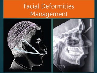
Facial Deformities Management: Orthognathic Surgery From Diagnosis to Treatment
- 2. Definition Surgical repositioning of maxilla and/or mandible, and/or their segments, with or without orthodontic treatment, in order to improve dentofacial function and aesthetics (in a stable manner) and health-related quality of life. The combined orthodontic-surgical correction of dentofacial deformities: Genioplasty? Treatment of sleep apnea?
- 3. Orthognathic Surgery It is a procedure in which correction of dentofacial deformities and malocclusion are corrected by orthodontic and surgery of facial skeleton. The term orthognathic originates from the Greek words orthos, ‘straight’, and gnathos ‘jaw’. It is possible to correct, or “straighten”, deformities separately in either the maxilla or the mandible with many types of surgical techniques or to do procedures concurrently on both jaws (bimaxillary operations, or Bimax). The treatment does not change only the bony relations of the facial structures, but soft tissues as well, and by doing so, may alter the patient’s appearance. Contemporary orthognathic techniques can be applied in many fields of surgery: apart from correcting congenital and posttraumatic malocclusions, they can be used in the treatment of the obstrucive sleep apnea syndrome (Riley et al. 1993), to improve phonetics (Vallino 1990) or even in tumour resections (Grime et al. 1991, Sailer et al. 1999).
- 4. Aetiology of Dentofacial Deformities Significant deviation from normal proportions of the maxillomandibular complex that also negatively affect the relation of the teeth within each arch and the relationship of the arches with one another (occlusion). Nature Vs Nurture Genetics: C II div 2 C III Vertical facial dimension Environmental: Functional Matrix Effect Multifactorial: combined interaction of the two. Genotype Vs Phenotype
- 5. Family Study & Twin Study Hapsburge Jaw: Genetically determined profile. Monozygotic twins: Sir Francis Galton in 1875. Familial cephalometric studies: Harris..C II div I Kloeppel…genetic s is the main aetiological factor in C II div 2 Schultze et al…strong familial occurrence
- 6. Classification of Dentofacial Deformities Not an easy task Primary descriptor: Primary morphological parameter of the deformity. The most obvious and severe. Secondary descriptor: Highly relevant additional morphological relationship.
- 8. Terminology for Orthognathic Surgical Procedures Maxillary Surgery Sagittal plane: Anterior repositioning or advancement. Posterior repositioning or setback (pushback). Vertical plane: Superior repositioning or impaction. Inferior repositioning or setdown (downgraft). Transverse plane: Bodily translation/ to left or right. Transverse expansion. Mandibular Surgery Sagittal plane: Advancement Setback Mandibular autorotation: Follows vertical movement of maxilla. Forward (anticlockwise or counterclockwise). Backward (clockwise)
- 9. Terminology for Orthognathic Surgical Procedures Chin Surgery (Osseous genioplasty): Advancement-Setback Vertical augmentation/ reduction Transverse (lateral) expansion/ reduction Asymmetrical
- 10. Prevalence of Dentofacial Deformities There is no exact data of dentofacial deformities among adult population. 1960 Profit and White: 10% class II malocclusion with 3% severe enough to warrant surgery (70% mandibular surgery). C.III malocclusion 0.6% to 21% Severe open bite 0.6% and 16% Recently Profit et al (1998) from (NHANES III in USA): 20% deviation from normal bite: 2% with disfiguring and at the limit of orthodontic capacity. Severe class II (>6mm over jet) 4.3% while class III (>-3 mm O.J.)
- 11. In Scandinavia 40 to 75% - or even higher- of malocclusion has been reported in children (Heikinheimo 1989, Permert et al. 1998), and 10% of young would be of difinte need for orthodontic treatment. In the Netherlands: 28% Angel class II 23% maxillary O.J. More than 5 mm 39% objective need for orthodontic treatment (Burgersdijk L et al 1991). In Finland: 510 000 Finns with skeletal class II, and 15 000 need surgery. 1 million Finns (20%) have deviation from normal bite with 2% (20 000) need surgery. These figures are seem high in terms of resources and cost.
- 12. History of Orthognathic Surgery Treatment of malocclusions (historically) has been aimed at correction of dental abnormalities, with little attention to the accompanying facial deformity. (Now it is malpractice!!!) In the last 60 years, surgical techniques were developed to position the whole midface, mandible and dentoalveolar segments into new desire positions. Combining of surgery and orthodontic treatment for dentofacial deformities correction has become an integral part of nowadays practice. Orthognathic surgery was originally developed in the United States of America (Steinhäuser 1996).
- 13. The early-phase surgery was mainly limited to the mandible, while maxillary procedures were to come later. The first mandibular osteotomy is considered to be Hullihen´s procedure in 1849 to correct a protrusive malposition of a mandibular alveolar segment caused by a burn (Hullihen 1849). Osteotomy of the mandibular body for the correction of prognathism was first carried out in 1897 as so called ´St Louis operation´. Vilray Blair: The 1st to classify the facial deformities 1st to underline the importance of orthodontic treatment in orthognathic surgery. History of Orthognathic Surgery 1. Mandibular Osteotomy
- 14. Causes of Dentofacial Deformities Multifactorial nature: Inherited tendencies Prenatal problems Systemic conditions that occur during facial growth Trauma Environmental influences An understanding of basic principles of facial growth as they relate to the development of dentofacial deformities is essential.
- 15. Basic Principles of Facial Growth Complex procedure influenced by variant factors. Area with their intrinsic growth potential: Sphenoethmoidal synchondroses Sphenooccipital synchondroses Nasal septum Majority of craniofacial bones grow on response to adjacent soft tissue and functional demands placed on these bones. nose, oral, hypopharyngeal airway, facial muscles and muscles of mastication.
- 16. General direction of the normal growth of the face is downward and forward with lateral expansion. Maxilla and mandible grow by remodeling or differential apposition and resorption of bone, leads to changes in three dimensions. Area relocation is the concept given by Enlow and Han’s to describe the maxillary-mandibular complex enlarging in the forward and downward direction and an “enlarging pyramid” Direction and amount of growth, characterize an individual’s growth pattern. Alterations in the pattern of growth or in the rate at which this growth occurs may result in abnormal skeletal morphology with accompanying malocclusion.
- 19. Genetic Influences Has a certain role in dentofacial deformities development. Patterns of inheritance are seen in a patient with a dentofacial deformity.(??? Multifactorial) Sometimes associated with congenital syndromes such as: Related to embryonic abnormality of neural crest: Hemifacial microsomia Mandibulofacial dysostosis (Treacher Collins syndrome) Cleft lip and palate Craniosynostosis (premature fusion of craniofacial sutures). Fetal alcohol syndrome (maternal systemic influence)
- 21. Environmental Influence In early prenatal stage molding of the developing fetal head may result in severe mandibular deficiency. Postnatal period: Abnormal function may result in altered facial growth because of soft tissue and muscular function often influence the position of teeth and growth of jaw. Abnormal tongue position or size. Respiratory difficulty Mouth breathing Abnormal tongue and lip posture Trauma: Direct effect Late effect
- 22. Evaluation of Patients with Dentofacial Deformity For best possible result and because the patient care is number one, integral approach or team approach should be practiced through out the treatment period. The most important phase is the evaluation of the existed problems and the treatment goals. Interview the patient to explore the intentions. Examination of facial structure with consideration of frontal and profile esthetics should be done thoroughly to: Evaluate the proportions of the face. Evaluate the symmetry of the face. Evaluation of the facial esthetics and balance.
- 24. Soft tissue of the throat should be evaluated. Photograph documentation is mandatory. A complete dental examination should include: A dental arch form Symmetry Tooth alignment Occlusal abnormalities in all 3D Masticatory muscles and TMJ should be evaluated. A screening periodontal examination. Impressions and bite registration. Lateral cephalometric and panorama radiographs are important in assessment phase.
- 25. Cephalometric analyses can be done by several technique to diagnose the exact problem and the cause of it. Sterelithic 3D model constructed from CT data. Computerized digital technology role. Now it is the time to fabricate the problem list and the treatment plan that combine opinions from all specialties providing the care.
- 26. Presurgical Treatment Phase Periodontal Treatment Phase Poor oral hygiene is a bad indicator. (Why?) Time of doing mucogingival surgery ex. Doing grafting of tissue that withstand better the trauma of orthodontic and surgical treatment. Restorative considerations: Full thorough examination Long term function of the restoration Final restoration till the end of surgical treatment and finishing time of orthodontic treatment.
- 27. Orthodontic Roles in Orthognathic Surgery When the case of malocclusion should go for surgery? When should we start the treatment and why? Favorable Vs. unfavorable growth. Orthognathic surgery should be delayed until growth is ceased in patients with growth excess, although can be done earlier for patients with growth deficiency. Can be simple teeth movement to 12-18 months of appliance therapy for severe crowding and incisor malposition. Retention is so important after finishing the orthodontic treatment, that can be done by using large stabilizing arch wire to withstand the forces resulting from IMF and surgical manipulation.
- 28. Decompensation of Compensation Movement
- 29. Presurgical Orthodontics Objectives ● Alignment of crowded arches ● Leveling of the curve of Spee ● Decompensation of compensated incisors ● Transverse arch co-ordination
- 30. Before surgery Crum-babble hooks should be placed in the upper and lower arch to be used for fixation
- 31. Final Treatment Planning Re-evaluation of the evaluation finished at the initial phase. Facial structures and malocclusion re-examination Presurgical photographs and radiographs are obtained. Pre-surgical models, a centric relation bite registration, and face bow recording are completed. Mock surgery is done to determine the exact surgical movement to accomplish the intended occlusion. Using computerized program to predict the facial profile after correction of the deformity: Better prediction of the result (+) Sharing the patient in treatment plan (+) Inability to predict every surgical technique for every patient(-)
- 32. Orthognathic Surgery Stages of treatment: 1- Examination and record taking 2- Treatment planning with Surgeon 3- Patient consent 4- Presurgical orthodontics 5- Surgical planning and model surgery 6- Orthognathic surgery 7- Postsurgical orthodontics 8- Retention
- 33. Evaluation of the Patient Clinical examination Radiographic examination Analysis of study models Psychological examination where appropriate
- 34. Clinical Examination Comfortably seated patient with Frankfort plane horizontal. Frontal assessment: Facial proportions: Three equal vertical components: Distance from hairline-to soft tissue bridge of the nose From soft tissue bridge of the nose to alar base From alar base to the chin, Determine whether or not there is a relative deficiency or excess in the vertical height of either maxillary or mandibular thirds.
- 36. The Alar Base Width This, as measured from the lateral aspects of alar cartilage of the nose, should equal the intercanthal distance as measured between the inner canthi of the eyes. Important when planning a maxillary impaction.
- 37. Incisor exposure (The lip-incisor relationship) The average upper lip length of 20-25 mm, 2-4 mm of incisor crown should be exposed. Increase the exposure with smiling to level of the gingival margin. This is crucial when planning the ultimate vertical height of the midface. This measurement should be done with face at rest. Sn to upper lip vermillion border should be a third of the total (half of the lower lip vermillion border to the menton).
- 38. Facial Asymmetry Important to note any asymmetry of the middle or lower third of the face. Marking the midline on patient’s face. Dental midline may not be coincident with skeletal ones. Deviation of maxillary midline than skeletal, there is an indication of ortho correction rather than surgery, while mandibular one in relation to upper midline, determine the cause. Surgery is necessary if asymmetry is skeletal.
- 40. Profile Assessment Relative protrusion of the maxilla and mandible Position of the infra-orbital margin Nasal morphology Morphology of the ears Chin depth Chin-throat angel
- 42. Temporomandibular Joint Examination There is no evidence of malocclusion or jaw deformity causing TMJ symptoms. In surgical patients, it is important to record any abnormalities. Path of opening and closure, clicking or crepitation and also the extent of maximum opening should be recorded.
- 43. Intraoral Examination Full examination with the study models and radiographs. Soft tissues General periodontal condition Tongue size, position and activity Mentalis muscle activity Finger or thumb sucking Hard tissue Dental assessment
- 44. Periodontal evaluation Adequate attached gingiva Maintain bone around the necks of each of the teeth at the interdental osteotomy sites
- 46. Dentition Vertical Overbite Plane of occlusion Curve of Spee Horizontal Anatomical variation Crowding / spacing Overjet Transverse Cross-bites
- 47. Cephalometric Analysis 1) Describe the subject’s dento-facial morphology 2) Quantitative description of morphological deviations 3) Make diagnostic and treatment planing decisions 2) Evaluate change over time - treatment induced and growth process. Evaluating relationships, both horizontal and vertical of 5 major functional components of the face: the cranial base; the maxilla; the mandible, the maxillary and mandibular dento-alveolus
- 48. Cephalometric Analysis Skeletal and dental relationships are measured by reference to a landmark or plane drawn on the lateral cephalogram. These can be either ‘ hand traced’ or more commonly now digitalized using specialized cephalometric software (e.g. QuickCeph (Mac), Dolphin Imaging (Windows)).
- 49. METHODS OF CEPHALOMETRIC ANALYSIS Metric approach - use of selected linear and angular measures: The analysis is usually given in tabular form with data expressed either as a linear measurement (in mm or a proportion (%)) or as an angle (degrees). The advantage of angular measurements is that they are not influenced by image magnification or patient size. Standard deviation for each measurement allows the clinician to easily see where their patient differs most significantly from the norm.
- 50. Graphic Approach An alternative presentation of normative data is to express it graphically in the form of a template. “Overlay” of individual’s tracing on a reference template and visual inspection of degree of variation. This is superimposed on the patient’ s cephalogram to see where the patient varies from the norm. An example is the Proportionate Template, which is useful in determining the degree of anteroposterior (AP) and vertical skeletal dysplasia present in adult patients.
- 51. Cephalometric Analyses Down’s(1948) Wylie(1947,1952) Rediel(1952) Steiner’s(1953) Tweed’s(1954) Sassouni(1955) Bjork (1961) Eastman(1970) Jaraback(1972) Harvold(1974) Wits(1975) Ricketts(1979) Pancherz(1982) McNamara’s(1983) Holdaway(soft tissue)1983 Bass(aesthetic)1991
- 52. ANATOMIC LANDMARKS Sella (S) Nasion (N) Glabella (G) Orbitale ( O ) Articulare (Ar) Pterygomaxillary fissure (PTM) Posterior nasal spine (PNS)
- 53. ANATOMIC LANDMARKS Anterior nasal spine (ANS) Menton (Me) Gnathion (Gn) Pogonion (Pg) Mandibular plane (MP)
- 54. Cepahlo. Tracing
- 55. Limitations of Cephalometric Analysis Individual variability Ethnic variability Gender variability
- 56. Maxillary retrusion Mandibular retrusion Clockwise mandibular rotation Upper and lower denture base retrusion Overjet increase Findings of Cephalometric Analysis
- 57. Model Surgery Requirement 1- Upper and lower impressions. 2- Wax bite 3- Face bow registration 4- Semi adjustable articulator
- 58. Model Surgery Curtsey of Dr Yanal Nusair and Dr Bader Burgan
- 59. Model Surgery Curtsey of Dr Yanal Nusair and Dr Bader Burgan Curtsey of Dr Yanal Nusair and Dr Bader Burgan
- 60. Model Surgery Curtsey of Dr Yanal Nusair and Dr Bader Burgan
- 61. Model Surgery Curtsey of Dr Yanal Nusair and Dr Bader Burgan
- 62. Surgical Splints (wafers) Curtsey of Dr Yanal Nusair and Dr Bader Burgan
- 63. Mock Surgery
- 64. Surgical Techniques Single jaw Bimaxillary surgery
- 65. Surgical Treatment Phase Mandibular Excess C III molar and canine relation and reverse O.J. Obvious facial deformity associated with mandibular prominence including lower lip and chin in AP and Vertical dimensions. Incompetent lips (but with abnormal strain of O.O.) One of 1st dentofacial deformities recognized to be treated with both ortho and surgery. Removing section of the body and moving the anterior segment posteriorly. Sup-apical osteotomy of anterior mandible if reverse O.J. is isolated with normal molar. Rarely used
- 67. Vertical Ramus Osteotomy Caldwell and Letterman (1950s): Extraoral approach Condylar seg. overlap the dental segment. Can be done via intraoral approach with angulated oscillating saw: Elimination the need for submandibular incision Reduce the risk of damaging the marginal mandibular nerve.
- 69. Post-Operative
- 70. Pre-Operative Ceph. Post-Operative Ceph.
- 71. B.S.S.O. First described by Trauner and Obwegeser and later modified by Daplont, Hunsick and Epker. Osteotomy split the ramus and the posterior part of mandibular body in a sagittal fashion. Telescoping effect allows moving the mandible in multiple directions. Treating both deficiency as well as excess. Demerits: Risk of ID damage. Segment fracture.
- 72. Mandibular Deficiency The most obvious clinical feature is retruded position of the chin on lateral profile. Excess labiomental fold with a procumbent lower lip. Abnormal posture of upper lip. Poor throat form. Intraorally is associated with class II molar and canine relationship and increased O.J. in incisor area.
- 73. Surgical Correction As early as 1909 with disappointing results till 1950s. 1957 Robinson used extraoral approach for correction by doing vertical osteotomy with iliac crest bone graft in osteotomy site: Several modifications done Rarely used in cases with severe abnormal anatomy or for revision surgery. Demerits: Facial scar Damaging the facial nerve branches
- 74. B.S.S.O. with Advancement the mandible Currently most commonly used technique. Intraoral incision with modified 3rd molar incision. Overlapping of the condylar segment (proximal segment) and dental segment (distal segment) allows for better healing and stability. Rigid fixation is an advantage to eliminate the need for IMF. If chin position in AP dimension is good, a total subapical osteotomy may be good for advancing the mandible to correct CII relationship. Reduced lower facial height can be increased by interpositional bone graft in the osteotomy site.
- 78. Genioplasty To correct the position and projection of the chin. Intraoral incision. Anterior, posterior, vertical reduction or augmentation or correction of asymmetry can be done. Alloplastic materials can be used for augmentation the deficient chin.
- 80. Maxillary Excess In AP, vertical and transverse dimensions. 1970 total single jaw surgery become popular: Segmental surgery Two stage surgery Bell et al, 1970: Total maxillary surgery in one stage Most common procedure for correction of maxillary deformity in all 3D.
- 81. Vertical Maxillary Excess Characteristic facial appearance: Elongation of the lower facial third Narrow nose (especially in base) Excessive gingiva and incisor show (gummy smile) Lip incompetent May exhibit C I, II or III dental malocclusion. Usually associated with anterior open bite. Treatment: Maxillary impaction (total or segmental osteotomy)
- 84. Anterior-Posterior Maxillary Excess Convex facial profile: C II div I Corrected by total maxillary surgery (Single jaw) Segmental osteotomy is the procedure of choice (Why?)
- 87. Maxillary and Midface Deficiency Occurs in anterioposterior, vertical or transverse D. Patient’s facial appearance depends on the location and severity of deformity. Retruded upper lip Deficiency of paranasal and infraorbital rim areas Inadequate tooth exposure during smile Prominent chin (relative to middle third of the face) C III with reversed overjet.
- 91. Correction Le Fort I osteotomy with down fracture of maxilla can be used to correct this deformity. Depending on the degree of advancement: Bone graft used to: Enhance the healing Improve the postoperative stability Vertical deficiency can be managed with downward placement of maxilla (with or without bone graft) to elongate the lower third of the face: Improves the overall facial proportion. Normalizes exposure of the incisors during smile. Proper diagnosis is important as most blame the lower jaw in C III skeletal relation while the cause is deficient maxilla.
- 92. Midface Deficiency Zygomatic bone and infra-orbital rim deficiency a Le Fort III is necessary for correction. Apert ‘s syndrom Crouzon’s syndrom
- 94. Transverse Maxillary Deficiency Narrow arch Posterior cross-bite Constricted palate
- 95. Bimaxillary Surgery To treat multiple deformities in both jaws. Enhance the stability by dividing the amount of single jaw movement over both jaws, to achieve the best occlusal, functional and esthetic results. Facial asymmetry in more than two spaces is one of the most challenging cases for correction.
- 109. Orthognathic Surgery and Obstructed Sleep Apnea Cessation of airflow for more than 10 seconds leads to occurring of apneic events during sleeping, leading to: Sleep disturbances/ deprivation Daytime somnolence Severe hypoxia during sleep with potential risk of respiratory and cardiac problems or even death. Collapsed airway during sleep due to decreased muscle tone on palate, tongue or pharyngeal musculature that is accentuated during supine position. Alcohol, obesity and sedative drugs during sleep aggravate the problem.
- 110. O.S.A. Diagnosis based on: A comperhensive physical evaluation. Nasophryngoscopy Dentofacial evaluation Polysomnography sleep study.
- 111. Treatment Nonsurgical Measure Weight loss Positional changes during sleep Jaw positioning devices Continuous positive airway pressure (CPAP): Using mouth or nasal mask during sleep. Surgical Correction Uvulopalatoplaty (UPP) Uvulupharyngeoplasty (UPP) Both (UPPP) Maxillary-mandibular advancement surgery.