
Recommended
More Related Content
What's hot
What's hot (20)
Similar to Management of spontaneous intracerebral hemorrhage
Similar to Management of spontaneous intracerebral hemorrhage (20)
More from Ahmed Mohamed
Recently uploaded
Recently uploaded (20)
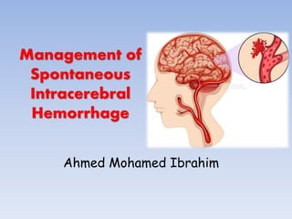
Management of spontaneous intracerebral hemorrhage
- 2. • Spontaneous, non-traumatic intra-cerebral hemorrhage (ICH) remains a significant cause of morbidity and mortality throughout the world.
- 3. Causes CAUSEs PIMARY MEANS OF DIAGNOSIS CHARACTERISTICS Hypertension Clinical history Rupture of small arterioles related to degenerative changes induced by uncontrolled hypertension Amyloid angiopathy Clinical history Rupture of small and medium-sized arteries, with deposition of b-amyloid protein; presents as lobar hemorrhages in persons older than 70 years of age Arteriovenous malformation Imaging studies such as magnetic resonance imaging and conventional angiography Rupture of abnormal small vessels connecting arteries and veins Intracranial aneurysm Imaging studies such as magnetic resonance angiography and conventional angiography Rupture of saccular dilatation from a medium- sized artery that is usually associated with subarachnoid hemorrhage
- 4. Causes CAUSEs PIMARY MEANS OF DIAGNOSIS CHARACTERISTICS Cavernous angioma Imaging studies such as magnetic resonance imaging Rupture of abnormal capillary-like vessels with intermingled connective tissue Venous angioma Imaging studies such as magnetic resonance imaging and conventional angiography Rupture of abnormal dilatation of venules Dural venous sinus thrombosis Imaging studies such as magnetic resonance venography and conventional angiography Result of hemorrhagic venous infarction; anticoagulation and, in rare cases, transvenous thrombolytic agents can improve outcome Intracranial neoplasm Imaging studies such as magnetic resonance imaging Results of necrosis and bleeding within hypervascular neoplasms; long-term outcome determined by the characteristics of the underlying neoplasm
- 5. Causes CAUSES PIMARY MEANS OF DIAGNOSIS CHARACTERISTICS Coagulopathy Clinical history Most commonly associated with use of anticoagulants or thrombolytic agents; rapid correction of underlying abnormality important to avert continued bleeding Vasculitis Measurement of serologic and cerebrospinal fluid markers; brain biopsy Rupture of small or medium-sized arteries with inflammation and degeneration; immunosuppressive medications may be indicated Cocaine or alcohol use Clinical history Underlying vascular abnormalities may be present Hemorrhagic ischemic stroke Imaging studies such as magnetic resonance imaging and conventional angiography Hemorrhage in region of cerebral infarction as a result of ischemic damage to blood–brain barrier
- 6. Most Common Sites and Sources of Intracerebral Hemorrhage. • Intracerebral hemorrhages most commonly involve cerebral lobes, originating from penetrating cortical branches of the anterior, middle, or posterior cerebral arteries (A); • basal ganglia, originating from ascending lenticulostriate branches of the middle cerebral artery (B); • the thalamus, originating from ascending thalmogeniculate branches of the posterior cerebral artery (C); t • he pons, originating from paramedian branches of the basilar artery (D); • and the cerebellum, originating from penetrating branches of the posterior inferior, anterior inferior, or superior cerebellar arteries (E).
- 7. Emergency Diagnosis and Assessment • ICH is a medical emergency. • Rapid diagnosis and attentive management of patients with ICH is crucial, because early deterioration is common in the first few hours after ICH onset.
- 8. Pre-hospital Management • The primary objective is to provide airway management if needed, provide cardiovascular support, and transport the patient to the closest facility prepared to care for patients with acute stroke • Secondary priorities for EMS providers 1- Obtaining a focused history regarding the timing of symptom onset (or the time the patient was last normal); information about medical history, medication, and drug use; and contact information for family. 2- Provide advance notice to the ED of the impending arrival of a potential stroke patient so that critical pathways can be initiated and consulting services alerted. Advance notice by EMS has been demonstrated to significantly shorten time to computed tomography (CT) scanning in the ED
- 9. Emergency Diagnosis and Assessment • The crucial resources necessary to manage patients with ICH include neurology, neuroradiology, neurosurgery, and critical care facilities that include adequately trained nurses and physicians. • Consultation via telemedicine can be a valuable tool for hospitals without on-site presence of consultants.
- 10. Emergency Diagnosis and Assessment • A baseline severity score should be performed as part of the initial evaluation of patients with ICH (Class I; Level of Evidence B). • Rapid neuroimaging with CT or MRI is recommended to distinguish ischemic stroke from ICH (Class I; Level of Evidence A).
- 11. Assessment
- 12. Assessment
- 13. Assessment
- 14. Neuroimaging • The abrupt onset of focal neurological symptoms is presumed to be vascular in origin until proven otherwise. • However, it is impossible to know whether symptoms are caused by ischemia or hemorrhage on the basis of clinical characteristics alone. Vomiting, systolic BP (SBP) >220 mm Hg, severe headache, coma or decreased level of consciousness, and symptom progression over minutes or hours all suggest ICH, although none of these findings are specific; neuroimaging is thus mandatory.
- 15. Neuroimaging • CT and magnetic resonance imaging (MRI) are both reasonable for initial evaluation. • CT is very sensitive for identifying acute hemorrhage and is considered the “gold standard”; gradient echo and T2* susceptibility- weighted MRI are as sensitive as CT for detection of acute hemorrhage and are more sensitive for identification of prior hemorrhage. • Time, cost, proximity to the ED, patient tolerance, clinical status, and MRI availability may, however, preclude emergent MRI in many cases
- 16. Neuroimaging • The high rate of early neurological deterioration after ICH is related in part to active bleeding that may proceed for hours after symptom onset → Hematoma expansion → increases risk of poor functional outcome and death. • CT angiography (CTA) and contrast-enhanced CT may identify patients at high risk of ICH expansion based on the presence of contrast within the hematoma, often termed a spot sign. • A larger number of contrast spots suggests even higher risk of expansion.
- 17. Neuroimaging • Early diagnosis of underlying vascular abnormalities can both influence clinical management and guide prognosis in ICH patients. • Risk factors for underlying vascular abnormalities are: age <65 years, female sex, nonsmoker, lobar ICH, intraventricular extension, and absence of a history of hypertension or coagulopathy.
- 18. Neuroimaging • MRI, magnetic resonance angiography, magnetic resonance venography, and CTA or CT venography can identify specific causes of hemorrhage, including arteriovenous malformations, tumors, moyamoya, and cerebral vein thrombosis. • A catheter angiogram may be considered if clinical suspicion is high or noninvasive studies are suggestive of an underlying lesion
- 19. Neuroimaging • Radiological evidence suggestive of vascular abnormalities as causative for ICH can include: 1) Presence of subarachnoid hemorrhage 2) Enlarged vessels or calcifications along the margins of the ICH 3) Hyperattenuation within a dural venous sinus or cortical vein along the presumed venous drainage path 4) Unusual hematoma shape 5) Presence of edema out of proportion to the time of presumed ICH 6) An unusual hemorrhage location 7) Presence of other abnormal structures in the brain (like a mass).
- 20. BP: Recommendations • For ICH patients presenting with SBP between 150 and 220 mm Hg and without contraindication to acute BP treatment, acute lowering of SBP to 140 mm Hg is safe (Class I; Level of Evidence A) and can be effective for improving functional outcome (Class IIa; Level of Evidence B). • For ICH patients presenting with SBP >220 mm Hg, it may be reasonable to consider aggressive reduction of BP with a continuous intravenous infusion and frequent BP monitoring (Class IIb; Level of Evidence C).
- 21. BP: Recommendations • NICE Giudelines
- 22. Seizures and Antiseizure Drugs • Clinical seizures should be treated with antiseizure drugs (Class I; Level of Evidence A). • Patients with a change in mental status who are found to have electrographic seizures on EEG should be treated with antiseizure drugs (Class I; Level of Evidence C). • Continuous EEG monitoring is probably indicated in ICH patients with depressed mental status that is out of proportion to the degree of brain injury (Class IIa; Level of Evidence C). • Prophylactic antiseizure medication is not recommended (Class III; Level of Evidence B).
- 23. ICP Monitoring and Treatment • Ventricular drainage as treatment for hydrocephalus is reasonable, especially in patients with decreased level of consciousness (Class IIa; Level of Evidence B). • Patients with a GCS score of ≤8, those with clinical evidence of transtentorial herniation, or those with significant IVH or hydrocephalus might be considered for ICP monitoring and treatment. A CPP of 50 to 70 mm Hg may be reasonable to maintain depending on the status of cerebral autoregulation (Class IIb; Level of Evidence C). • Corticosteroids should not be administered for treatment of elevated ICP in ICH (Class III; Level of Evidence B). because they are not effective in ICH and increase complications.
- 24. Intra-ventricular Hemorrhage • IVH occurs in ≈45% of patients with spontaneous ICH and is an independent factor associated with poor outcome • Most IVH is secondary and related to hypertensive hemorrhages involving the basal ganglia and thalamus. • Animal studies and clinical series have reported that intraventricular administration of fibrinolytic agents, including urokinase, streptokinase, and recombinant tissue-type plasminogen activator (rtPA), in IVH may reduce morbidity and mortality by accelerating blood clearance and clot lysis.
- 25. Intra-ventricular Hemorrhage • Although intraventricular administration of rtPA in IVH appears to have a fairly low complication rate, the efficacy and safety of this treatment are uncertain (Class IIb; Level of Evidence B). • The efficacy of endoscopic treatment of IVH is uncertain (Class IIb; Level of Evidence B).
- 26. Surgical Treatment Surgical Treatment of ICH (Clot Removal) • The role of surgery for most patients with spontaneous ICH remains controversial. The theoretical rationale for hematoma evacuation revolves around the concepts of preventing herniation, reducing ICP, and decreasing the pathophysiological impact of the hematoma on surrounding tissue by decreasing mass effect or the cellular toxicity of blood products. • Randomized trials comparing surgery to conservative management have not demonstrated a clear benefit for surgical intervention. Moreover, the generalizability of the results of these trials can be questioned, because patients at risk for herniation were likely excluded and the largest and most recent studies had high rates of treatment group crossover from conservative management to surgery.
- 27. Surgical Treatment Craniotomy for Supratentorial Hemorrhage • 2 largest randomized trials STICH and STICH II trials focused on early surgery (<24 hours of randomization) to study role of surgery versus initial conservative treatment • early hematoma evacuation has not been shown to be beneficial. *STICH: Surgical Trial for Intracerebral Haemorrhage
- 28. Surgical Treatment Craniotomy for Posterior Fossa Hemorrhage • Because of the narrow confines of the posterior fossa, deterioration can occur quickly in cerebellar hemorrhage caused by obstructive hydrocephalus or local mass effect on the brainstem. • Several nonrandomized studies have suggested that patients with cerebellar hemorrhages >3 cm in diameter or patients in whom cerebellar hemorrhage is associated with brainstem compression or hydrocephalus have better outcomes with surgical decompression. • Attempting to control ICP via means other than hematoma evacuation, such as ventricular catheter (VC) insertion alone, is considered insufficient, is not recommended, and may actually be harmful, particularly in patients with compressed cisterns
- 29. Surgical Treatment • Patients with cerebellar hemorrhage who are deteriorating neurologically or who have brainstem compression and/or hydrocephalus from ventricular obstruction should undergo surgical removal of the hemorrhage as soon as possible (Class I; Level of Evidence B). • Initial treatment of these patients with ventricular drainage rather than surgical evacuation is not recommended (Class III; Level of Evidence C).
- 30. Surgical Treatment • For most patients with supratentorial ICH, the usefulness of surgery is not well established (Class IIb; Level of Evidence A). • A policy of early hematoma evacuation is not clearly beneficial compared with hematoma evacuation when patients deteriorate (Class IIb; Level of Evidence A). • Supratentorial hematoma evacuation in deteriorating patients might be considered as a life-saving measure (Class IIb; Level of Evidence C).
- 31. Surgical Treatment • DC with or without hematoma evacuation might reduce mortality for patients with supratentorial ICH who are in a coma, have large hematomas with significant midline shift, or have elevated ICP refractory to medical management (Class IIb; Level of Evidence C). • The effectiveness of minimally invasive clot evacuation with stereotactic or endoscopic aspiration with or without thrombolytic usage is uncertain (Class IIb; Level of Evidence B).
- 32. Cases
- 33. ARTERIOVENOUS MALFORMATION (AVM) • Is a tangle of abnormal blood vessels connecting arteries and veins in the brain.
- 34. ARTERIOVENOUS MALFORMATION (AVM) CLINICAL PICTURE 1. HEADACHE 2. SEIZURES 3. FOCAL NEUROLOGICAL DEFECIT 4. LEARNING DISABILITIES AND COGNITIVE DYSFUNCTION 5. HEMORRHAGE
- 35. Imaging • CT/CTA • MRI • DSA: Angiography remains the gold standard for the evaluation of AVMs.
- 37. MODALITIES OF TREATMENT • ENDOVASCULAR • SURGERY • RADIOSURGERY
- 38. Endovascular
- 39. Radiosurgery
- 40. Case 1 • AS, Male patient, 32 years old presented by sudden onset severe headache. • ED: Blood pressure was normal, neck pain
- 41. • CT brain was done: 4th ventricle hemorrhage. • CTA: free Case 1
- 42. • DSA: Cerebellar AVM supplied by left SCA & left PICA + Venous drainage via straight sinus and left transverse sinus Case 1
- 43. Cerebellar AVM supplied by left SCA & left PICA + Venous drainage via straight sinus and left transverse sinus Case 1
- 44. • Plan of Care: AVM embolization with Onyx Case 1
- 45. Case 2 • AM, Male Patient 20 years old , Presented with Rt eye lid swelling. MRI : Brain + Orbit
- 46. DSA through ICA In ICA, the visible here is the Ophthalmic Artery
- 48. Angiography thru ECA • The AVM
- 50. TTT Plan • The patient will have AVM embolization with Onyx then surgically removed with plastic surgery to the eye lid after that. Before After
- 51. Intracranial Aneurysms • Clinical Picture - SAH - Compression
- 52. Hunt and hess scale
- 55. Case 3 • AA, 70 years old, male patient, hypertensive presented by sudden sever headache. • Brain CT:
- 56. • CTA:
- 57. • DSA:
- 60. Case 4 • WM, Female patient 33 years old, with no previous history of chronic illness. • Presented by Sudden falling attack with disturbed conscious level, then she was taken to the ER but she was diagnosed as a conversion case as she regained consciousness but with severe headache an difficluty in moving her head. • 1 week later: She developed acute onset Left sided weakness
- 62. • The patient was prescribed to Xalerto • Multiple CT Brain was done.
- 63. • But there was hyper-dense lesion
- 64. However the Radiologist asked for more verification of that lesion, her neurologist told her that it is not necessary
- 65. • CTA
- 67. • Till its total Occlusion
- 68. Case 5 • Female patient 57 years old, not diabetic or hypertensive presented by sudden severe headache of 3 days duration.
- 69. • CT Brain
- 70. CTA
- 71. Before Coiling After Coiling