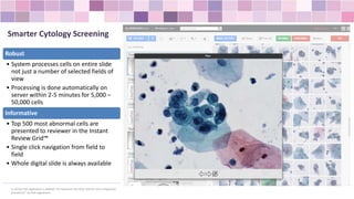
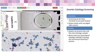
SMART IMAGING TECHNOLOGIES web-pathology.net
Smart Cytology Screening
Gyn and Non-Gyn Appliations
Smarter Cytology Screening
Robust
• System processes cells on entire slide
not just a number of selected fields of
view
• Processing is done automatically on
server within 2-5 minutes for 5,000 –
50,000 cells
Informative
• Top 500 most abnormal cells are
presented to reviewer in the Instant
Review Grid™
• Single click navigation from field to
field
• Whole digital slide is always available
In US this IVD application is labeled "For Research Use Only. Not for use in diagnostic
procedures“ by FDA regulations.
Smarter Cytology Screening
Convenient
• Screening can be done
remotely via any web browser
• System works with any digital
slide scanner
Versatile
• System can process Gyn and
Non-Gyn Cytology samples
• Works with any Liquid-Based
Cytology methods including
the economical ClearPrep™
process
How it works
New slides are processed automatically
on server. 2-5 minutes per slide
depending on cell density
HIPPA-compliant notification is routed to
a user when analysis is complete and
slide is ready for review
User quickly reviews abnormal cells with
Instant View Grid™ and completes
diagnosis in a few clicks
Smart Cytology™ screening application processes new slides
automatically in the background and sends notification to the user
when analysis is complete
1
2
3
Application Setup
Validation and training of Smart IHC™ scoring application is quick and
easy. System is trained automatically from slides with known diagnosis
• Upload slides with known diagnosis
• 10 slides per biomarker is usually
sufficient
1
• Annotate patterns with known score on some
slides for training
• Keep other slides without annotations (for
validation)
2
3
• Validate that application scoring is correct for
annotated areas
• Validate application scoring for non-annotated slides
• Keep validation sides, records and reports
In US Smart Cytology IVD application is labeled for "For Research Use Only. Not for
use in diagnostic procedures“ by FDA regulation.
Analyzing Cytology Slides with
Machine Learning Methods
Machine Learning algorithms learn to
recognize images and patterns the same way
humans do – by example, rather than by
human-derived “handcrafted features” such
as shape, size, brightness etc.
Adding Machine Learning methods to image
analysis adds a number of benefits:
• No need to formalize complex
“handcrafted features”, cytologist can just
point to patterns they need to recognize
• No dependency on image analysis
engineers (almost).
• System can be trained on variety of
samples to achieve robust recognition
• New data samples can easily be added to
a model to increase accuracy
The Science
© 2015 Smart Imaging Technologies. Co.
Web-pathology.net sales@simagis.us

Using Artificial Intelligence For Cytology Screening

  • 1.
    SMART IMAGING TECHNOLOGIESweb-pathology.net Smart Cytology Screening Gyn and Non-Gyn Appliations
  • 2.
    Smarter Cytology Screening Robust •System processes cells on entire slide not just a number of selected fields of view • Processing is done automatically on server within 2-5 minutes for 5,000 – 50,000 cells Informative • Top 500 most abnormal cells are presented to reviewer in the Instant Review Grid™ • Single click navigation from field to field • Whole digital slide is always available In US this IVD application is labeled "For Research Use Only. Not for use in diagnostic procedures“ by FDA regulations.
  • 3.
    Smarter Cytology Screening Convenient •Screening can be done remotely via any web browser • System works with any digital slide scanner Versatile • System can process Gyn and Non-Gyn Cytology samples • Works with any Liquid-Based Cytology methods including the economical ClearPrep™ process
  • 4.
    How it works Newslides are processed automatically on server. 2-5 minutes per slide depending on cell density HIPPA-compliant notification is routed to a user when analysis is complete and slide is ready for review User quickly reviews abnormal cells with Instant View Grid™ and completes diagnosis in a few clicks Smart Cytology™ screening application processes new slides automatically in the background and sends notification to the user when analysis is complete 1 2 3
  • 5.
    Application Setup Validation andtraining of Smart IHC™ scoring application is quick and easy. System is trained automatically from slides with known diagnosis • Upload slides with known diagnosis • 10 slides per biomarker is usually sufficient 1 • Annotate patterns with known score on some slides for training • Keep other slides without annotations (for validation) 2 3 • Validate that application scoring is correct for annotated areas • Validate application scoring for non-annotated slides • Keep validation sides, records and reports In US Smart Cytology IVD application is labeled for "For Research Use Only. Not for use in diagnostic procedures“ by FDA regulation.
  • 6.
    Analyzing Cytology Slideswith Machine Learning Methods Machine Learning algorithms learn to recognize images and patterns the same way humans do – by example, rather than by human-derived “handcrafted features” such as shape, size, brightness etc. Adding Machine Learning methods to image analysis adds a number of benefits: • No need to formalize complex “handcrafted features”, cytologist can just point to patterns they need to recognize • No dependency on image analysis engineers (almost). • System can be trained on variety of samples to achieve robust recognition • New data samples can easily be added to a model to increase accuracy The Science © 2015 Smart Imaging Technologies. Co. Web-pathology.net sales@simagis.us