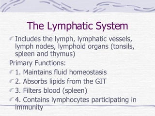
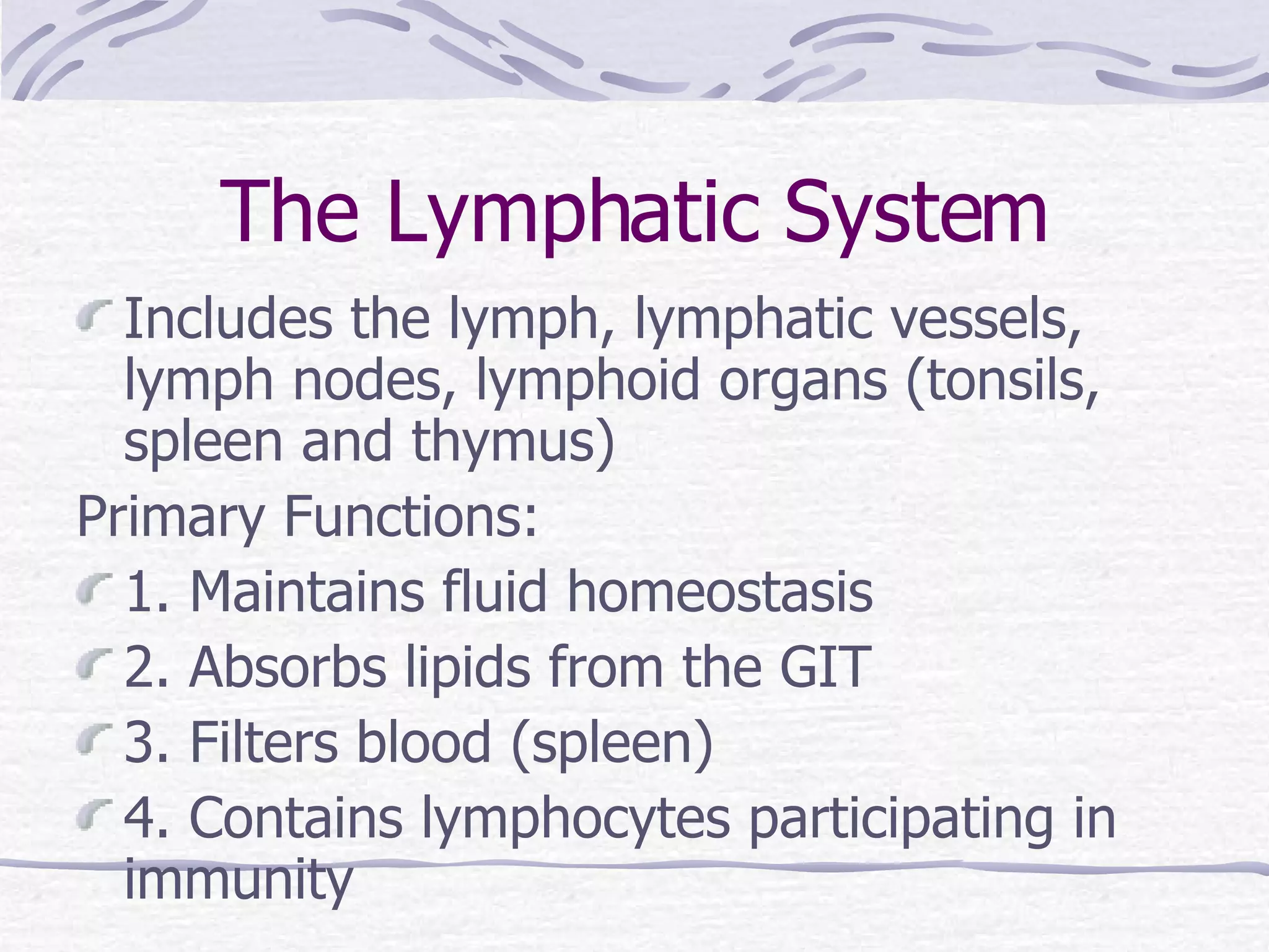
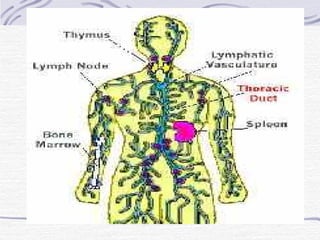
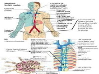
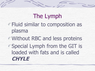
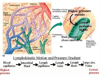
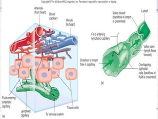
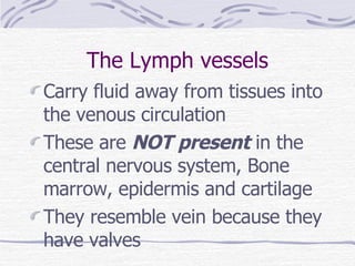
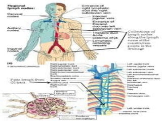
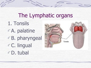
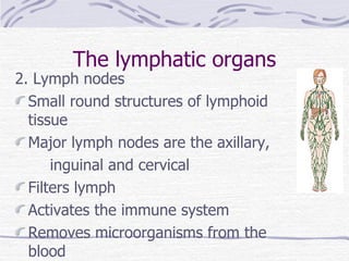
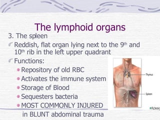
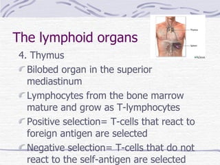
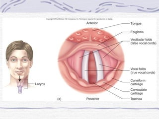
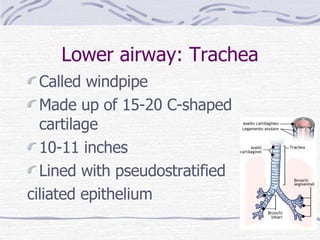
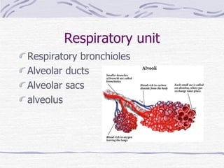
The Lymphatic System Includes the lymph, lymphatic vessels, lymph nodes, lymphoid organs (tonsils, spleen and thymus) Primary Functions: 1. Maintains fluid homeostasis 2. Absorbs lipids from the GIT 3. Filters blood (spleen) 4. Contains lymphocytes participating in immunity
 
 
The Lymph Fluid similar to composition as plasma Without RBC and less proteins Special Lymph from the GIT is loaded with fats and is called  CHYLE
 
Fig. 14.2
The Lymph vessels Carry fluid away from tissues into the venous circulation These are  NOT present  in the central nervous system, Bone marrow, epidermis and cartilage They resemble vein because they have valves
3 mechanisms of edema formation 1. Decrease oncotic pressure   cirrhosis and kwashiorkor 2. Increased hydrostatic pressure   CHF and prolonged standing 3. Lymphatic obstruction or destruction   filiariasis, lymph node dissection
Lymphatic duct 1. Right lymphatic duct- drains the right side of the head and the upper right side of the thorax and the right extremities 2. Thoracic duct- drains the rest of the body
 
The Lymphatic organs 1. Tonsils A. palatine B. pharyngeal C. lingual D. tubal
The lymphatic organs 2. Lymph nodes Small round structures of lymphoid tissue Major lymph nodes are the axillary, inguinal and cervical Filters lymph Activates the immune system Removes microorganisms from the blood
The lymphoid organs 3. The spleen Reddish, flat organ lying next to the 9 th  and 10 th  rib in the left upper quadrant Functions: Repository of old RBC Activates the immune system Storage of Blood Sequesters bacteria MOST COMMONLY INJURED  in BLUNT abdominal trauma
The lymphoid organs 4. Thymus Bilobed organ in the superior mediastinum Lymphocytes from the bone marrow mature and grow as T-lymphocytes Positive selection= T-cells that react to foreign antigen are selected Negative selection= T-cells that do not react to the self-antigen are selected
 
IMMUNITY Innate or Non-specific 1. Mechanical- skin, mucus, saliva, urine 2. Chemical- enzymes, lysozymes 3. Vascular Blood cells- Neutrophils and macrophages 4. Inflammation Specific Immunity 1. Cellular Immunity- T-cell system 2. Humoral Immunity- B-cell system
Non-specific Immunity Inflammatory response Reaction of the vacularized living tissues to injury Classic signs: 1. Rubor- redness 2. Calor- heat 3. Tumor- swelling 4. Dolor- pain 5. Functio laesa- loss of function
Inflammation Initial reaction   Vasoconstriction! VC   VD   VP (due to HISTAMINE) VD   Redness and Heat VP   Swelling Bradykinin, Prostaglandin, compression of tissues   pain
Specific Immunity: T cell T-cells originate in the bone marrow and mature in the thymus 4 types of T-cells 1. Cytotoxic T cells- kill infected cells, cancer cells and transplanted cells 2. Helper T cells- help the humoral immunity 3. Suppressor T cells- suppress that actions of cytotoxic and Helper cells 4. Memory T cells- for recall and specificity
Specific Immunity: B cells B cells are produced and mature in the bone marrow B cells   helped by Helper cells   turn into Plasma cells   secrete ANTIBODIES
ANTIBODIES Are proteins that can combine with antigens and function for: 1. Opsonization 2. Neutralization 3. Activation of complement system
ANTIBODIES types Five classes 1. Ig M- priMary response, pentaMer 2. Ig G- secondary response, most abundant, can cross placenta 3. Ig A- secreted by the body (sIgA) 4. Ig D- receptor for B cells 5. Ig E- allergic and parasitic reactions
TYPES OF IMMUNITY 1. Natural Immunity Active natural Passive natural 2. Artificial Immunity Active artificial Passive artificial
Fig. 14.18
End of Immune System
THE RESPIRATORY SYSTEM
The Respiratory System Composed of the air conducting system and the respiratory unit (lungs) Major function is RESPIRATION Commonly divided into UPPER RESPIRATORY tract and LOWER RESPIRATORY tract
Ventilation and Respiration VENTILATION- movement of air from the atmosphere into the lungs and out of the lungs EXTERNAL RESPIRATION- exchange of gases between the alveoli and the blood INTERNAL RESPIRATION- exchange of gases between the blood and the tissues
The Respiratory System Commonly divided into  UPPER RESPIRATORY tract and LOWER RESPIRATORY tract
The UPPER Respiratory tract The nose, pharynx and larynx
The LOWER respiratory tract Composed of The trachea down to the Lungs
The NOSE Made up of nasal bones and cartilages The NASAL cavity is made up of bones, cartilages and turbinates or conchae The nostril is the external opening The choanae is the internal opening
Functions of the nose 1 . Hairs or vibrissae filter large particles 2. Blood vessels warm the air 3. Mucus serves to humidify the air 4. Phonation
The Pharynx Musculo-membranous tube from behind the nasal cavity to the level of the cricoid cartilage (C6)
The Pharynx 3 component parts 1. Nasopharynx 2. Oropharynx 3. Laryngopharynx
The Larynx Upper expanded portion of the trachea Made up of cartilages 3 Unpaired- Cricoid, Thyroid and Epiglottis ( cUte) 6 Paired- Cuneiform, Corniculate and Arytenoid (pACC) Function: air passageway and phonation
 
The vocal cords 1.  False vocal cords 2. True vocal cords 3. Glottis- the space between the true vocal cords and is the narrowest portion of the adult airway
Fig. 15.4
Lower airway: Trachea Called windpipe Made up of 15-20 C-shaped cartilage 10-11 inches Lined with pseudostratified  ciliated epithelium
Bronchus RIGHT Bronchus Wider Shorter More vertical Left bronchus Narrower Longer More horizontal
Bronchioles Primary bronchus  secondary bronchus  tertiary bronchus   terminal bronchioles Respiratory bronchioles belong to the respiratory unit
Respiratory unit Respiratory bronchioles Alveolar ducts Alveolar sacs alveolus
The Pleura Surrounds the lungs and provide protection 1. Parietal pleura- in the chest wall 2. Visceral pleura- intimately attached to the lungs 3. Pleural space- in between the two
 
Diaphragm
Respiratory Physiology 1. Ventilation and gas exchange 2. Mechanics of breathing 3. Gas transport 4. Pulmonary volumes and capacities 5. Respiratory control
Ventilation Ventilation is the movement of air into the lungs Air (oxygen) moves by the process of diffusion from the higher concentration in the alveoli to the pulmonary capillaries
Mechanics of breathing
Gas exchange Respiratory membrane is thin which facilitates gas exchange Rate of diffusion depends on the thickness of the membrane, surface area and partial pressure of gases
Gas transport 1. OXYGEN- majority is transported in the blood loosely bound to hemoglobin- oxyhemoglobin 2. CARBON DIOXIDE- majority is transported in the blood in the RBC as BICARBONATE
Fig. 15.8
LUNG VOLUMES 1. Tidal volume – TV 2. Inspiratory Reserve Volume- IRV 3. Expiratory Reserve Volume- ERV 4. Residual volume- RV
LUNG CAPACITIES Lung volume + another lung volume 1. Inspiratory Capacity- IC 2. Functional Residual Capacity- FRC 3. Vital capacity- VC 4. Total Lung capacity- TLC
 
 
NOT CLINICALLY measured 1. Residual volume 2. Functional residual volume 3. Total lung capacity
Control of Respiration: Central Respiratory center in  the medulla Controls the rate and depth of respiration Increased CO2 is the most potent stimulus
Control of Respiration: Peripheral 1. Chemoreceptors in the carotid and aortic bodies Sensitive to changes in pH and O2 Decreased O2   increase respiration Decreased pH (acidosis)   increase respiration 2. Herring-Breurer reflex Stretch receptors in the lungs limit the inspiration
ASSESSMENT Normal Breathing pattern: 12-21 respiratory rate Active inspiration with contraction of diaphragm Passive expiration with relaxation of diaphragm  Steady rhythm and regular rate and size I:E ratio is 1:2
Abnormal breathing pattern 1. Cheyne-Stoke’s 2. Biot’s 3. Kussmaul’s 4. Agoral
Breathing rates 1. Eupnea- 12-21 2. Bradypnea- 11 and below 3. Tachypnea- 22 and above 4. Dyspnea- difficulty of breathing
Breathing Position 1. Platypnea- inability to breath in an upright position 2. Orthopnea- difficulty in breathing in any position except upright
Breath sounds: Normal 1. Bronchial or Tracheal- in the sternum 2. Bronchovesicular- in the interscapular area 3. Vesicular- lung periphery
Adventitious Breath sounds 1. Rales or crackles 2. Stridor 3. Wheeze 4. Rhonchi 5. Friction rub
 

Lymphatics And Respiratory System

  • 1.
    The Lymphatic SystemIncludes the lymph, lymphatic vessels, lymph nodes, lymphoid organs (tonsils, spleen and thymus) Primary Functions: 1. Maintains fluid homeostasis 2. Absorbs lipids from the GIT 3. Filters blood (spleen) 4. Contains lymphocytes participating in immunity
  • 2.
  • 3.
  • 4.
    The Lymph Fluidsimilar to composition as plasma Without RBC and less proteins Special Lymph from the GIT is loaded with fats and is called CHYLE
  • 5.
  • 6.
  • 7.
    The Lymph vesselsCarry fluid away from tissues into the venous circulation These are NOT present in the central nervous system, Bone marrow, epidermis and cartilage They resemble vein because they have valves
  • 8.
    3 mechanisms ofedema formation 1. Decrease oncotic pressure  cirrhosis and kwashiorkor 2. Increased hydrostatic pressure  CHF and prolonged standing 3. Lymphatic obstruction or destruction  filiariasis, lymph node dissection
  • 9.
    Lymphatic duct 1.Right lymphatic duct- drains the right side of the head and the upper right side of the thorax and the right extremities 2. Thoracic duct- drains the rest of the body
  • 10.
  • 11.
    The Lymphatic organs1. Tonsils A. palatine B. pharyngeal C. lingual D. tubal
  • 12.
    The lymphatic organs2. Lymph nodes Small round structures of lymphoid tissue Major lymph nodes are the axillary, inguinal and cervical Filters lymph Activates the immune system Removes microorganisms from the blood
  • 13.
    The lymphoid organs3. The spleen Reddish, flat organ lying next to the 9 th and 10 th rib in the left upper quadrant Functions: Repository of old RBC Activates the immune system Storage of Blood Sequesters bacteria MOST COMMONLY INJURED in BLUNT abdominal trauma
  • 14.
    The lymphoid organs4. Thymus Bilobed organ in the superior mediastinum Lymphocytes from the bone marrow mature and grow as T-lymphocytes Positive selection= T-cells that react to foreign antigen are selected Negative selection= T-cells that do not react to the self-antigen are selected
  • 15.
  • 16.
    IMMUNITY Innate orNon-specific 1. Mechanical- skin, mucus, saliva, urine 2. Chemical- enzymes, lysozymes 3. Vascular Blood cells- Neutrophils and macrophages 4. Inflammation Specific Immunity 1. Cellular Immunity- T-cell system 2. Humoral Immunity- B-cell system
  • 17.
    Non-specific Immunity Inflammatoryresponse Reaction of the vacularized living tissues to injury Classic signs: 1. Rubor- redness 2. Calor- heat 3. Tumor- swelling 4. Dolor- pain 5. Functio laesa- loss of function
  • 18.
    Inflammation Initial reaction Vasoconstriction! VC  VD  VP (due to HISTAMINE) VD  Redness and Heat VP  Swelling Bradykinin, Prostaglandin, compression of tissues  pain
  • 19.
    Specific Immunity: Tcell T-cells originate in the bone marrow and mature in the thymus 4 types of T-cells 1. Cytotoxic T cells- kill infected cells, cancer cells and transplanted cells 2. Helper T cells- help the humoral immunity 3. Suppressor T cells- suppress that actions of cytotoxic and Helper cells 4. Memory T cells- for recall and specificity
  • 20.
    Specific Immunity: Bcells B cells are produced and mature in the bone marrow B cells  helped by Helper cells  turn into Plasma cells  secrete ANTIBODIES
  • 21.
    ANTIBODIES Are proteinsthat can combine with antigens and function for: 1. Opsonization 2. Neutralization 3. Activation of complement system
  • 22.
    ANTIBODIES types Fiveclasses 1. Ig M- priMary response, pentaMer 2. Ig G- secondary response, most abundant, can cross placenta 3. Ig A- secreted by the body (sIgA) 4. Ig D- receptor for B cells 5. Ig E- allergic and parasitic reactions
  • 23.
    TYPES OF IMMUNITY1. Natural Immunity Active natural Passive natural 2. Artificial Immunity Active artificial Passive artificial
  • 24.
  • 25.
  • 26.
  • 27.
    The Respiratory SystemComposed of the air conducting system and the respiratory unit (lungs) Major function is RESPIRATION Commonly divided into UPPER RESPIRATORY tract and LOWER RESPIRATORY tract
  • 28.
    Ventilation and RespirationVENTILATION- movement of air from the atmosphere into the lungs and out of the lungs EXTERNAL RESPIRATION- exchange of gases between the alveoli and the blood INTERNAL RESPIRATION- exchange of gases between the blood and the tissues
  • 29.
    The Respiratory SystemCommonly divided into UPPER RESPIRATORY tract and LOWER RESPIRATORY tract
  • 30.
    The UPPER Respiratorytract The nose, pharynx and larynx
  • 31.
    The LOWER respiratorytract Composed of The trachea down to the Lungs
  • 32.
    The NOSE Madeup of nasal bones and cartilages The NASAL cavity is made up of bones, cartilages and turbinates or conchae The nostril is the external opening The choanae is the internal opening
  • 33.
    Functions of thenose 1 . Hairs or vibrissae filter large particles 2. Blood vessels warm the air 3. Mucus serves to humidify the air 4. Phonation
  • 34.
    The Pharynx Musculo-membranoustube from behind the nasal cavity to the level of the cricoid cartilage (C6)
  • 35.
    The Pharynx 3component parts 1. Nasopharynx 2. Oropharynx 3. Laryngopharynx
  • 36.
    The Larynx Upperexpanded portion of the trachea Made up of cartilages 3 Unpaired- Cricoid, Thyroid and Epiglottis ( cUte) 6 Paired- Cuneiform, Corniculate and Arytenoid (pACC) Function: air passageway and phonation
  • 37.
  • 38.
    The vocal cords1. False vocal cords 2. True vocal cords 3. Glottis- the space between the true vocal cords and is the narrowest portion of the adult airway
  • 39.
  • 40.
    Lower airway: TracheaCalled windpipe Made up of 15-20 C-shaped cartilage 10-11 inches Lined with pseudostratified ciliated epithelium
  • 41.
    Bronchus RIGHT BronchusWider Shorter More vertical Left bronchus Narrower Longer More horizontal
  • 42.
    Bronchioles Primary bronchus secondary bronchus  tertiary bronchus  terminal bronchioles Respiratory bronchioles belong to the respiratory unit
  • 43.
    Respiratory unit Respiratorybronchioles Alveolar ducts Alveolar sacs alveolus
  • 44.
    The Pleura Surroundsthe lungs and provide protection 1. Parietal pleura- in the chest wall 2. Visceral pleura- intimately attached to the lungs 3. Pleural space- in between the two
  • 45.
  • 46.
  • 47.
    Respiratory Physiology 1.Ventilation and gas exchange 2. Mechanics of breathing 3. Gas transport 4. Pulmonary volumes and capacities 5. Respiratory control
  • 48.
    Ventilation Ventilation isthe movement of air into the lungs Air (oxygen) moves by the process of diffusion from the higher concentration in the alveoli to the pulmonary capillaries
  • 49.
  • 50.
    Gas exchange Respiratorymembrane is thin which facilitates gas exchange Rate of diffusion depends on the thickness of the membrane, surface area and partial pressure of gases
  • 51.
    Gas transport 1.OXYGEN- majority is transported in the blood loosely bound to hemoglobin- oxyhemoglobin 2. CARBON DIOXIDE- majority is transported in the blood in the RBC as BICARBONATE
  • 52.
  • 53.
    LUNG VOLUMES 1.Tidal volume – TV 2. Inspiratory Reserve Volume- IRV 3. Expiratory Reserve Volume- ERV 4. Residual volume- RV
  • 54.
    LUNG CAPACITIES Lungvolume + another lung volume 1. Inspiratory Capacity- IC 2. Functional Residual Capacity- FRC 3. Vital capacity- VC 4. Total Lung capacity- TLC
  • 55.
  • 56.
  • 57.
    NOT CLINICALLY measured1. Residual volume 2. Functional residual volume 3. Total lung capacity
  • 58.
    Control of Respiration:Central Respiratory center in the medulla Controls the rate and depth of respiration Increased CO2 is the most potent stimulus
  • 59.
    Control of Respiration:Peripheral 1. Chemoreceptors in the carotid and aortic bodies Sensitive to changes in pH and O2 Decreased O2  increase respiration Decreased pH (acidosis)  increase respiration 2. Herring-Breurer reflex Stretch receptors in the lungs limit the inspiration
  • 60.
    ASSESSMENT Normal Breathingpattern: 12-21 respiratory rate Active inspiration with contraction of diaphragm Passive expiration with relaxation of diaphragm Steady rhythm and regular rate and size I:E ratio is 1:2
  • 61.
    Abnormal breathing pattern1. Cheyne-Stoke’s 2. Biot’s 3. Kussmaul’s 4. Agoral
  • 62.
    Breathing rates 1.Eupnea- 12-21 2. Bradypnea- 11 and below 3. Tachypnea- 22 and above 4. Dyspnea- difficulty of breathing
  • 63.
    Breathing Position 1.Platypnea- inability to breath in an upright position 2. Orthopnea- difficulty in breathing in any position except upright
  • 64.
    Breath sounds: Normal1. Bronchial or Tracheal- in the sternum 2. Bronchovesicular- in the interscapular area 3. Vesicular- lung periphery
  • 65.
    Adventitious Breath sounds1. Rales or crackles 2. Stridor 3. Wheeze 4. Rhonchi 5. Friction rub
  • 66.