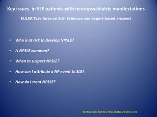
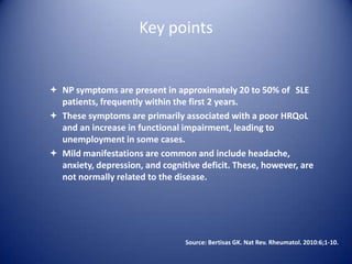
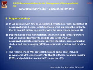
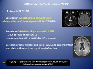
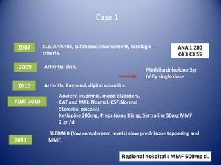
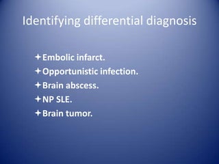
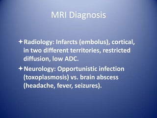
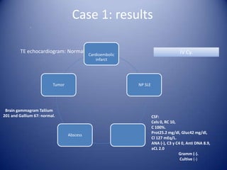
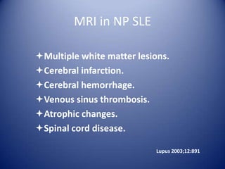
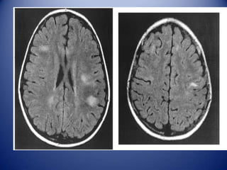
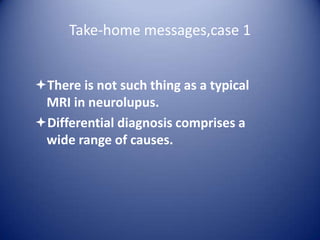
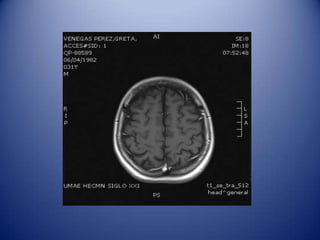
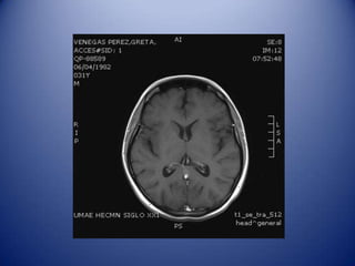
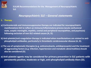
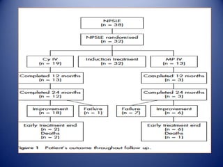
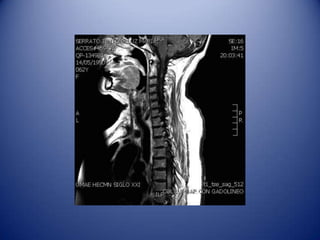
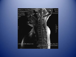
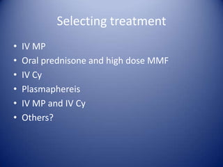
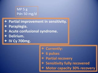
The 10th International Congress on SLE
Buenos Aires, Argentina

Neuro-psychiatric SLE
Leonor A. Barile-Fabris, MD, PhD
Professor of Rheumatology
Chair, Rheumatology Department
Centro Médico Nacional Siglo XXI
Mexico City, Mexico
Key points

NP manifestations have been
increasingly recognized.
Both attribution and diagnosis
remain clinical challenges.
Selection of optimum treatment is
complex due to scarce and
heterogeneous clinical data.
Key issues in SLE patients with neuropsychiatric manifestations
EULAR Task force on SLE- Evidence and expert-based answers

• Who is at risk to develop NPSLE?
• Is NPSLE common?
• When to suspect NPSLE?
• How can I attribute a NP event to SLE?
• How do I treat NPSLE?

Bertisas GK.Nat Rev Rheumatol.2010:6;1-10
Key points

 NP symptoms are present in approximately 20 to 50% of SLE
patients, frequently within the first 2 years.
 These symptoms are primarily associated with a poor HRQoL
and an increase in functional impairment, leading to
unemployment in some cases.
 Mild manifestations are common and include headache,
anxiety, depression, and cognitive deficit. These, however, are
not normally related to the disease.

Source: Bertisas GK. Nat Rev. Rheumatol. 2010:6;1-10.
EULAR Recommendations for the
Management of Neuropsychiatric SLE

eular

Neuropsychiatric SLE – General statements
1. When do they occur?
-May precede, coincide, or follow the diagnosis of SLE but commonly (40-50%) occur
within the first year after SLE diagnosis,
-Usually (50-60%) in the presence of generalized disease activity (B).
2. Cumulative incidence of neuropsychiatric manifestations:
- common (10-20%): cerebrovascular disease, seizures
- relatively uncommon (3-10%): severe cognitive dysfunction, major depression,
acute confusional state and peripheral nervous disorders

- rare (<3%): psychosis, myelitis, chorea, cranial neuropathies, aseptic meningitis (B)

Bertisas GK. Ann Rheum Dis: 69.2074-82
NP events at the time of diagnosis

Hanly JG. Ann Rheum Dis
2101;3:529-35.
NP Manifestations in 88 SLE patients at the Centro Médico
Nacional “La Raza”, Mexico City

Manifestation

Number

Seizures

32

Delirium

21

Stroke

15

Pheripheral neuropathy

12

Optic neuritis

10

Transverse myelitis

4

Barile et al. Lupus 1988;7:S 107
GLADEL
Seizures

99

30.9

Headeache

54

16.8

Psychosis

49

15.3

Delirium

47

14.6

Stroke

34

10.6

Sensitive neuropathy

33

10.3

Motor neuropathy

31

9.6

Coma

10

3.13

Aseptic meningitis

7

2.19

Transverse myelitis

7

1.7

Chorea

5

1.5

Ataxia

4

1.2

Pseudo tumor cerebri

2

0.63

Organic brain syndrome

2

0.63

Barile et al. Lupus 1998 (Suppl);7:53
Differing prevalences in LSE
Highly heterogeneous clinical manifestations.
Some are not specific or “subclinical”.
Manifestations may be present despite the
absence of other disease activity signs.
Attribution is difficult to establish.
There might be differences between inception
and survival cohorts.
Bertisas GK.Nat Rev Rheumatol.2010:6;1-10
EULAR Recommendations for the Management of Neuropsychiatric
SLE

Neuropsychiatric SLE – General statements

4. Diagnostic work-up

a) In SLE patients with new or unexplained symptoms or signs suggestive of
neuropsychiatric disease, initial diagnostic work-up should be similar to
that in non-SLE patients presenting with the same manifestations (D).
b) Depending upon the manifestation, this may include lumbar puncture
and CSF analysis (primarily to exclude CNS infection), EEG,
neuropsychological assessment of cognitive function, nerve conduction
studies, and neuro-imaging (MRI) to assess brain structure and function
(D).
c) The recommended MRI protocol (brain and spinal cord) includes
conventional MRI sequences (T1/T2 FLAIR), diffusion-weighted imaging
(DWI), and gadolinium-enhanced T1 sequences (B).

Bertisas GK. Ann Rheum Dis: 69.2074-82

eular
MRI white-matter lesions in NPSLE
•

↑ signal in Τ2 / FLAIR

• Localized in subcortical and periventricular
white matter and frontal-parietal lobe (70–80%)

• Prevalence 50–60% of all patients with NPSLE
…but 18–40% of non-NPSLE
…no correlation with a particular NP syndrome
• Cerebral atrophy, number and size of WML and cerebral infarcts
correlate with severity of cognitive dysfunction

In young SLE patients new MRI WMLs (especially if ≥5, ≥6-8mm, and
bilateral may suggest active NPSLE
Case 1

2007

SLE: Arthritis, cutaneous involvement, serologic
criteria.

2009

Arthritis, skin.

2010

ANA 1:280
C4 3 C3 55

Arthritis, Raynaud, digital vasculitis.

Abril 2010

2011

Methilprdnisolone 3gr
IV Cy single dose

Anxiety, insomnia, mood disorders.
CAT and MRI: Normal. CSF:Normal
Steroidal psicoisis
Ketiapine 200mg, Prednisone 35mg, Sertraline 50mg MMF
2 gr /d.
SLEDAI 0 (low complement levels) slow prednisone tappering and
MMF.

Regional hospital : MMF 500mg d.
Case 1 (Readmission)

24/02/13:
Seizures.
Increased reflexes.
DFH Levertiracetam, Metilprednisolone 3 gr.
03/03/13:
Seizures.
Increased reflexes.
DFH Levertiracetam, Metilprednisolone 3 gr.
Abnormal movements.
Topiramate and lumbar puncture.
06/03/13:
Anxiety-depression disorder.
IRM
Identifying differential diagnosis
Embolic infarct.
Opportunistic infection.
Brain abscess.
NP SLE.
Brain tumor.
MRI Diagnosis
Radiology: Infarcts (embolus), cortical,
in two different territories, restricted
diffusion, low ADC.
Neurology: Opportunistic infection
(toxoplasmosis) vs. brain abscess
(headache, fever, seizures).
Case 1: results
.

TE echocardiogram: Normal

Tumor

IV Cy.

Cardioembolic
infarct

NP SLE

Brain gammagram Taliium
201 and Gallium 67: normal.

Abscess

CSF:
Cels 0, RC 10,
C 100%.
Prot25.2 mg/dl, Gluc42 mg/dl,
Cl 127 mEq/L.
ANA (-), C3 y C4 0, Anti DNA 8.9,
aCL 2.0
Gramm (-).
Cultive (-)
MRI in NP SLE
Multiple white matter lesions.
Cerebral infarction.
Cerebral hemorrhage.
Venous sinus thrombosis.
Atrophic changes.
Spinal cord disease.
Lupus 2003;12:891
Saggital T1 image: Clot in the Stright sinus
Take-home messages,case 1
There is not such thing as a typical
MRI in neurolupus.
Differential diagnosis comprises a
wide range of causes.
Prognosis and treatment
Prognosis
Poor prognosis factors:
• Caucasians?
• Active disease
• aRO, LA, IgG aCl.

Rheumatology 2004; 43:1555-1560.
Prognosis in 2 referral hospitals in
Mexico City

29%

71%

improvement

no response
Poor prognosis, cont’d.
Male gender.
neuroSLICC ≥1 p = 0.0001.
↑antiDNA p= 0.21.
Low complement levels p= 0.05.
↑ESR p= 0.036.
Poor prognosis factors
Consolidation analysis:
Normal ESR and complement: 85.7%
improved.
Low complement and high ESR : 69.2%
worsened.
P= 0.001
Treatment

Barile L. Reumatol Clin. 2005;1 Supl 2: S42-5
eular
EULAR Recommendations for the Management of Neuropsychiatric
SLE

Neuropsychiatric SLE – General statements
5. Therapy
a) Corticosteroids and immunosuppressive therapy are indicated for neuropsychiatric
manifestations felt to reflect an immune/inflammatory process (acute confusional
state, aseptic meningitis, myelitis, cranial and peripheral neuropathies, and psychosis)
following exclusion of non-SLE related causes (A, D).
b) Anti-platelet/anti-coagulation therapy is indicated when manifestations are related to antiphospholipid antibodies, particularly in thrombotic cerebrovascular disease (A, D).
c) The use of symptomatic therapies (e.g. anticonvulsants, antidepressants) and the treatment
of aggravating factors (e.g. infection, hypertension and metabolic abnormalities) should
also be considered (D).
d) Anti-platelet agents may be considered for primary prevention in SLE patients with

persistently positive, moderate or high, anti-phospholipid antibody titers (D).
•

Induction Metilprednisolone (MP)
1 g/d for 3 d.

•

MP 1 g/d por 3 d, monthly for 4 m,
then bimonthly for 6 m, and
subsequently every 3 m for 1 y.
or

•

Ciclophosphamide (Cy) 0.75 g/m2
bs monthly for 1 y, and every 3 m
for another y.

Ann Rheum Dis 2005;64:620–625.
Allocation
Median monthly values of visual analogue scale ratings for changes in
muscular strenght in NP and TM patients

P=0.04

0= No changes from basal conditions; 10= the best possible
improvement
Seizures
CFM
MPD
Average prednisone intake/ day
Response to treatment
MPD

CY

p

Failure

7

1

<0.003

Improvement

11

18

<0.05

Response
Trevisani et al Cochrane 2008
Case: Female, 62 years old.
 2001: Optic Neuritis in right eye.
 2010: Non Hodgkin lymphoma, QxTx RxRx.
 May 2010: Optic Neuritis in left eye.
Hypotiroidism. ANA 1:640 H, lymphopenia,
leukopenia, Neurolupus: Pdn 50mg/d and
Mycophenolate.
 Oct 3 2010: Hyperstetic sensitive level C5
and T7, medular discharges, hyporeflexia.
Oct 3 2010: Hyperstetic sensitive level C5 and
T7, medular discharges, hyporeflexia.

MRI: Hyper intensity with T1
enhancement, suggestive of
longitudinal myelopathy
From C2 to T12 with high activity in
neuro-imaging
Selecting treatment
•
•
•
•
•
•

IV MP
Oral prednisone and high dose MMF
IV Cy
Plasmaphereis
IV MP and IV Cy
Others?
MP 5 g
Pdn 50 mg/d







Partial improvement in sensitivity.
Paraplegia.
Acute confusional syndrome.
Delirium.
IV Cy 700mg.







Currently:
6 pulses
Partial recovery
Sensitivity fully recovered
Motor capacity 30% recovery
Take-home messages: Case 2
Despite published evidence, response to
treatment even within the same clinical
manifestation may be heterogeneous.
Transverse myelopathy has a better
prognosis than longitudinal myelopathy.
Final considerations
There are different clinical subgroups in
neurolupus.
Etiopathogenic mechanisms might be
different, but they all seem to be related to
vascular endothelium.
NP SLE has a profound impact in
prognosis, HRQoL and damage.

Lupus neuropsiquiatria

  • 1.
    The 10th InternationalCongress on SLE Buenos Aires, Argentina Neuro-psychiatric SLE Leonor A. Barile-Fabris, MD, PhD Professor of Rheumatology Chair, Rheumatology Department Centro Médico Nacional Siglo XXI Mexico City, Mexico
  • 2.
    Key points NP manifestationshave been increasingly recognized. Both attribution and diagnosis remain clinical challenges. Selection of optimum treatment is complex due to scarce and heterogeneous clinical data.
  • 3.
    Key issues inSLE patients with neuropsychiatric manifestations EULAR Task force on SLE- Evidence and expert-based answers • Who is at risk to develop NPSLE? • Is NPSLE common? • When to suspect NPSLE? • How can I attribute a NP event to SLE? • How do I treat NPSLE? Bertisas GK.Nat Rev Rheumatol.2010:6;1-10
  • 4.
    Key points  NPsymptoms are present in approximately 20 to 50% of SLE patients, frequently within the first 2 years.  These symptoms are primarily associated with a poor HRQoL and an increase in functional impairment, leading to unemployment in some cases.  Mild manifestations are common and include headache, anxiety, depression, and cognitive deficit. These, however, are not normally related to the disease. Source: Bertisas GK. Nat Rev. Rheumatol. 2010:6;1-10.
  • 5.
    EULAR Recommendations forthe Management of Neuropsychiatric SLE eular Neuropsychiatric SLE – General statements 1. When do they occur? -May precede, coincide, or follow the diagnosis of SLE but commonly (40-50%) occur within the first year after SLE diagnosis, -Usually (50-60%) in the presence of generalized disease activity (B). 2. Cumulative incidence of neuropsychiatric manifestations: - common (10-20%): cerebrovascular disease, seizures - relatively uncommon (3-10%): severe cognitive dysfunction, major depression, acute confusional state and peripheral nervous disorders - rare (<3%): psychosis, myelitis, chorea, cranial neuropathies, aseptic meningitis (B) Bertisas GK. Ann Rheum Dis: 69.2074-82
  • 6.
    NP events atthe time of diagnosis Hanly JG. Ann Rheum Dis 2101;3:529-35.
  • 7.
    NP Manifestations in88 SLE patients at the Centro Médico Nacional “La Raza”, Mexico City Manifestation Number Seizures 32 Delirium 21 Stroke 15 Pheripheral neuropathy 12 Optic neuritis 10 Transverse myelitis 4 Barile et al. Lupus 1988;7:S 107
  • 8.
    GLADEL Seizures 99 30.9 Headeache 54 16.8 Psychosis 49 15.3 Delirium 47 14.6 Stroke 34 10.6 Sensitive neuropathy 33 10.3 Motor neuropathy 31 9.6 Coma 10 3.13 Asepticmeningitis 7 2.19 Transverse myelitis 7 1.7 Chorea 5 1.5 Ataxia 4 1.2 Pseudo tumor cerebri 2 0.63 Organic brain syndrome 2 0.63 Barile et al. Lupus 1998 (Suppl);7:53
  • 9.
    Differing prevalences inLSE Highly heterogeneous clinical manifestations. Some are not specific or “subclinical”. Manifestations may be present despite the absence of other disease activity signs. Attribution is difficult to establish. There might be differences between inception and survival cohorts.
  • 10.
    Bertisas GK.Nat RevRheumatol.2010:6;1-10
  • 11.
    Bertisas GK.Nat RevRheumatol.2010:6;1-10
  • 12.
    EULAR Recommendations forthe Management of Neuropsychiatric SLE Neuropsychiatric SLE – General statements 4. Diagnostic work-up a) In SLE patients with new or unexplained symptoms or signs suggestive of neuropsychiatric disease, initial diagnostic work-up should be similar to that in non-SLE patients presenting with the same manifestations (D). b) Depending upon the manifestation, this may include lumbar puncture and CSF analysis (primarily to exclude CNS infection), EEG, neuropsychological assessment of cognitive function, nerve conduction studies, and neuro-imaging (MRI) to assess brain structure and function (D). c) The recommended MRI protocol (brain and spinal cord) includes conventional MRI sequences (T1/T2 FLAIR), diffusion-weighted imaging (DWI), and gadolinium-enhanced T1 sequences (B). Bertisas GK. Ann Rheum Dis: 69.2074-82 eular
  • 13.
    MRI white-matter lesionsin NPSLE • ↑ signal in Τ2 / FLAIR • Localized in subcortical and periventricular white matter and frontal-parietal lobe (70–80%) • Prevalence 50–60% of all patients with NPSLE …but 18–40% of non-NPSLE …no correlation with a particular NP syndrome • Cerebral atrophy, number and size of WML and cerebral infarcts correlate with severity of cognitive dysfunction In young SLE patients new MRI WMLs (especially if ≥5, ≥6-8mm, and bilateral may suggest active NPSLE
  • 14.
    Case 1 2007 SLE: Arthritis,cutaneous involvement, serologic criteria. 2009 Arthritis, skin. 2010 ANA 1:280 C4 3 C3 55 Arthritis, Raynaud, digital vasculitis. Abril 2010 2011 Methilprdnisolone 3gr IV Cy single dose Anxiety, insomnia, mood disorders. CAT and MRI: Normal. CSF:Normal Steroidal psicoisis Ketiapine 200mg, Prednisone 35mg, Sertraline 50mg MMF 2 gr /d. SLEDAI 0 (low complement levels) slow prednisone tappering and MMF. Regional hospital : MMF 500mg d.
  • 15.
    Case 1 (Readmission) 24/02/13: Seizures. Increasedreflexes. DFH Levertiracetam, Metilprednisolone 3 gr. 03/03/13: Seizures. Increased reflexes. DFH Levertiracetam, Metilprednisolone 3 gr. Abnormal movements. Topiramate and lumbar puncture. 06/03/13: Anxiety-depression disorder.
  • 16.
  • 17.
    Identifying differential diagnosis Embolicinfarct. Opportunistic infection. Brain abscess. NP SLE. Brain tumor.
  • 18.
    MRI Diagnosis Radiology: Infarcts(embolus), cortical, in two different territories, restricted diffusion, low ADC. Neurology: Opportunistic infection (toxoplasmosis) vs. brain abscess (headache, fever, seizures).
  • 19.
    Case 1: results . TEechocardiogram: Normal Tumor IV Cy. Cardioembolic infarct NP SLE Brain gammagram Taliium 201 and Gallium 67: normal. Abscess CSF: Cels 0, RC 10, C 100%. Prot25.2 mg/dl, Gluc42 mg/dl, Cl 127 mEq/L. ANA (-), C3 y C4 0, Anti DNA 8.9, aCL 2.0 Gramm (-). Cultive (-)
  • 20.
    MRI in NPSLE Multiple white matter lesions. Cerebral infarction. Cerebral hemorrhage. Venous sinus thrombosis. Atrophic changes. Spinal cord disease. Lupus 2003;12:891
  • 24.
    Saggital T1 image:Clot in the Stright sinus
  • 26.
    Take-home messages,case 1 Thereis not such thing as a typical MRI in neurolupus. Differential diagnosis comprises a wide range of causes.
  • 29.
  • 30.
    Prognosis Poor prognosis factors: •Caucasians? • Active disease • aRO, LA, IgG aCl. Rheumatology 2004; 43:1555-1560.
  • 31.
    Prognosis in 2referral hospitals in Mexico City 29% 71% improvement no response
  • 32.
    Poor prognosis, cont’d. Malegender. neuroSLICC ≥1 p = 0.0001. ↑antiDNA p= 0.21. Low complement levels p= 0.05. ↑ESR p= 0.036.
  • 33.
    Poor prognosis factors Consolidationanalysis: Normal ESR and complement: 85.7% improved. Low complement and high ESR : 69.2% worsened. P= 0.001
  • 34.
    Treatment Barile L. ReumatolClin. 2005;1 Supl 2: S42-5
  • 35.
    eular EULAR Recommendations forthe Management of Neuropsychiatric SLE Neuropsychiatric SLE – General statements 5. Therapy a) Corticosteroids and immunosuppressive therapy are indicated for neuropsychiatric manifestations felt to reflect an immune/inflammatory process (acute confusional state, aseptic meningitis, myelitis, cranial and peripheral neuropathies, and psychosis) following exclusion of non-SLE related causes (A, D). b) Anti-platelet/anti-coagulation therapy is indicated when manifestations are related to antiphospholipid antibodies, particularly in thrombotic cerebrovascular disease (A, D). c) The use of symptomatic therapies (e.g. anticonvulsants, antidepressants) and the treatment of aggravating factors (e.g. infection, hypertension and metabolic abnormalities) should also be considered (D). d) Anti-platelet agents may be considered for primary prevention in SLE patients with persistently positive, moderate or high, anti-phospholipid antibody titers (D).
  • 36.
    • Induction Metilprednisolone (MP) 1g/d for 3 d. • MP 1 g/d por 3 d, monthly for 4 m, then bimonthly for 6 m, and subsequently every 3 m for 1 y. or • Ciclophosphamide (Cy) 0.75 g/m2 bs monthly for 1 y, and every 3 m for another y. Ann Rheum Dis 2005;64:620–625.
  • 38.
  • 39.
    Median monthly valuesof visual analogue scale ratings for changes in muscular strenght in NP and TM patients P=0.04 0= No changes from basal conditions; 10= the best possible improvement
  • 40.
  • 41.
  • 42.
  • 43.
    Trevisani et alCochrane 2008
  • 44.
    Case: Female, 62years old.  2001: Optic Neuritis in right eye.  2010: Non Hodgkin lymphoma, QxTx RxRx.  May 2010: Optic Neuritis in left eye. Hypotiroidism. ANA 1:640 H, lymphopenia, leukopenia, Neurolupus: Pdn 50mg/d and Mycophenolate.  Oct 3 2010: Hyperstetic sensitive level C5 and T7, medular discharges, hyporeflexia.
  • 45.
    Oct 3 2010:Hyperstetic sensitive level C5 and T7, medular discharges, hyporeflexia. MRI: Hyper intensity with T1 enhancement, suggestive of longitudinal myelopathy From C2 to T12 with high activity in neuro-imaging
  • 48.
    Selecting treatment • • • • • • IV MP Oralprednisone and high dose MMF IV Cy Plasmaphereis IV MP and IV Cy Others?
  • 49.
    MP 5 g Pdn50 mg/d      Partial improvement in sensitivity. Paraplegia. Acute confusional syndrome. Delirium. IV Cy 700mg.      Currently: 6 pulses Partial recovery Sensitivity fully recovered Motor capacity 30% recovery
  • 50.
    Take-home messages: Case2 Despite published evidence, response to treatment even within the same clinical manifestation may be heterogeneous. Transverse myelopathy has a better prognosis than longitudinal myelopathy.
  • 51.
    Final considerations There aredifferent clinical subgroups in neurolupus. Etiopathogenic mechanisms might be different, but they all seem to be related to vascular endothelium. NP SLE has a profound impact in prognosis, HRQoL and damage.

Editor's Notes

  • #16 2007 recibiendo tratamiento con diclofenaco, prednisona 5mg día, cloroquina 150mg, azatioprina 100mg día. Buena respuesta y lo dejo de tomar.2009 metotrexato 10mg y prednisona 5mg. 72 hrs. posterior a medicación.
  • #17 Infarto embolico, por ser cortical y 2 territorios diferentes., con restricción de la difusión y ADC bajo.Infección por microorganismo oportunista Toxoplasmosis por inmunosupresión o abceso cerebral por cefalea, fiebre y crisis convulsivas.Neurocirugía: Imagen isointensa en T1 e hiperintensa en T2 a nivel frontal derecho, parietal izquierdo y occipital derecho cortico subcortical con edema perilesional leve, que no refuerzan con medio de contraste.