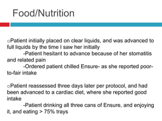
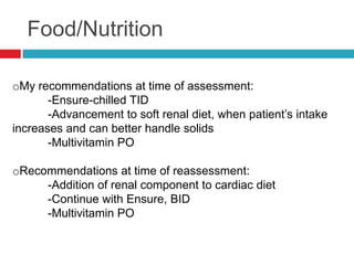
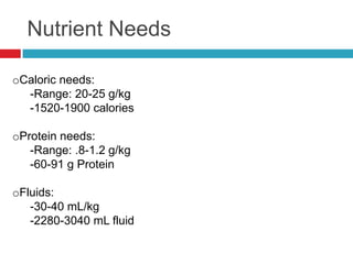
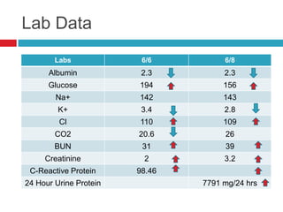
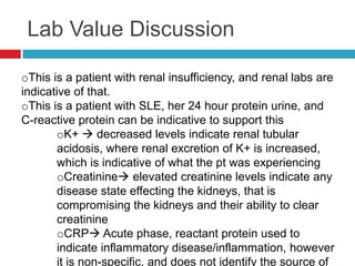
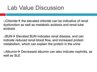
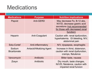
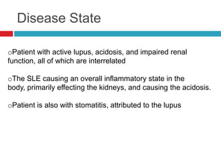
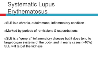
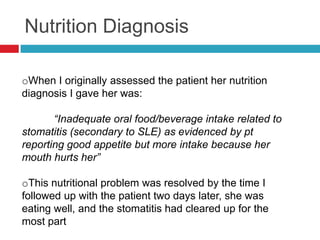
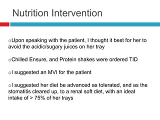
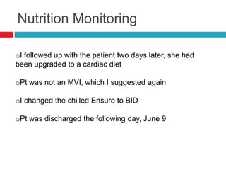
This patient was admitted with symptoms of nausea, vomiting, abdominal pain and black stools. He has a history of hypertension, gallbladder removal and GERD. Diagnosis includes GERD, GI bleeding and cirrhosis of the liver. A jejunostomy tube was placed for nutrition due to malabsorption from liver disease. The patient was started on TPN and stabilized. TPN was tapered and the patient advanced to a clear liquid diet and then oral diet as tolerated. He was discharged on a soft diet restricted to 2000ml fluids per day with sodium and protein recommendations.
This case study describes a 57-year-old male admitted to the hospital with nausea, vomiting, abdominal pain, jaundice, ascites, and black stools. He was diagnosed with GERD, gastrointestinal bleeding, and cirrhosis. During his hospital stay, he received various treatments including tube feeding and TPN. His condition deteriorated and he was transferred to the ICU. He later stabilized and was transitioned to an oral diet to prepare for discharge with diagnoses of chronic cirrhosis, GERD, and esophageal varices.
Applied nutrition 3 rd presentation - diseases of liver, gall bladder, and ...MD Specialclass
The document provides detailed information about diseases of the liver, gallbladder, and pancreas. It discusses the anatomy and functions of the liver, signs and symptoms of various hepatitis types, cirrhosis, and hepatic coma. It also covers cholecystitis, including causes, clinical manifestations, and dietary management for related conditions.
This patient has end stage liver disease due to chronic alcoholism. He has a history of gastrointestinal bleeding from esophageal varices. He was admitted with edema, ascites, and weight gain. His medications and conditions require careful management of nutrients. Tube feedings were started but caused intolerance, so a soft diet was ordered to prevent further bleeding. Nutrition interventions focus on managing malnutrition and gastrointestinal symptoms while supporting organ function and transition to an oral diet.
Crisis de hiperglicemia en pacientes adultos con diabetesMaria Enriquez
This document discusses the diagnosis and treatment of hyperglycemic crises in adult patients with diabetes mellitus. It addresses diabetic ketoacidosis (DKA) and hyperosmolar hyperglycemic state (HHS), defining their diagnostic criteria and typical clinical presentations. For DKA, symptoms develop rapidly and include abdominal pain and Kussmaul breathing. For HHS, symptoms develop more gradually over days and include neurological changes with dehydration. Laboratory findings for both conditions include high blood glucose, electrolyte abnormalities, and metabolic acidosis. Treatment involves fluid resuscitation, insulin administration, electrolyte replacement, and identifying and treating any precipitating factors.
Child with acute liver failure dr. kirtichandra kodaliSanjeev Kumar
A 3-year-old boy presented with fever, cough, loose stools and increasing drowsiness over the past few days. On examination, he showed signs of dehydration and liver enlargement. Laboratory tests found elevated liver enzymes and ammonia levels, indicating acute liver failure. The cause was determined to be paracetamol toxicity from an accidental overdose, as serum paracetamol levels were found to be toxic. The patient was treated with N-acetyl cysteine and supportive care, and his condition improved such that his liver function normalized and he was discharged.
1) The patient, a 70-year-old male, presented with abdominal distention, weakness, decreased appetite and weight loss. His history included alcoholism and smoking.
2) Laboratory tests showed abnormalities indicative of liver damage including low hemoglobin and albumin levels as well as elevated liver enzymes. Ultrasound found changes to the liver, gallbladder and spleen.
3) Based on the clinical presentation and test results, the patient was diagnosed with alcoholic liver cirrhosis. Treatment focused on cessation of alcohol consumption and supplementation to address malnutrition.
1. This case presentation summarizes a 55-year-old male farmer who presented with abdominal swelling and discomfort for 2 months and scanty urination and constipation for 1 month.
2. On examination, he had signs of chronic liver disease including jaundice, edema, and hepatic encephalopathy.
3. Investigations confirmed chronic hepatitis B infection, decompensated cirrhosis, and grade 2-4 esophageal varices.
4. He was diagnosed with decompensated chronic liver disease and grade 1 hepatic encephalopathy, and started on treatment including diuretics, beta-blockers, lactulose, and vitamin supplementation.
This document describes a case study of a 26-year-old female patient diagnosed with Hepatitis C. She presents with fatigue, anorexia, pale skin, and weakness. Lab tests show abnormalities indicating liver damage, vitamin K deficiency, anemia, and pre-diabetes. Her usual diet consists of juice, diet soda, and take-out providing only 57% of her estimated energy needs. The nutrition intervention aims to gradually increase calorie intake to meet requirements through a sample menu emphasizing protein, healthy fats, and carbohydrate control. Education focuses on nutrient needs, healthy choices, and monitoring will track outcomes like weight and blood glucose.
This case study describes a 57-year-old male patient with a diagnosis of cirrhosis of the liver and a history of alcoholism, hypertension, and cholecystectomy. Laboratory tests show abnormalities indicative of liver disease including elevated creatinine, PTT, triglycerides, and cholesterol as well as low albumin, glucose, sodium, and chloride levels. After admission, the patient's weight increased to 194 pounds due to fluid accumulation in the abdomen. A jejunostomy tube was placed for nutrition due to risk of esophageal bleeding from varices and impaired nutrient absorption from liver disease. Tube feedings were later not well tolerated. The patient was started on TPN but concerns included potential edema and
CN is a 41-year-old female on hemodialysis for stage 5 chronic kidney disease. Her current diet provides too much phosphorus and calcium compared to recommendations. She takes several medications to manage complications of kidney disease, including hyperparathyroidism. Her lab values show abnormalities consistent with kidney failure. Her diet needs modification to meet nutrient guidelines for dialysis patients and support medication treatment of her conditions.
Chronic Liver Disease in pediatric: a case presentation and discussionDr Abdalla M. Gamal
A presentation from a tutorial about an interesting case that came to the Pediatric Department of Sebha Medical Center and was imaged by the Radiology Department.
The tutorial was a joint effort between Dr Zeinab Salem Ali (from Pediatric Department) and me (from Radiology Department). In her slides, Dr Zeinab presented the case history, examination, investigations, differential diagnosis and discussed the clinical presentation, investigations and management for chronic liver diseases in pediatric patients.In my slides, I discussed the definition, etiology, natural history of this condition and explained the role of imaging in its diagnosis.
These are my slides after some modifications. I added an aknowlegement page to illustrate Dr Zeinab effort and to thank Dr Khaled Aljasem from Pediatric Department for his effort in revising the original presentations and the constructive feedback he provided which improved the quality of the presented material. Then I added a summary for the parts Dr Zeinab has presented to make this powerpoint presentation complete.
This presentation was presented by Dr Zeinab Salem (from Pediatric Department) and me in a joint tutorial between Pediatric Department and Radiology Department of Sebha Medical Center.
This case presentation discusses a 39-year-old male patient who presented with abdominal distension, leg pains, general weakness, and a history of portal vein thrombosis. Laboratory tests and ultrasound results led to a diagnosis of alcoholic pancreatitis. The patient had a history of alcohol use for 10 years. He was treated with IV fluids, analgesics, antibiotics, pancreatic enzyme supplements, and vitamins. His condition normalized with treatment and medications prior to discharge.
A 35-year-old male auto driver was admitted with decreased urine output for 3 days and abdominal pain and fever for 10 days. Examination found pallor and abdominal tenderness. Tests showed acute kidney injury and a urine culture grew gram-negative bacilli. He was diagnosed with acute pyelonephritis likely caused by E. coli infection. He received IV and oral antibiotics and underwent hemodialysis. His kidney function and other lab values gradually improved with treatment.
This document discusses jaundice in multiple patients and the underlying causes. It begins by summarizing different causes of jaundice, including unconjugated hyperbilirubinemia from excessive production or failure of conjugation, and conjugated hyperbilirubinemia from hepatocellular damage or obstruction of bile flow.
It then analyzes several case studies: three with unconjugated hyperbilirubinemia from megaloblastic anemia, hereditary spherocytosis, and an unknown cause; two with conjugated hyperbilirubinemia, one from hepatocellular damage and one from obstruction; and one with malaria-induced cholestasis.
The document emphasizes different
This document presents a case study of a 41-year-old male patient (SW) with chronic kidney disease who is undergoing hemodialysis 3 times per week. It provides details on his medical history, social history, physical exam findings, laboratory values, current diet, and medication regimen. It finds that his current diet is low in calories and protein and high in phosphorus and potassium. It develops an intervention plan focused on increasing calorie intake, reducing phosphorus intake, and educating on limiting high potassium and phosphorus foods given his chronic kidney disease and medications. It discusses how the patient is at risk for secondary hyperparathyroidism due to low calcium levels and provides a recent literature reference on managing this condition in
Mahmuda, a 2.5 year old girl, presented with swelling of the body for 15 days, decreased urine output, and red urine for 9 days. On examination, she had puffy face and swelling of the body. Tests found protein in the urine and low albumin levels. She was diagnosed with initial nephrotic syndrome and a urine infection. She received antibiotics, albumin infusion, diuretics to reduce swelling, and prednisolone for 6 weeks followed by a gradual taper, with calcium and vitamin supplements. Her parents were counseled and she will follow up every 2 weeks then monthly to monitor her response and for any side effects or infections.
Case presentation on Alcoholic liver diseaseHAMMADKC
A 60-year old male patient presented with complaints of yellowish skin discoloration, swelling in the lower legs, constipation, abdominal distension, and fatigue. He had a history of alcohol use for 8 years and smoking for 12 years. Laboratory tests showed elevated bilirubin levels and liver enzymes. An ultrasound revealed a fatty liver with surface nodules. He was diagnosed with alcoholic liver disease and treated with medications, lifestyle counseling, and advised to abstain from alcohol and smoking. His symptoms improved over three days of treatment and he was discharged.
The document discusses various gastrointestinal disorders, classifying them into categories such as impaired digestion/absorption, altered secretion/transit, immune dysregulation, and neoplastic degeneration. Specific disorders of the esophagus are then discussed in more detail, including reflux esophagitis, infections, webs/rings, and motility disorders like achalasia and diffuse esophageal spasm. Common symptoms and clinical features of esophageal diseases are also outlined.
CASE PRESENTATION ONCIRRHOSIS OF LIVER WITH PORTAL HYPERTENSION, HEPATIC EN...Akhil Joseph
A DETAIL CASE PRESENTATION ON CIRRHOSIS OF LIVER WITH PORTAL HYPERTENSION, HEPATIC ENCEPHALOPATHY AND GRADE II OESOPHAGEAL VARICES WITH CONGESTIVE GASTROPATHY. LIVER CIRRHOSIS AND ALL ITS COMPLICATION IN A PATIENT.
Discus the scenario of my case base selective patient
Explain the patient’s present and previous health history
Discus the chronic liver disease
Explain the causes of chronic liver disease
Review the psychopathology of chronic liver disease
Prepare the nursing management
1) A 3-year-old boy presented with recurrent jaundice since infancy and itching for 6 months. Liver tests showed elevated bilirubin and alkaline phosphatase.
2) Liver biopsy found intrahepatic cholestasis with fibrosis suggestive of progressive familial intrahepatic cholestasis (PFIC). Immunostaining revealed an absence of multidrug resistance protein 3 (MDR3) expression.
3) Genetic testing confirmed PFIC type 3, which causes cholestasis due to mutations affecting bile salt export pump function. The patient was started on ursodeoxycholic acid and rifampicin to manage symptoms.
Diarrhea is defined as an increase in stool frequency or liquidity. For infants it is considered diarrhea if there are more than 3 watery stools per day, while for older children it is 3 or more loose stools per day. The causes of diarrhea include viral, bacterial, and parasitic infections. Rotavirus is the most common cause of acute diarrhea in children. Treatment involves oral rehydration with solutions like ORS as well as continued feeding. For some cases antibiotics or zinc may be used. Prevention strategies include vaccines, handwashing, safe water, and breastfeeding.
Mr. Biju, age 44, was admitted to the hospital with acute on chronic liver failure and severe alcoholic hepatitis. He developed lower respiratory tract infections and ascites during his hospital stay. His liver function tests and renal function tests were monitored over the course of two months, showing abnormalities consistent with liver and kidney damage. He was discharged with medications to treat his conditions and instructions to follow a restricted diet and avoid alcohol in order to manage his liver failure.
Nutrition management in systemic lupus erythematosus (SLE) was explored. A search found few randomized trials on nutrition for SLE. The best results were from trials on dehydroepiandrosterone (DHEA), which reduced SLE activity, and N-acetylcysteine (NAC), which reduced SLE disease activity. However, trials on omega-3 fatty acids found no clear benefits and some potential adverse effects. Overall, the evidence is limited and inconclusive on specific nutrition interventions for SLE due to small sample sizes and limitations of current studies. Further large randomized controlled trials are still needed.
* SLE can affect all components of the respiratory system, with clinical presentations ranging from asymptomatic to life-threatening.
* Pleural effusion is the most common pulmonary manifestation in SLE and usually presents as bilateral, small or moderate exudative effusions that resolve spontaneously without residual damage.
* Diffuse alveolar hemorrhage is a serious pulmonary complication characterized by hemoptysis, rapid drop in hemoglobin, and new infiltrates on imaging, requiring aggressive immunosuppression.
Pathogenesis systemic lupus erythematosus by dr bashir ahmed dar associate pr...Prof Dr Bashir Ahmed Dar
Systemic lupus erythematosus is a chronic, multisystem, inflammatory disorder of autoimmune etiology, occurring predominantly in young women. Common manifestations may include arthralgias and arthritis; malar and other skin rashes; pleuritis or pericarditis; renal or CNS involvement; and hematologic cytopenias.
Systemic lupus erythematosus (SLE) is an autoimmune disease where the immune system attacks its own tissues, causing inflammation and damage. It is characterized by periods of disease flares and remission. Common symptoms include joint pain, rashes, and fatigue. SLE can affect many organs like the skin, lungs, heart, and kidneys. Diagnosis involves evaluating symptoms, lab tests like antinuclear antibodies, and sometimes biopsies. Treatment aims to reduce symptoms during flares and prevent organ damage using medications like corticosteroids, antimalarials, and immunosuppressants. SLE affects mostly women of childbearing age and has no known cure.
Nutrition in Cancer Prevention and TreatmentTim Crowe
This presentation will help you to understand the influence that dietary and lifestyle factors play in the prevention and causation of cancer. It outlines the important nutritional considerations for patients undergoing treatment for cancer and reviews procedures to improve patient safety by knowing the risks and benefits of antioxidant supplementation during cancer treatment
Obstructive jaundice (OJ) is a common problem in daily clinical practice. However, completely understanding the pathophysiological changes in OJ remains a challenge for planning current and future management. The effects of OJ are widespread, affecting the biliary tree, hepatic cells, liver function, and causing systemic complications. The lack of bile in the intestine, destruction of the intestinal mucosal barrier, and increased absorption of endotoxins can lead to endotoxemia, production of proinflammatory cytokines, and induce systemic inflammatory response syndrome, ultimately leading to multiple organ dysfunction syndrome. Proper management of OJ includes adequate water supply and electrolyte replacement, nutritional support, preventive antibiotics, pain relief, and itching relief. The surgical treatment of OJ depends on the cause, location, and severity of the obstruction. Biliary drainage, surgery, and endoscopic intervention are potential treatment options depending on the patient's condition. In addition to modern medical treatments, Traditional Chinese medicine may offer therapeutic benefits for OJ. A comprehensive search was conducted on PubMed for relevant articles published up to August 1970
Acute renal failure and chronic renal failure are discussed. Acute renal failure can be prerenal, renal, or postrenal and is characterized by a sudden reduction in urine output. Chronic renal failure is a permanent loss of kidney function that progresses to end stage renal disease. It has multiple etiologies including glomerular diseases. Both present with electrolyte imbalances, fluid retention, and other complications. Treatment focuses on fluid management, diet modification, and dialysis or transplantation as needed.
A 57-year-old male was admitted with nausea, vomiting and abdominal pain due to cirrhosis and bleeding esophageal varices. He has a history of alcoholism. Initial labs showed hyponatremia and low albumin. A jejunostomy tube was placed for nutrition but caused intolerance. He was started on TPN with Hepatamine which was later tapered as he transitioned to an oral soft diet.
This document discusses obstructive jaundice, providing definitions, pathophysiology, effects on various body systems, etiology, history and examination findings, laboratory investigations, imaging modalities, and causes of biliary obstruction. It defines obstructive jaundice as a failure of bile to reach the intestine due to mechanical obstruction. Pathophysiological changes include bile duct dilation, hepatic fibrosis, and portal hypertension. Causes include gallstones, strictures, tumors, and congenital anomalies. A thorough history, physical exam, and lab tests can localize the level and cause of obstruction, while imaging modalities like ultrasound and MRCP can identify and characterize obstructive lesions.
This document presents a case study of a 55-year-old female patient admitted with abdominal distention and leg swelling over the past month. Her history and examination findings are detailed. Investigations show signs of cirrhosis and portal hypertension likely related to hepatitis B. She develops renal impairment and a renal biopsy reveals diffuse proliferative glomerulonephritis. Further tests confirm hepatitis B virus infection is the cause of her kidney disease. She is diagnosed with hepatitis B virus related diffuse proliferative glomerulonephritis and treated accordingly.
The document contains a problem list, nursing care plan, and drug study for a 70-year-old patient admitted with acute gastroenteritis and severe dehydration who was experiencing frequent diarrhea, vomiting, and weakness. The nursing care plan focuses on monitoring the patient's vital signs and fluid intake/output to manage their dehydration and restoring normal bowel function. The drug study examines medications prescribed including furosemide, ceftriaxone, ranitidine, and metronidazole to treat the infection, control fluid levels, and reduce stomach acid.
Medical nutrition therapy status post whipple procedureValerie Agyeman
SG, a 72-year-old female, underwent a Whipple procedure for pancreatic adenocarcinoma. She experienced significant weight loss pre-operatively and nutritional complications post-operatively including delayed gastric emptying and electrolyte abnormalities. The registered dietitian's nutrition diagnoses included malnutrition, unintentional weight loss, and altered GI function. The RD prescribed a carbohydrate-controlled diet and thickened liquids, provided nutrition education, and collaborated with other providers to optimize SG's recovery.
Renal failure occurs when the kidneys cannot remove waste or regulate fluids and electrolytes. There are two main types: acute renal failure, which develops rapidly over hours to days; and chronic renal failure, which is progressive and irreversible. Acute renal failure causes a sudden loss of kidney function and can result from prerenal issues, direct kidney damage, or urinary tract obstruction. Chronic renal failure is treated through diet, medication, and often dialysis to remove waste when kidney function declines. Nursing focuses on managing complications, nutrition, fluid balance, and educating patients.
INTRODUCTION
Cancer is a general term used to refer to a condition where the body’s cells begin to grow and reproduce in an uncontrollable way. Lung cancers are the fourth most common cancer reported in the Indian males.
DEFINITION
Lung carcinoma is a malignant lung tumor characterized by uncontrolled cell growth in tissues of the lung. If left untreated, this growth can spread beyond the lung by the process of metastasis into nearby tissue or other parts of the body.
CAUSES
The most common causes of fracture include,
I. Tobacco smoke
Tobacco use is responsible for more than one of every six deaths. The younger a person is when he or she starts smoking, the greater the risk of developing lung cancer.
II. Secondhand smoke
Passive smoking has been identified as a possible cause of lung cancer in nonsmokers. People who are involuntarily exposed to tobacco smoke in a closed environment (house, automobile, and building) have an increased risk of lung cancer when compared with unexposed nonsmokers.
III. Environmental and occupational exposure
Various carcinogens have been identified in the atmosphere, including motor vehicle emissions and pollutants fromrefineries and manufacturing plants. High levels of radon have been associated with the development of lung cancer, especially when combined with cigarette smoking. Chronic exposure to industrial carcinogens, such as arsenic, asbestos, mustard gas, chromates, coke oven fumes, nickel, oil, and radiation has been associated with the development of lung cancer.
IV. Genetics
Some familial predisposition to lung cancer seems apparent, because the incidence of lung cancer in close relatives of patients with lung cancer appears to be two to three times that in the general population regardless of smoking status.
TYPES OF LUNG CANCER:
1. Small cell lung carcinoma
• Accounts for 15%-25% of lung cancers
• It is most malignant form
• Tends to spread early via lymphatic and bloodstream
• Is frequently associated with endocrine disturbances
• Predominantly central and can cause bronchial obstruction and pneumonia.
2. Non-small cell lung carcinoma
Is further classified by cell type,
Adenocarcinoma
• Most common type
• Accounts for approximately 30%-40% of lung cancers
• More common in women
• Often gas no clinical manifestations until widespread metastasis is present
• Usually begins in mucous glandular tissue, is most commonly located in peripheral portions of lungs.
Squamous cell carcinoma
• Second most common type of lung cancer
• Accounts for 30%-35% of lung cancers
• Is more common in men
• Arises from the bronchial epithelium of the lungs or bronchus, slow-growing cancer that usually begins in the bronchial tubes.
Large cell carcinoma
• The least common form
• Accounts for 5%-15% of lung cancers
• Composed of large sized cells that are anaplastic and often arise in the bronchi, commonly causes cavitation
• Is highly metastatic via lymphatic and blood.
STAGING OF NON-SMALL CELL LUNG C
1) The patient is a 53-year-old woman seeking pharmacotherapy for weight loss in addition to lifestyle modifications. She has a history of anxiety treated with paroxetine and past narcotic abuse.
2) Orlistat is recommended as it inhibits fat absorption and has beneficial effects on lipids, important for this patient with dyslipidemia.
3) Lorcaserin is not recommended due to potential interaction with paroxetine. Phentermine is not recommended due to risk of addiction given her history of abuse.
Chronic pancreatitis is a long-term inflammation of the pancreas that results in permanent damage. The patient, AH, underwent a Whipple procedure for chronic pancreatitis caused by hypertriglyceridemia and possible bile duct stones. Over her 7 day hospital stay, AH's diet was advanced from NPO to regular meals. She was educated on GI and diabetes diets and discharged tolerating a regular diet.
Acute Kidney Injury (AKI) is a common complication, affecting 5-7% of hospital admissions and 30% of intensive care unit patients. The top causes of AKI in India are diarrheal diseases, sepsis, malaria, drug toxicity, and hospital-acquired injuries. Biomarkers like cystatin C and kidney injury molecule 1 can help detect AKI earlier than creatinine. Treatment involves fluid resuscitation, eliminating nephrotoxins, and renal replacement therapy for complications like electrolyte imbalances or uremia. Outcomes depend on the underlying cause, with pre-renal and post-renal AKI having a better prognosis than intrinsic renal injury.
11 Turman Management Of Acute Renal Failure In PicuDang Thanh Tuan
This document provides an overview of the definition, causes, risk factors, evaluation, and management of acute renal failure (ARF) in pediatric intensive care unit patients. It discusses the importance of differentiating between pre-renal, renal, and post-renal causes of ARF. Key factors that influence outcomes like oliguria, multi-organ failure, and late initiation of dialysis are outlined. Fluid management and treatment of complications such as hypertension, electrolyte abnormalities, and anemia are also reviewed. Emerging potential new treatments for ARF are described but many have not proven effective in clinical trials.
The document discusses abnormal liver function tests (LFTs). It describes that hepatocellular injury patterns usually show elevated AST and ALT, and can be caused by drugs, alcohol, hepatitis viruses, steatohepatitis, autoimmune hepatitis, and genetic disorders like Wilson's disease and hereditary hemochromatosis. Cholestatic patterns are characterized by elevated alkaline phosphatase and can be from intrahepatic causes like primary biliary cirrhosis or extrahepatic causes like gallstones, primary sclerosing cholangitis, malignancy, chronic pancreatitis, or HIV cholangiopathy. Isolated hyperbilirubinemia can indicate hemolysis, Dubin-Johnson syndrome, or Gilbert
This document provides an overview of acute pancreatitis including its:
- Pathophysiology involving trypsin activation within acinar cells
- Risk factors such as gallstones, alcohol use, anatomical obstructions
- Clinical manifestations like severe abdominal pain
- Diagnosis using blood tests, imaging, and scoring systems to determine severity
- Management approaches depending on severity, including antibiotics for infected necrosis
This document discusses primary biliary cirrhosis (PBC), a chronic disease that slowly destroys bile ducts in the liver. It begins by describing a 45-year-old patient presenting with fatigue, itching, jaundice, and an enlarged spleen. Test results and liver biopsy are needed to diagnose PBC, which is characterized by antibodies and inflammation of bile ducts. Ursodeoxycholic acid is the standard treatment and improves liver tests, though transplantation may be needed for advanced cases. The document then provides further details on symptoms, causes, complications, diagnostic tests, treatment goals, and managing specific complications of PBC.
1. Diarrhea is defined as an increased frequency, fluidity, and volume of stool. It can be acute (<14 days), persistent (14-30 days), or chronic (>30 days).
2. The document discusses the pathophysiology, etiology, indications for referral to a gastroenterologist, initial evaluation, and recommendations for evaluating patients with chronic diarrhea.
3. The differential diagnosis and workup of inflammatory bowel diseases like Crohn's disease and ulcerative colitis are outlined, including relevant history, physical exam findings, diagnostic studies, endoscopy, histopathology, and radiology findings.
The document discusses acute renal failure (ARF), defining it as a rapid reduction in kidney function over days or weeks. It describes ARF as oliguric (<500mL urine/day), anuric (<100mL/day), or non-oliguric. Causes include pre-renal (e.g. dehydration), post-renal (e.g. blockages), and intrinsic renal failure. ARF diagnosis involves labs, ultrasound, and treating underlying causes while preventing complications like electrolyte abnormalities.
Similar to Lupus & The Kidneys Case Study (20)
1. LUPUS & THE KIDNEYS
Sarah Thelen
Dietetic Intern
Benedictine University
2. The Patient
oPatient data:
-65”
-167 lbs
-BMI: 28
oAfrican American female:
-lives with family
oAdmitted with c/o: fatigue, weakness, and
decreased intake
oAllergies: Sulfa
3. The Patient
oPatient with relevant medical hx:
-Systematic Lupus Erythematosus (SLE)
-Impaired renal function (Nephrotic
syndrome??)
-HTN
oClinical Impression:
-Active SLE
-Stomatitis
-Impaired renal function
4. Food/Nutrition
oPt experiencing stomatitis inferior to tongue, impinging
patient’s po intake
oPatient experiencing diarrhea, decreased intake, but still
very hungry
oPatient also did not have her dentures with her
5. Food/Nutrition
oPatient initially placed on clear liquids, and was advanced to
full liquids by the time I saw her initially
-Patient hesitant to advance because of her stomatitis
and related pain
-Ordered patient chilled Ensure- as she reported poor-
to-fair intake
oPatient reassessed three days later per protocol, and had
been advanced to a cardiac diet, where she reported good
intake
-Patient drinking all three cans of Ensure, and enjoying
it, and eating > 75% trays
6. Food/Nutrition
oMy recommendations at time of assessment:
-Ensure-chilled TID
-Advancement to soft renal diet, when patient’s intake
increases and can better handle solids
-Multivitamin PO
oRecommendations at time of reassessment:
-Addition of renal component to cardiac diet
-Continue with Ensure, BID
-Multivitamin PO
7. Nutrient Needs
oCaloric needs:
-Range: 20-25 g/kg
-1520-1900 calories
oProtein needs:
-Range: .8-1.2 g/kg
-60-91 g Protein
oFluids:
-30-40 mL/kg
-2280-3040 mL fluid
8. Lab Data
Labs 6/6 6/8
Albumin 2.3 2.3
Glucose 194 156
Na+ 142 143
K+ 3.4 2.8
Cl 110 109
CO2 20.6 26
BUN 31 39
Creatinine 2 3.2
C-Reactive Protein 98.46
24 Hour Urine Protein 7791 mg/24 hrs
9. Lab Value Discussion
oThis is a patient with renal insufficiency, and renal labs are
indicative of that.
oThis is a patient with SLE, her 24 hour protein urine, and
C-reactive protein can be indicative to support this
oK+ decreased levels indicate renal tubular
acidosis, where renal excretion of K+ is increased,
which is indicative of what the pt was experiencing
oCreatinine elevated creatinine levels indicate any
disease state effecting the kidneys, that is
compromising the kidneys and their ability to clear
creatinine
oCRP Acute phase, reactant protein used to
indicate inflammatory disease/inflammation, however
it is non-specific, and does not identify the source of
10. Lab Value Discussion
oChloride the elevated chloride can be indicative of renal
dysfunction as well as metabolic acidosis and renal tube
acidosis
oBUN Elevated BUN indicates renal disease, and can
indicate reduced renal blood flow, and increased protein
metabolism, which can explain the protein in the urine
oAlbumin Decreased albumin can also indicate nephritis, as
well as SLE
11. Medications
Medications Purpose Nutrition Implications
Pepcid Anti-GERD May decrease Fe, B-12 abs,
N/V/D, decrease gastric acid
secretions (not recommended
for those with decreased renal
function)
Heparin Anti-Coagulant Caution with: renal dysfunction,
hyperkalemia. GI bleeding, N/V,
abdominal pain
Solu-Cortef Anti-inflammatory N/V, dyspepsia, esophagitis
Sodium Antacid/Alkalizing Agent Increase in thirst, distension,
Bicarbonate caution with renal dysfunction,
cramps, flatulence
Vancomycin Antibiotic Bitter taste, nausea
Zosyn Antibiotic Dry mouth, taste changes,
N/V/D, flatulence, caution with
impaired renal funcion
12. Disease State
oPatient with active lupus, acidosis, and impaired renal
function, all of which are interrelated
oThe SLE causing an overall inflammatory state in the
body, primarily effecting the kidneys, and causing the acidosis.
oPatient is also with stomatitis, attributed to the lupus
13. Systematic Lupus
Erythematosus
oSLE is a chronic, autoimmune, inflammatory condition
oMarked by periods of remissions & exacerbations
oSLE is a “general” inflammatory disease but it does tend to
target organ systems of the body, and in many cases (~40%)
SLE will target the kidneys
14. Nephrotic Syndrome
Hypothesis
o40% of all people with SLE, and as many as two-thirds of
all children will develop kidney complications
oNephrotic syndrome can be caused by SLE, and her renal
labs, and proteinuria corroborate that even though it was
medically diagnosed or charted, there is a very good chance
this patient was experiencing Nephrotic symdrome
secondary to SLE
oThe acidosis, I also hypothesize originated with Renal
Tubular Acidosis, that occurred from the impaired renal
function due to the inflammation of the kidneys secondary
from the lupus
15. Nutrition Diagnosis
oWhen I originally assessed the patient her nutrition
diagnosis I gave her was:
“Inadequate oral food/beverage intake related to
stomatitis (secondary to SLE) as evidenced by pt
reporting good appetite but more intake because her
mouth hurts her”
oThis nutritional problem was resolved by the time I
followed up with the patient two days later, she was
eating well, and the stomatitis had cleared up for the
most part
16. Nutrition Intervention
oUpon speaking with the patient, I thought it best for her to
avoid the acidic/sugary juices on her tray
oChilled Ensure, and Protein shakes were ordered TID
oI suggested an MVI for the patient
oI suggested her diet be advanced as tolerated, and as the
stomatitis cleared up, to a renal soft diet, with an ideal
intake of > 75% of her trays
17. Nutrition Monitoring
oI followed up with the patient two days later, she had
been upgraded to a cardiac diet
oPt was not an MVI, which I suggested again
oI changed the chilled Ensure to BID
oPt was discharged the following day, June 9
18. Resources
oNutrition Care Manual, American Dietetic Association
o"Nephrotic Syndrome in Adults." National Kidney and Urologic Diseases
Information Clearinghouse. Web. 18 June 2011.
http://kidney.niddk.nih.gov/kudiseases/pubs/nephrotic/
oStaff, Mayo Clinic. "Nephrotic Syndrome: Tests and Diagnosis -
MayoClinic.com." Mayo Clinic. Web. 18 June 2011.
http://www.mayoclinic.com/health/nephrotic-syndrome/DS01047/DSECTION=tests-
and-diagnosis
o"Nephrotic Syndrome - PubMed Health." Pub Med Health. Web. 18 June 2011.
http://www.ncbi.nlm.nih.gov/pubmedhealth/PMH0001520/
o"Nephrotic Syndrome: MedlinePlus Medical Encyclopedia." National Library of
Medicine - National Institutes of Health. Web. 18 June 2011.
http://www.nlm.nih.gov/medlineplus/ency/article/000490.htm
19. Resources
o"Renal Tubular Acidosis." National Kidney and Urologic Diseases Information
Clearinghouse. Web. 18 June 2011.
http://kidney.niddk.nih.gov/kudiseases/pubs/tubularacidosis/
o"LUPUS FOUNDATION OF AMERICA - Understanding Lupus." Lupus
Foundation of America. Web. 18 June 2011.
http://www.lupus.org/webmodules/webarticlesnet/templates/new_learnunderstandi
ng.aspx?articleid=2231