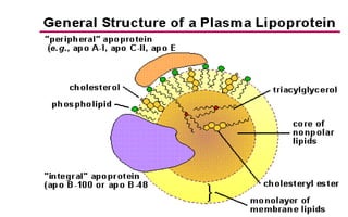
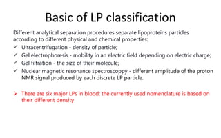
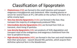
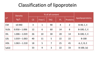
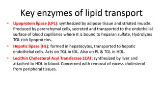
Basic of LP classification
Different analytical separation procedures separate lipoproteins particles
according to different physical and chemical properties:
 Ultracentrifugation - density of particle;
 Gel electrophoresis - mobility in an electric field depending on electric charge;
 Gel filtration - the size of their molecule;
 Nuclear magnetic resonance spectroscoppy - different amplitude of the proton
NMR signal produced by each discrete LP particle.
 There are six major LPs in blood; the currently used nomenclature is based on
their different density
• Chylomicrons (CM) are formed in the small intestine and transport
exogenous triacylglycerols and cholesterol. After standing plasma or
serum for several hours at 4°C, CM settle on the surface in the form of a
white creamy layer.
• Very low-density lipoproteins (VLDL) are formed in the liver, they
transport the majority of endogenously produced TAGs.
• Intermediate-density lipoproteins (IDL) are formed from VLDL as
transient, short-living LP particles.
• Low-density lipoproteins (LDL) are products of VLDL degradation and
transport most of the endogenous and exogenous cholesterol from the
liver to peripheral tissues.
• High-density lipoproteins (HDL) are formed in the liver and small intestine
from their precursors - nascent discoid HDL particles, which acquire their
definitive spherical form in the bloodstream.
Classification of lipoprotein
LP
Density
kg/L
% of all content
Apolipoproteins
CE Free C TAG PL Proteins
CM ≤0.940 3 1 90 4 2 B-48, C, E
VLDL 0.950 – 1.006 12 6 60 14 8 B-100, C, E
IDL 1.006 – 1.019 26 10 30 20 14 B-100, C, E
LDL 1.019 – 1.063 40 11 5 22 22 B-100
HDL 1.063 – 1.210 18 5 7 25 45 A, C, D, E
Lp(a) 33 9 3 22 33 B-100, (a)
Oil-drop of mixed micelle model of lipoprotein structure
APOLIPOPROTEINS
Apolipoproteins can be divided into integrals, which are a permanent part of
the lipoprotein particle (e.g. apoB) and freely associated, which LP particles
can exchange with each other (apoAI, AII, AIV, CI, CII, CIII, and E). Basic
functions of apolipoproteins are:
1. Structural:
– In CMs the structure is stabilized by the molecule apoB-48, which remains in
CM remnants until their uptake in the liver.
– ApoB-100 has a structural function in VLDL particles, which is also present in all
lipoprotein particles derived from VLDL (VLDL-remnants, IDL, LDL). In HDL
particles,
– apoprotein A (apo A) performs this function.
2. Specific receptor ligand: Part of the amino acid sequence of apoproteins is
the ligand for the receptor through which the LP particle enters the cell.
ApoB-100 and apoE have this function.
3. Activation or inhibition of the activity of enzymes involved in lipoprotein
metabolism: ApoA and apoC have this function concerning lecithin-
cholesterol acyl transferase (LCAT) and lipoprotein lipase (LPL).
Name Association
with LP
Function
Apo AI HDL
Structural protein for HDL; ligand for ABCA1 transporter,
activator of lecithin-cholesterol acyltransferase (LCAT).
Apo AII HDL Structural protein for HDL; inhibits cellular cholesterol efflux .
Apo IV HDL Activator of lipoprotein lipase (LPL) and LCAT .
Apo B48 CM Required for assembly and secretion of chylomicrons.
Apo
B100
VLDL, LDL
Structural protein for VLDL, IDL, LDL, and Lp(a); ligand for the
LDL receptor; required for assembly and secretion of VLDL.
Apo CI VLDL, HDL Activator of LCAT
Apo CII
CM, VLDL,
HDL
Essential cofactor for LPL.
Apo CIII
CM, VLDL,
HDL
Inhibition LPL and hepatic lipase.
Apo E
CM, VLDL,
HDL
Ligand for hepatic chylomicron and VLDL remnant receptor,
ligand for LDL receptor
• There are three different apoE alleles in humans: E-2, the most frequent E-
3, and E-4.
• Compared to apoE3, apoE2 has reduced affinity and apoE4 has enhanced
affinity for the LDL (apoB/E) receptor.
• ApoE-2 phenotype is associated with familiar dysbetalipoproteinemia with
an accelerated atherosclerosis.
• Homozygotes for E-4 have higher risk of Alzheimer´s diseases.
ApoE phenotypes
Key enzymes of lipid transport
• Lipoprotein lipase (LPL): synthesized by adipose tissue and striated muscle.
Produced by parenchymal cells, secreted and transported to the endothelial
surface of blood capillaries where it is bound to heparan sulfate. Hydrolyzes
TGL rich lipoproteins.
• Hepatic lipase (HL): formed in hepatocytes, transported to hepatic
endothelial cells. Acts on TGL in IDL. Also on PL & TGL in HDL.
• Lecithin Cholesterol Acyl Transferase LCAT: synthesized by liver and
attached to HDL in blood. Concerned with removal of excess cholesterol
from peripheral tissues.
Lipid metabolism
• occurs in three major areas
– Intestine
– Liver
– Extrahepatic tissues (Muscle and adipose tissue)
• In a normal diet, 95% of lipids are triacylglycerols (TAG), the remaining 5%
are all other dietary fats, including cholesterol (CH).
• Gastric lipase partially hydrolyzes TAG and released fatty acids (FA) activate
contraction of the gallbladder with the subsequent expulsion of bile
and pancreatic juice into the duodenum.
Absorption of dietary lipid
• Bile emulsifies TAG to smaller droplets
which are more accessible to digestion
by pancreatic lipase.
• Glycerol, FAs, and partly also 2-monoacylglycerol enter the enterocyte,
together with free cholesterol. Short- and medium-chain FAs can be
absorbed from the enterocyte into the bloodstream directly and circulate
bound to albumin. The longer fatty acids in the enterocyte are used for TAG
re-synthesis and cholesterol esterification.
Chylomicron metabolism
• Dietary lipids, including fat-soluble
vitamins, are resorbed in the small
intestinal mucosa and incorporated into
lipoprotein particles - chylomicrons
(CM). The major apolipoprotein in CM is
apolipoprotein B-48 (apoB-48) ), which
is synthesized in enterocytes.
• Chylomicrons enter the bloodstream
through the lymph. The highest
concentration of CM in the blood
occurs 3–6 hours after eating and after
10 – 12 hours of fasting are no longer
present in the blood of a healthy
person.
TAG
FA+GLYCEROL
TG
CHOLESTEROL
NASCENT CHYLOMICRON
A1
B48
CE
Chylomicron metabolism
• In circulation, CMs receive additional apoE and apoC from HDL particles. ApoC-II
is an activator of lipoprotein lipase (LPL), which hydrolyzes TAG from the core of
CM to form free fatty acids (FFA) and glycerol. FFAs are used by muscle cells
mostly as a source of energy, while TAGs are resynthesized in adipocytes
• FFAs are used by muscle cells mostly as a source of energy, while TAGs are
resynthesized in adipocytes.
• After hydrolysis of TAG, the CM core shrinks and smaller chylomicron
remnants (CMR) are formed. When the amount of TAG in the nucleus drops to
∽20%, CM submits apoC-II to the HDL particle losing the ability to further
hydrolyse TAG.
• Excess envelope phospholipids together with apoA are incorporated into HDL
particles.
• CMRs containing the rest of TAG and all dietary cholesterol are taken up by the
liver via apoE binding receptors.
absorption of dietary lipids
formation of nascent
chylomicrons
plasma hydrolysis by LPL
loss of ApoA and ApoC
and formation of
chylomicron ramenant
hepatic sequestration by
interaction of Apo-E with LDL
receptor
VLDL metabolism
• VLDL particles synthesized in the liver transport TAG (and partially cholesterol) from the
liver to peripheral tissues.
• The rate of VLDL synthesis is limited by the rate of hepatic synthesis of apoB-100, which is
their essential structural apoprotein.
• Increased hepatic TAG synthesis from FA or carbohydrate precursors, accelerated in
metabolic syndrome, diabetes, obesity, produces larger VLDL1 particles, while
physiological metabolism produces smaller VLDL2 particles.
• Similarly to CM, VLDL particles receive apoC and apoE from HDL particles upon entry into
the circulation.
• Both apoC-II and insulin activate endothelial LPL, which hydrolyzes TAG in the particle core.
If the content of TAG in VLDL decreases to about 30%, a transient particle - intermediate-
density lipoprotein (IDL) is formed.
• IDLs bind with the receptor on hepatocytes via apoE and they are subsequently
degraded, or they lose additional TAG by activity HL and change to LDL particles.
• IDL particles have a very short biological half-life under physiological circumstance, but are
highly atherogenic
FFA
TAG
VLDL
Nascent VLDL
APO B100
APO E,C (HDL)
PLASMA
In the liver: synthesis of VLDL by
incorporation of triglycerides with Apo B100,
and small amount of Apo C and Apo E.
After their secretion into plasma VLDL will
be enriched with Apo-E and Apo-C
transferred from HDL
VLDL
IDL
TG FFA
LPL
LPL TG
APO C,E(HDL)
LDL
CELL MEMBRANE
VIT D
MYELIN SHEATH
STEROID HORMONE
CHOLESTEROL
APO-B100
1
2
3
Transformation of
VLDL to IDL by loosing
Triglycerides and FFA IDL are normally transient in plasma and will be
rapidly eliminated
- by fixation on hepatic receptors and degradation
- or by plasma hydrolysis with loss of the
triglycerides, Apo-E and Apo-C and formation of
LDL rich in cholesterol
Interaction with LDL cell
receptor and Apo-B100
than release of cholesterol
into the cell
Metabolism of LDL
• LDL particles originate from the last stage of VLDL metabolism. They contain
only 10% TAG from the original VLDL particles, but all cholesterol and
structural apoB-100, the ligand for LDL receptors (LDL-R) both on the surface
of liver cells and cells of various peripheral tissues.
• The size of the LDL particles allows them to pass through the endothelium
into the interstitial space and can theoretically come into contact with any
cell.
• Approximately 30 – 40% of LDL are catabolized within 24 hours,
predominantly by LDL-R. The LDL particle bound to LDL-R is internalized into
the cell and, after cleavage of LDL-R, is hydrolyzed to cholesterol and amino
acids.
• The released LDL-R returns to the cell membrane surface.
• Intracellular cholesterol is a signal for the subsequent regulation of the
amount of LDL- R on the cell membrane so that the cell is not overfilled with
cholesterol. The entry of cholesterol into cells via LDL-R does not depend on
the level of cholesterol in the blood but the number of LDL-R on the cell
surface
• LDL particles are currently considered to be the most
pathogenic population of lipoproteins.
• LDL particles in the bloodstream are heterogeneous in
composition and size.
– Large, lighter LDL particles containing more TAGs are less atherogenic.
They are eliminated from the circulation by LDL-R; the amount
entering the cell is strictly regulated.
– The smaller and heavier LDL particle are more atherogenic. Slowed
catabolism, prolonged circulation of particles, and various
pathological conditions lead to the modification of LDL particles (e.g.
glycation, oxidation).
Metabolism of LDL
Formation of sdLDL particles
sdLDL and process of atherogenesis
• Small dense LDLs (sdLDL)
are not recognized by LDL-
R, due to their size they
enter the subendothelial
spaces by transcytosis,
where they aggregate and
easily oxidize.
Oxidized LDL particles are internalized into macrophages (as well as
other cells) via scavenger receptors (SR). A macrophage in the
subendothelial space crowded with LDL particles is known as a foam cell,
which is the first step in the process of atherogenesis.
Lp(a)
• Lp(a) binding to apo B100 has a structural similarity to plasminogen,
• Lp(a) interferes with fibrinolysis by competing with plasminogen
binding to plasminogen receptors on fibrinogen, and fibrin, leading
to decreased thrombolysis.
• Lp(a) has often been considered as an independent non modifiable
cardiovascular risk factor with atherogenic and also
thrombogenic potential
• The most quantity of cholesterol not used by cells is returned to the
liver via a process called reverse transport of cholesterol mediated by
HDL particles.
• The first particle of HDL is formed in an immature form (nascent
HDL) from phospholipids and apo A-I, and minimal cholesterol content.
• In the bloodstream they are enriched for the enzyme LCAT. These
small, discoid particles enter the subendothelial spaces of cells with
excess cholesterol by transcytosis, remove cholesterol from the cells,
and after its esterification with the LCAT enzyme, they move it to the
core of LP particle, thus acquiring a spherical shape (HDL2 and HDL3
particles)
• Excess cholesterol is transported by HDL particles back to the liver
• Due to the involvement of HDL particles in reverse cholesterol
transport, it has long been assumed that the higher the HDL-C level, the
lower the cardiovascular risk.
HDL metabolism
• FUNCTIONS
– Serves as a circulating reservoir for apo-C and apo-E.
– Delivering-chol. esters to liver for uptake by SR-B1
(reverse cholesterol transport).
– Protective effect - anti-inflammatory, anti-oxidative,
platelet anti-aggregatory, anticoagulant, and
profibrinolytic activities
HDL metabolism
• HDL and revers cholesterol transport
Lipid profile
Systematic lipid profile
• Total cholesterol
• HDL cholesterol
• LDL cholesterol
• Triglycerides
Extended lipid profile
• Apo A and Apo B
• Lipoprotein
electrophoresis
• Lp(a)
Preparation:
 Blood should be collected after a 12-hour fast (no food or drink,
except water).
 For the most accurate results, wait at least 2 months after a heart
attack, surgery, infection, injury or pregnancy to check cholesterol
levels.
 Cholesterol level is elevated during pregnancy (till 6 weeks after
delivery)
 Some drugs are known to increase cholesterol levels as anabolic
steroids, beta blockers, epinephrine, oral contraceptives and vit, D.
Specimen
• Serum
• Heparinized plasma
Normal rare common common rare common rare
Normal CM LDL LDL+VLDL IDL VLDL CM+VLDL
Aspect of fasting serum
Normal I IIa IIb III
IV V
1 2 3 4 4 5 6
Triglycerides determination
• GOP method
Enzymatic hydrolysis of triglycerides with subsequent
determination of glycerol by colorimetric method.
Triglycerides + H2O glycerol + fatty acids
Lipase
glycerol + ATP glycerol-3 P + ADP
Glycerol Kinase
Glycerol-3 P + O2 dihydroxyacetone phosphate + H2O2
Glycerol P-Oxydase
H2O2 + 4-AAP + 4-chlorophenol quinoneimine dye + 4 H2O
Peroxydase (POD)
The intensity of the red color produced is directly proportional to
the concentration of triglycerides in the sample when measured
at 510nm. Normal value < 1.5 g/l
Cholesterol determination
• GOP method
Enzymatic hydrolysis and oxydation of cholesterol with
subsequent determination by colorimetric method.
Esterified Cholesterol NE cholesterol + fatty acids
Cholesterol Esterase
NE chloesterol Cholestenon + H2O2
Cholesterol oxydase
H2O2 + 4-AAP + 4-chlorophenol quinoneimine dye + 4 H2O
Peroxydase (POD)
The intensity of the red color produced is directly proportional to
the concentration of cholesterol in the sample when measured at
510nm.
Normal value < 2 g/l
• Precipitation method
– after selective precipitation of LDL and VLDL with a polyanion cation
complex or phosphotungstic acid in the presence of divalent cations. HDL
cholesterol is assayed on the supernatant after centrifugation by
enzymatic technique.
• Directly determination
– The method depends on the properties of a detergent which solubilizes
only the HDL so that the HDL-c is released to react with the cholesterol
esterase, cholesterol oxidase and Chromogens to give color .The non HDL
lipoproteins LDL, VLDL and chylomicrons are inhibited from reacting with
the enzymes due to absorption of the detergents on their surfaces . The
intensity of the color formed is proportional to the HDL-c concentration in
the sample .
HDL-c determination
Referance range:
Men > 0.45 g/l
Women > 0.55g/l
TC/HDL-C= (atherogenicity index) is
used as a cardiovascular risk factor:
< 4.9 for Men
< 4.2 for Women.
• calculation method
LDL chol. = Total Chol. – (Trig /5 + HDL chol)
Not valid if triglycerides level is > 400 mg/dl
• Direct determination
after selective destruction of non LDL-cholesterol, LDL Cholesterol is
evaluated by the enzymatic couple Oxydase/peroxidase.
LDL-c determination
LDL Cholesterol reference value < 140 mg /l.
the pathological threshold is different depending on the number of risk
factors presented by the patient: HDL cholesterol< 0.35 g/l, high blood
pressure, ongoing smoking, diabetes, family coronary history or arterial
diseases, age (≥ 45 years in men, ≥ 55 years in women), early menopause .
Apoproteins AI and B determination
– Different immunological techniques are used for determination
of these two main apoproteins with specific antiserums and
purified standards (immunonephelometricy,
immunoturbidimetry, electro-immuno-diffusion….).
– Reference value: apoB = 0.6 -1.40 g
apoA1 > 1.10 g / l
By immunological techniques (immunonephelometry,
immunoturbidimetry).
Reference value: < 0.30 g / l.
Lp(a) derermination
LIPOPROTEIN ELECTROPHORESIS
this test is practical on agarose or
polyacrylamide gel.
After electrophoretic migration of LP and
staining, it allows to visualize qualitatively:
- an overload of chylomicron
- an increase in VLDL and LDL
- the presence of IDL or Lpa
- a relative decrease in HDL
Dyslipoproteinemias
• Dyslipoproteinemias (DLP) is a group of metabolic diseases that are
manifested by changes in the quality or quantity of serum lipoprotein
particles. They have a significant impact on cardiovascular and
cerebrovascular mortality, especially in developed countries.
• Classification of dyslipidemia
– The first classification was the Fredrickson classification based on
phenotypes observed in electrophoresis.
– Etiological classification : congenital / acquired
– European Atherosclerosis Society (EAS) classification, based on levels
of total cholesterol and triacylglycerols.
FREDRICKSON CLASSIFICATION OF HYPERLIPIDEMIA
Type I IIa IIb III IV V
Lipoprotein
pattern
↑CM ↑LDL
↑LDL
↑VLDL
↑IDL or
remnants ↑VLDL ↑VLDL
Cholesterol N/↑ ↑↑↑ ↑↑ ↑ ↑ ↑↑
TAG ↑↑↑ N/↑ ↑ ↑ ↑↑
↑↑
↑
Risk of AS - increased
Sings of MS - - central obesity, DM, hypertension, hyperuricemia
Pancreatitis,
abdominal
symptoms
+ - - - + +
Etiological classification
• The development of genetics and molecular biology makes it
possible to divide DLP into two basic groups:
– Primary : congenital, genetically determined
– Secondary: arising from other acute and chronic diseases,
drugs or nutritional factors.
• Most cases of lipoprotein disorders are due to an interaction
between genetic and environmental factors. The same disease
may not cause DLP in all patients and may lead to different DLP
phenotypes in different individuals.
Primitive hyperlipidemia
• Fredrickson's classification defined 6 classes of hyperlipidemia. It is
a phenotypic classification, based on abnormalities of lipid
metabolism objectified by lipoprotein electrophoresis.
 Type I : hyperchylomicronemia
 Type IIa : essential high cholesterol.
 Type IIb : Mixed hyperlipidemia
 Type III: dysbetalipidemia
 Type IV: endogenous hypertriglyceridemia
 Type V: mixed hypertriglyceridemia.
endothelial cell
transporter for
lipoprotein
lipase
Pathologie
Métabolique
Type selon
Fredrickson
Caractéristiques
Diabetes (type I ou II ) IV ou IIb ↓ Activité de la LPL (↓ insuline )
↑Synthèse des VLDL (↓ insuline )
Obesity IV ↑ VLDL
L’association : Obésité, hypertension,
diabète type caractérisé par :↓
cholestérol HDL
↑ risque cardiovasculaire
Hyperuricemia, gouts IV ou IIb
Cholestasis
intra- or extrahepatic
IIa ou IIb Lipoprotéines anormales : lipoprotéine X
↓ activité LCAT (présence des sels
biliaires ?
SECONDARY HYPERLIPIDEMIA
Pathologie
Métabolique
Type selon
Fredrickson
Caractéristiques
Insuffisance
rénale chronique
IV ↑ Synthèse VLDL
↓ Catabolisme des VLDL (↑ apoCIII ,
déficit de la lipase hépatique )
Syndrome
néphrotique
IV ou IIb ↑ Synthèse VLDL et ↓ Catabolisme des
VLDL (↓activité de la lipase hépatique)
Hypothyroïdie IIa ou IIb ↓ catabolisme des LDL et du cholestérol
Traitement par b-
bloquants
IV ↓ activité LPL
Traitement par des
corticoïdes
IV ou IIb ↓ activité LPL
SECONDARY HYPERLIPIDEMIA

lpp.pptx

  • 2.
    Basic of LPclassification Different analytical separation procedures separate lipoproteins particles according to different physical and chemical properties:  Ultracentrifugation - density of particle;  Gel electrophoresis - mobility in an electric field depending on electric charge;  Gel filtration - the size of their molecule;  Nuclear magnetic resonance spectroscoppy - different amplitude of the proton NMR signal produced by each discrete LP particle.  There are six major LPs in blood; the currently used nomenclature is based on their different density
  • 3.
    • Chylomicrons (CM)are formed in the small intestine and transport exogenous triacylglycerols and cholesterol. After standing plasma or serum for several hours at 4°C, CM settle on the surface in the form of a white creamy layer. • Very low-density lipoproteins (VLDL) are formed in the liver, they transport the majority of endogenously produced TAGs. • Intermediate-density lipoproteins (IDL) are formed from VLDL as transient, short-living LP particles. • Low-density lipoproteins (LDL) are products of VLDL degradation and transport most of the endogenous and exogenous cholesterol from the liver to peripheral tissues. • High-density lipoproteins (HDL) are formed in the liver and small intestine from their precursors - nascent discoid HDL particles, which acquire their definitive spherical form in the bloodstream. Classification of lipoprotein
  • 4.
    Classification of lipoprotein LP Density kg/L %of all content Apolipoproteins CE Free C TAG PL Proteins CM ≤0.940 3 1 90 4 2 B-48, C, E VLDL 0.950 – 1.006 12 6 60 14 8 B-100, C, E IDL 1.006 – 1.019 26 10 30 20 14 B-100, C, E LDL 1.019 – 1.063 40 11 5 22 22 B-100 HDL 1.063 – 1.210 18 5 7 25 45 A, C, D, E Lp(a) 33 9 3 22 33 B-100, (a)
  • 5.
    Oil-drop of mixedmicelle model of lipoprotein structure
  • 6.
    APOLIPOPROTEINS Apolipoproteins can bedivided into integrals, which are a permanent part of the lipoprotein particle (e.g. apoB) and freely associated, which LP particles can exchange with each other (apoAI, AII, AIV, CI, CII, CIII, and E). Basic functions of apolipoproteins are: 1. Structural: – In CMs the structure is stabilized by the molecule apoB-48, which remains in CM remnants until their uptake in the liver. – ApoB-100 has a structural function in VLDL particles, which is also present in all lipoprotein particles derived from VLDL (VLDL-remnants, IDL, LDL). In HDL particles, – apoprotein A (apo A) performs this function. 2. Specific receptor ligand: Part of the amino acid sequence of apoproteins is the ligand for the receptor through which the LP particle enters the cell. ApoB-100 and apoE have this function. 3. Activation or inhibition of the activity of enzymes involved in lipoprotein metabolism: ApoA and apoC have this function concerning lecithin- cholesterol acyl transferase (LCAT) and lipoprotein lipase (LPL).
  • 7.
    Name Association with LP Function ApoAI HDL Structural protein for HDL; ligand for ABCA1 transporter, activator of lecithin-cholesterol acyltransferase (LCAT). Apo AII HDL Structural protein for HDL; inhibits cellular cholesterol efflux . Apo IV HDL Activator of lipoprotein lipase (LPL) and LCAT . Apo B48 CM Required for assembly and secretion of chylomicrons. Apo B100 VLDL, LDL Structural protein for VLDL, IDL, LDL, and Lp(a); ligand for the LDL receptor; required for assembly and secretion of VLDL. Apo CI VLDL, HDL Activator of LCAT Apo CII CM, VLDL, HDL Essential cofactor for LPL. Apo CIII CM, VLDL, HDL Inhibition LPL and hepatic lipase. Apo E CM, VLDL, HDL Ligand for hepatic chylomicron and VLDL remnant receptor, ligand for LDL receptor
  • 8.
    • There arethree different apoE alleles in humans: E-2, the most frequent E- 3, and E-4. • Compared to apoE3, apoE2 has reduced affinity and apoE4 has enhanced affinity for the LDL (apoB/E) receptor. • ApoE-2 phenotype is associated with familiar dysbetalipoproteinemia with an accelerated atherosclerosis. • Homozygotes for E-4 have higher risk of Alzheimer´s diseases. ApoE phenotypes
  • 9.
    Key enzymes oflipid transport • Lipoprotein lipase (LPL): synthesized by adipose tissue and striated muscle. Produced by parenchymal cells, secreted and transported to the endothelial surface of blood capillaries where it is bound to heparan sulfate. Hydrolyzes TGL rich lipoproteins. • Hepatic lipase (HL): formed in hepatocytes, transported to hepatic endothelial cells. Acts on TGL in IDL. Also on PL & TGL in HDL. • Lecithin Cholesterol Acyl Transferase LCAT: synthesized by liver and attached to HDL in blood. Concerned with removal of excess cholesterol from peripheral tissues.
  • 10.
    Lipid metabolism • occursin three major areas – Intestine – Liver – Extrahepatic tissues (Muscle and adipose tissue)
  • 12.
    • In anormal diet, 95% of lipids are triacylglycerols (TAG), the remaining 5% are all other dietary fats, including cholesterol (CH). • Gastric lipase partially hydrolyzes TAG and released fatty acids (FA) activate contraction of the gallbladder with the subsequent expulsion of bile and pancreatic juice into the duodenum. Absorption of dietary lipid • Bile emulsifies TAG to smaller droplets which are more accessible to digestion by pancreatic lipase. • Glycerol, FAs, and partly also 2-monoacylglycerol enter the enterocyte, together with free cholesterol. Short- and medium-chain FAs can be absorbed from the enterocyte into the bloodstream directly and circulate bound to albumin. The longer fatty acids in the enterocyte are used for TAG re-synthesis and cholesterol esterification.
  • 13.
    Chylomicron metabolism • Dietarylipids, including fat-soluble vitamins, are resorbed in the small intestinal mucosa and incorporated into lipoprotein particles - chylomicrons (CM). The major apolipoprotein in CM is apolipoprotein B-48 (apoB-48) ), which is synthesized in enterocytes. • Chylomicrons enter the bloodstream through the lymph. The highest concentration of CM in the blood occurs 3–6 hours after eating and after 10 – 12 hours of fasting are no longer present in the blood of a healthy person. TAG FA+GLYCEROL TG CHOLESTEROL NASCENT CHYLOMICRON A1 B48 CE
  • 14.
    Chylomicron metabolism • Incirculation, CMs receive additional apoE and apoC from HDL particles. ApoC-II is an activator of lipoprotein lipase (LPL), which hydrolyzes TAG from the core of CM to form free fatty acids (FFA) and glycerol. FFAs are used by muscle cells mostly as a source of energy, while TAGs are resynthesized in adipocytes • FFAs are used by muscle cells mostly as a source of energy, while TAGs are resynthesized in adipocytes. • After hydrolysis of TAG, the CM core shrinks and smaller chylomicron remnants (CMR) are formed. When the amount of TAG in the nucleus drops to ∽20%, CM submits apoC-II to the HDL particle losing the ability to further hydrolyse TAG. • Excess envelope phospholipids together with apoA are incorporated into HDL particles. • CMRs containing the rest of TAG and all dietary cholesterol are taken up by the liver via apoE binding receptors.
  • 15.
    absorption of dietarylipids formation of nascent chylomicrons plasma hydrolysis by LPL loss of ApoA and ApoC and formation of chylomicron ramenant hepatic sequestration by interaction of Apo-E with LDL receptor
  • 16.
    VLDL metabolism • VLDLparticles synthesized in the liver transport TAG (and partially cholesterol) from the liver to peripheral tissues. • The rate of VLDL synthesis is limited by the rate of hepatic synthesis of apoB-100, which is their essential structural apoprotein. • Increased hepatic TAG synthesis from FA or carbohydrate precursors, accelerated in metabolic syndrome, diabetes, obesity, produces larger VLDL1 particles, while physiological metabolism produces smaller VLDL2 particles. • Similarly to CM, VLDL particles receive apoC and apoE from HDL particles upon entry into the circulation. • Both apoC-II and insulin activate endothelial LPL, which hydrolyzes TAG in the particle core. If the content of TAG in VLDL decreases to about 30%, a transient particle - intermediate- density lipoprotein (IDL) is formed. • IDLs bind with the receptor on hepatocytes via apoE and they are subsequently degraded, or they lose additional TAG by activity HL and change to LDL particles. • IDL particles have a very short biological half-life under physiological circumstance, but are highly atherogenic
  • 17.
    FFA TAG VLDL Nascent VLDL APO B100 APOE,C (HDL) PLASMA In the liver: synthesis of VLDL by incorporation of triglycerides with Apo B100, and small amount of Apo C and Apo E. After their secretion into plasma VLDL will be enriched with Apo-E and Apo-C transferred from HDL
  • 18.
    VLDL IDL TG FFA LPL LPL TG APOC,E(HDL) LDL CELL MEMBRANE VIT D MYELIN SHEATH STEROID HORMONE CHOLESTEROL APO-B100 1 2 3 Transformation of VLDL to IDL by loosing Triglycerides and FFA IDL are normally transient in plasma and will be rapidly eliminated - by fixation on hepatic receptors and degradation - or by plasma hydrolysis with loss of the triglycerides, Apo-E and Apo-C and formation of LDL rich in cholesterol Interaction with LDL cell receptor and Apo-B100 than release of cholesterol into the cell
  • 19.
    Metabolism of LDL •LDL particles originate from the last stage of VLDL metabolism. They contain only 10% TAG from the original VLDL particles, but all cholesterol and structural apoB-100, the ligand for LDL receptors (LDL-R) both on the surface of liver cells and cells of various peripheral tissues. • The size of the LDL particles allows them to pass through the endothelium into the interstitial space and can theoretically come into contact with any cell. • Approximately 30 – 40% of LDL are catabolized within 24 hours, predominantly by LDL-R. The LDL particle bound to LDL-R is internalized into the cell and, after cleavage of LDL-R, is hydrolyzed to cholesterol and amino acids. • The released LDL-R returns to the cell membrane surface. • Intracellular cholesterol is a signal for the subsequent regulation of the amount of LDL- R on the cell membrane so that the cell is not overfilled with cholesterol. The entry of cholesterol into cells via LDL-R does not depend on the level of cholesterol in the blood but the number of LDL-R on the cell surface
  • 21.
    • LDL particlesare currently considered to be the most pathogenic population of lipoproteins. • LDL particles in the bloodstream are heterogeneous in composition and size. – Large, lighter LDL particles containing more TAGs are less atherogenic. They are eliminated from the circulation by LDL-R; the amount entering the cell is strictly regulated. – The smaller and heavier LDL particle are more atherogenic. Slowed catabolism, prolonged circulation of particles, and various pathological conditions lead to the modification of LDL particles (e.g. glycation, oxidation). Metabolism of LDL
  • 22.
  • 23.
    sdLDL and processof atherogenesis • Small dense LDLs (sdLDL) are not recognized by LDL- R, due to their size they enter the subendothelial spaces by transcytosis, where they aggregate and easily oxidize. Oxidized LDL particles are internalized into macrophages (as well as other cells) via scavenger receptors (SR). A macrophage in the subendothelial space crowded with LDL particles is known as a foam cell, which is the first step in the process of atherogenesis.
  • 24.
    Lp(a) • Lp(a) bindingto apo B100 has a structural similarity to plasminogen, • Lp(a) interferes with fibrinolysis by competing with plasminogen binding to plasminogen receptors on fibrinogen, and fibrin, leading to decreased thrombolysis. • Lp(a) has often been considered as an independent non modifiable cardiovascular risk factor with atherogenic and also thrombogenic potential
  • 25.
    • The mostquantity of cholesterol not used by cells is returned to the liver via a process called reverse transport of cholesterol mediated by HDL particles. • The first particle of HDL is formed in an immature form (nascent HDL) from phospholipids and apo A-I, and minimal cholesterol content. • In the bloodstream they are enriched for the enzyme LCAT. These small, discoid particles enter the subendothelial spaces of cells with excess cholesterol by transcytosis, remove cholesterol from the cells, and after its esterification with the LCAT enzyme, they move it to the core of LP particle, thus acquiring a spherical shape (HDL2 and HDL3 particles) • Excess cholesterol is transported by HDL particles back to the liver • Due to the involvement of HDL particles in reverse cholesterol transport, it has long been assumed that the higher the HDL-C level, the lower the cardiovascular risk. HDL metabolism
  • 26.
    • FUNCTIONS – Servesas a circulating reservoir for apo-C and apo-E. – Delivering-chol. esters to liver for uptake by SR-B1 (reverse cholesterol transport). – Protective effect - anti-inflammatory, anti-oxidative, platelet anti-aggregatory, anticoagulant, and profibrinolytic activities HDL metabolism
  • 27.
    • HDL andrevers cholesterol transport
  • 28.
    Lipid profile Systematic lipidprofile • Total cholesterol • HDL cholesterol • LDL cholesterol • Triglycerides Extended lipid profile • Apo A and Apo B • Lipoprotein electrophoresis • Lp(a)
  • 29.
    Preparation:  Blood shouldbe collected after a 12-hour fast (no food or drink, except water).  For the most accurate results, wait at least 2 months after a heart attack, surgery, infection, injury or pregnancy to check cholesterol levels.  Cholesterol level is elevated during pregnancy (till 6 weeks after delivery)  Some drugs are known to increase cholesterol levels as anabolic steroids, beta blockers, epinephrine, oral contraceptives and vit, D. Specimen • Serum • Heparinized plasma
  • 30.
    Normal rare commoncommon rare common rare Normal CM LDL LDL+VLDL IDL VLDL CM+VLDL Aspect of fasting serum Normal I IIa IIb III IV V 1 2 3 4 4 5 6
  • 31.
    Triglycerides determination • GOPmethod Enzymatic hydrolysis of triglycerides with subsequent determination of glycerol by colorimetric method. Triglycerides + H2O glycerol + fatty acids Lipase glycerol + ATP glycerol-3 P + ADP Glycerol Kinase Glycerol-3 P + O2 dihydroxyacetone phosphate + H2O2 Glycerol P-Oxydase H2O2 + 4-AAP + 4-chlorophenol quinoneimine dye + 4 H2O Peroxydase (POD) The intensity of the red color produced is directly proportional to the concentration of triglycerides in the sample when measured at 510nm. Normal value < 1.5 g/l
  • 32.
    Cholesterol determination • GOPmethod Enzymatic hydrolysis and oxydation of cholesterol with subsequent determination by colorimetric method. Esterified Cholesterol NE cholesterol + fatty acids Cholesterol Esterase NE chloesterol Cholestenon + H2O2 Cholesterol oxydase H2O2 + 4-AAP + 4-chlorophenol quinoneimine dye + 4 H2O Peroxydase (POD) The intensity of the red color produced is directly proportional to the concentration of cholesterol in the sample when measured at 510nm. Normal value < 2 g/l
  • 33.
    • Precipitation method –after selective precipitation of LDL and VLDL with a polyanion cation complex or phosphotungstic acid in the presence of divalent cations. HDL cholesterol is assayed on the supernatant after centrifugation by enzymatic technique. • Directly determination – The method depends on the properties of a detergent which solubilizes only the HDL so that the HDL-c is released to react with the cholesterol esterase, cholesterol oxidase and Chromogens to give color .The non HDL lipoproteins LDL, VLDL and chylomicrons are inhibited from reacting with the enzymes due to absorption of the detergents on their surfaces . The intensity of the color formed is proportional to the HDL-c concentration in the sample . HDL-c determination Referance range: Men > 0.45 g/l Women > 0.55g/l TC/HDL-C= (atherogenicity index) is used as a cardiovascular risk factor: < 4.9 for Men < 4.2 for Women.
  • 34.
    • calculation method LDLchol. = Total Chol. – (Trig /5 + HDL chol) Not valid if triglycerides level is > 400 mg/dl • Direct determination after selective destruction of non LDL-cholesterol, LDL Cholesterol is evaluated by the enzymatic couple Oxydase/peroxidase. LDL-c determination LDL Cholesterol reference value < 140 mg /l. the pathological threshold is different depending on the number of risk factors presented by the patient: HDL cholesterol< 0.35 g/l, high blood pressure, ongoing smoking, diabetes, family coronary history or arterial diseases, age (≥ 45 years in men, ≥ 55 years in women), early menopause .
  • 35.
    Apoproteins AI andB determination – Different immunological techniques are used for determination of these two main apoproteins with specific antiserums and purified standards (immunonephelometricy, immunoturbidimetry, electro-immuno-diffusion….). – Reference value: apoB = 0.6 -1.40 g apoA1 > 1.10 g / l By immunological techniques (immunonephelometry, immunoturbidimetry). Reference value: < 0.30 g / l. Lp(a) derermination
  • 36.
    LIPOPROTEIN ELECTROPHORESIS this testis practical on agarose or polyacrylamide gel. After electrophoretic migration of LP and staining, it allows to visualize qualitatively: - an overload of chylomicron - an increase in VLDL and LDL - the presence of IDL or Lpa - a relative decrease in HDL
  • 38.
    Dyslipoproteinemias • Dyslipoproteinemias (DLP)is a group of metabolic diseases that are manifested by changes in the quality or quantity of serum lipoprotein particles. They have a significant impact on cardiovascular and cerebrovascular mortality, especially in developed countries. • Classification of dyslipidemia – The first classification was the Fredrickson classification based on phenotypes observed in electrophoresis. – Etiological classification : congenital / acquired – European Atherosclerosis Society (EAS) classification, based on levels of total cholesterol and triacylglycerols.
  • 39.
    FREDRICKSON CLASSIFICATION OFHYPERLIPIDEMIA Type I IIa IIb III IV V Lipoprotein pattern ↑CM ↑LDL ↑LDL ↑VLDL ↑IDL or remnants ↑VLDL ↑VLDL Cholesterol N/↑ ↑↑↑ ↑↑ ↑ ↑ ↑↑ TAG ↑↑↑ N/↑ ↑ ↑ ↑↑ ↑↑ ↑ Risk of AS - increased Sings of MS - - central obesity, DM, hypertension, hyperuricemia Pancreatitis, abdominal symptoms + - - - + +
  • 40.
    Etiological classification • Thedevelopment of genetics and molecular biology makes it possible to divide DLP into two basic groups: – Primary : congenital, genetically determined – Secondary: arising from other acute and chronic diseases, drugs or nutritional factors. • Most cases of lipoprotein disorders are due to an interaction between genetic and environmental factors. The same disease may not cause DLP in all patients and may lead to different DLP phenotypes in different individuals.
  • 41.
    Primitive hyperlipidemia • Fredrickson'sclassification defined 6 classes of hyperlipidemia. It is a phenotypic classification, based on abnormalities of lipid metabolism objectified by lipoprotein electrophoresis.  Type I : hyperchylomicronemia  Type IIa : essential high cholesterol.  Type IIb : Mixed hyperlipidemia  Type III: dysbetalipidemia  Type IV: endogenous hypertriglyceridemia  Type V: mixed hypertriglyceridemia.
  • 45.
  • 46.
    Pathologie Métabolique Type selon Fredrickson Caractéristiques Diabetes (typeI ou II ) IV ou IIb ↓ Activité de la LPL (↓ insuline ) ↑Synthèse des VLDL (↓ insuline ) Obesity IV ↑ VLDL L’association : Obésité, hypertension, diabète type caractérisé par :↓ cholestérol HDL ↑ risque cardiovasculaire Hyperuricemia, gouts IV ou IIb Cholestasis intra- or extrahepatic IIa ou IIb Lipoprotéines anormales : lipoprotéine X ↓ activité LCAT (présence des sels biliaires ? SECONDARY HYPERLIPIDEMIA
  • 47.
    Pathologie Métabolique Type selon Fredrickson Caractéristiques Insuffisance rénale chronique IV↑ Synthèse VLDL ↓ Catabolisme des VLDL (↑ apoCIII , déficit de la lipase hépatique ) Syndrome néphrotique IV ou IIb ↑ Synthèse VLDL et ↓ Catabolisme des VLDL (↓activité de la lipase hépatique) Hypothyroïdie IIa ou IIb ↓ catabolisme des LDL et du cholestérol Traitement par b- bloquants IV ↓ activité LPL Traitement par des corticoïdes IV ou IIb ↓ activité LPL SECONDARY HYPERLIPIDEMIA

Editor's Notes

  • #2 Structure of lipoprotein
  • #5 There are 5 classes of lipoproteins which vary between them according to the density and their volume which go in opposite direction: the lightest (chylomicron) is the most bulky and very rich in triglycerides. Transports exogenous triglycerides. VLDL transports endogenous triglycerides (synthetized by the liver) and chlosterol. IDL intermediate form between VLDL and LDL less rich in triglycerides and richer in cholesterol than VLDL LDL rich in cholesterol. Carry cholesterol from liver to extra hepatic tissue (HDL) is the most dense, the smallest and the richest in protein.
  • #6 Comparative shape of different lipoproteins
  • #10 There are 3 essential enzymes involved in the metabolism of LP - Lipoprotein lipase (LPL) plays an essential role in the hydrolysis of triglycerides and clarification of serum (because a high triglyceride level makes the serum lactescent). LPL is present at the surface of endothelial blood capillaries. He is activated by Apo-CII transported by Chyl., VLDL, IDL, HDL. - Hepatic lipase (HL): plays the same role of LPL in the Liver. - Lecithin Cholesterol Acyl Transferase (LCAT) : ensures the esterification of free cholesterol captured in peripheral tissues by HDL to be thus transferred to the liver.
  • #13 Dietary lipids are emulsified by the bile in the small intestine. Lipids and bile interact to form micelles (like oil in water). Micelles are formed by fatty acid (FA) , monoglycerids, and cholesterol.
  • #16 Metabolism of chylomicron: 1- absorption of dietary lipids 2- formation of nascent chylomicrons 3- plasma hydrolysis by LPL 4- loss of ApoA and ApoC and formation of chylomicron ramenant 5- hepatic sequestration by interaction of Apo-E with LDL receptor.
  • #17 VLDLs are produced in the liver. They transport endogenous triglycerides synthesized by the liver from FFA of food origin or de novo synthesis VLDL in the liver before secretion into plasma contain ApoB-100, (protein synthesized by the liver) and small amounts of apoE & C (triglycerides represent environ 60% of the composition of VLDL). After their secretion into plasma, VLDL will be enriched with apoE & C released by plasma HDL.
  • #18 In the liver: synthesis of VLDL by incorporation of triglycerides with Apo B100, and small amount of Apo C and Apo E. After their secretion into plasma VLDL will be enriched with Apo-E and Apo-C transferred from HDL.
  • #19 1- Transformation of VLDL to IDL by loosing Triglycerides and FFA 2- IDL are normally transient in plasma and will be rapidly eliminated - by fixation on hepatic receptors and degradation - or by plasma hydrolysis with loss of the triglycerides, Apo-E and Apo-C and formation of LDL rich . 3- Interaction with LDL cell receptor and Apo-B100 than release of cholesterol into the cell.
  • #20 The VLDL triglycerides will be hydrolyzed by plasma LPL (like chylomicrons). This hydrolysis leads to the formation of IDL (Intermediate density lipoprotein) at lower triglyceride levels (<30%) 2 potential destinations for IDLs containing ApoB-100 - 40-60% will be eliminated from the plasma by the liver via the interaction with LDL receptors. Or LPL and HL convert the rest of the IDL to LDL [TG <10%]. Apo E, C, will be redistributed to HDL. LDL particles contain a core of cholesterol esters and a smaller amount of triglyceride.
  • #21 after fixation on the receptor site, LDL is internalized and degraded to free cholesterol, fatty acids and amino acids from Apo-B. Free cholesterol can: - be used for the structure of membranes - be stored in the form of esterified cholesterol after action of an enzyme, Acyl-Cholesterol-Acyl-Transferase (ACAT). inhibit  hydroxy  methylglutaryl CoA reductase, an enzyme regulating the synthesis of cholesterol. inhibit the synthesis of apo B-100 receptors
  • #27 HDL cholesterol have several functions intervene in the lipolysis of VLDL and chylomicron by transferring Apo-CII (activator of LPL) to them. And recover, after lipolysis of the particles , phospholipids, free cholesterol and apoproteins. bring back to the liver from tissues the cholesterol not exchanged with LDL triglycerides. Protective effect anti-inflammatory, antioxidant, anti-platelet aggregation, anticoagulant and profibrinolytic activities (true anti-atherogenic role)
  • #31 Aspect of the serum; for a 12-hour fasting serum, the serum must be clear, that is to say with a low level of VLDL and without chylomicron if it is opalescent there is an excess of VLDL, if it is lactescent, chylomicrons are present to confirm the actual presence of the chylomicrons, the serum is kept 24 h at + 4o and the microns then make a cream on the surface of the serum. 1- Clair normal 2- presence of chylomicron only 3- Clair with hyper cholesterol only 4- opalescent , presence of VLDL + cholesterol 5- lactesent presence of VLDL 6- lactesent + cream on surface presence of VLDL and Chylomicron
  • #32 Triglycerides in the sample are hydrolyzed by lipase to glycerol and fatty acids. The glycerol is then phosphorylated by ATP to glycerol-3-phosphate (G3P) and ADP in a reaction catalyzed by glycerol kinase (GK). G3P is then converted to dihydroxyacetone phosphate (DAP) and hydrogen peroxide(H2O2) by glycerophosphate oxidase (GPO). H2O2 then reacts with 4-aminoantipyrine (4-AAP) and 4-chlorophenol in a reaction catalyzed by peroxidase(POD) to yield a red colored quinoneimine dye. The intensity of the color produced is directly proportional to the concentration of triglycerides in the sample when measured at 510nm.
  • #33 cholesterol in the sample are hydrolyzed by cholesterol esterase to free cholesterol and fatty acids. The cholesterol is then oxidized to cholestenon and hydrogen peroxide(H2O2) by cholesterol oxidase . H2O2 then reacts with 4-aminoantipyrine (4-AAP) and 4-chlorophenol in a reaction catalyzed by peroxidase(POD) to yield a red colored quinoneimine dye. The intensity of the color produced is directly proportional to the concentration of triglycerides in the sample when measured at 510nm.
  • #37 it is a second-line examination which provides additional information in the case of serums with hypertriglyceridemia or mixed hyperlipemia. it allows a more precise typing of these hyperlipoproteinemies.
  • #43 Familial Hyperchylomiconemia (type I) characterized by a strong hypertriglyceridemia of exogenous origin with accumulation of chylomicrons. the genetic defect relates either to lipoprotein lipase or to its activator apo-CII. the enzyme is therefore either absent or inactive and the chylomicrons are not degraded.
  • #44 Familial hypercholesterolemia The main anomaly concerns the gene encoding the apo-B100 / E LDL receptor or is caused by mutations in the apo-B100 gene at its binding site on receptors. In all cases, LDL is not captured by the cells. The appearance of the serum is clear. Total cholesterol increases, LDL and apo-B increase. HDL and normal triglycerides
  • #45 Type III hyperlipoproteinemia. In this type the intermediate lipoproteins LDL and chylomicrons Remnant are not purified by hepatic lipase because the hepatic receptor sites with apo E (E1, E2, E3, E4) do not recognize the E2/E2 phenotype. The affected patient are homozygous E2/E2 type or heterozygote E2/E3 type. The accumulation of IDL and VLDL will be metabolized by the macrophage pathway generating atheroma plaques such as type IIa. therefore high cardiovascular risk
  • #46 Familial hypertriglyceridemia type IV Type IV or hypertriglyceridemia of endogenous origin is due to an increase in the hepatic synthesis of VLDL and to a slowing down of their catabolism. apo CII inhibitor of LPL can be increased. the serum is opalescent, cholesterol is normal or slightly increased, the triglycerides are very high and especially the triglycerides of VLDL. HDL and apo-A1 are decreasing. Familial hypertriglyceridemia type V type V is due to the increase in chylomicrons and VLDL . It’s a combination of type I and IV. the transmission mode is not determined. hypertriglyceridemia can be aggravated by a high consumption of carbohydrates alcohol and fats.