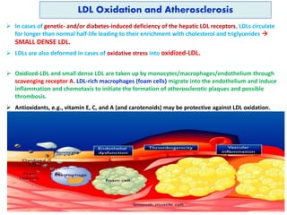
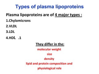
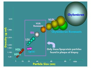
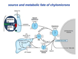
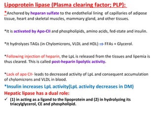
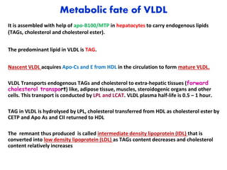
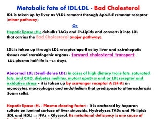
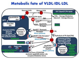
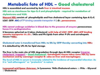
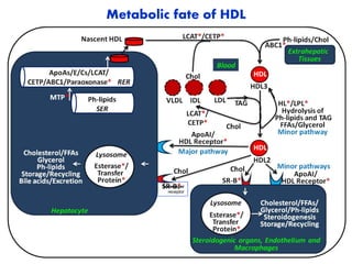
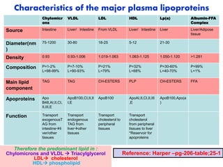
Dr. Umrana Mirza
Assistant Professor
Department of Biochemistry
AIMS
References:
1. Harper’s Biochemistry
2. Textbook of Biochemistry by
Vasudevan
Lipoproteins are molecular complexes that consist of lipids and a
specific carrier protein called apolipoprotein or apoprotein and
act as lipid transport molecules.
•
LIPIDS ARE TRANSPORTED IN THE PLASMA AS
LIPOPROTEINS
Lipoproteins are of two categories:
Structural (in Cellular/subcellular membranes).
Transport Functional (Plasma lipoproteins – P-LPs).
 Non-covalent forces stabilize the integration of protein and
lipid components of Plasma lipoproteins.
 The major function of Plasma lipoproteins is the transport and
inter-tissue homeostasis of triacylglycerols, cholesterol,
phospholipids and fat-soluble vitamins through the circulation
(blood and lymph).
Types of plasma lipoproteins
Plasma lipoproteins are of 4 major types :
1.Chylomicrons
2.VLDL
3.LDL
.1
4.HDL
They differ in the:
molecular weight
size
density
lipid and protein composition and
physiological role
Separation Methods resolve 4 major Types of
Plasma lipoproteins
Ultra-centrifugation: Depends on floatation
rate and density in NaCl solution:
Chylomicrons (<0.96),
VLDL (<1.006),
LDL (1.006-1.063),
HDL (1.063-1.21),
.
Paper or agarose gel Electrophoresis:
Depends on migration rate and net charge.
Chylomicrons,
-lipoproteins (LDL),
Pre--lipoproteins (VLDL),
-lipoproteins (HDL).
Structure of lipoproteins:
Lipoproteins Consist of a Nonpolar Core & a
Single surface layer of amphipathic Lipids.
The nonpolar lipid core consists of mainly
Triacylglycerol and cholesteryl ester and is
surrounded by a single surface layer of
Amphipathic phospholipid and Cholesterol
molecules.
These are oriented so that their polar groups face
outward to the aqueous medium, as in the cell
membrane.
The protein moiety of a lipoprotein is known as
An apolipoprotein or apoprotein, constituting
Nearly 70% of some HDL and as little as 1% of
chylomicrons.
Some apolipoproteins are:
1. integral- and cannot be removed (eg. Apo
B100 and Apo B 48), whereas others are
2. peripheral apoproteins- free to transfer to
other lipoproteins, (eg. Apo A ,apo Cs ect).
ApoA
ApoB
Types and Functions of Apolipoproteins (apoproteins)
Apolipoprotein A : is a peripheral protein (from intestine/liver): On HDL and
chylomicrons.
 ApoAI: Activates LCAT and is the ligand for HDL receptor. anti-atherogenic. It is
specific for HDL.
 ApoAII: Inhibits LCAT.
Apolipoprotein B: is an integral and structural component of VLDL and chylomicrons and
is a Ligand for LDL receptor.
 ApoB100: On VLDL, IDL and LDL (from liver).
 ApoB48: On Chylomicrons (from intestine).
Apolipoprotein C (from liver) : On HDL, chylomicrons,
VLDL.
 ApoCI: Possibly activates LCAT.
 ApoCII: Activates lipoprotein lipase (LPL).
 ApoCIII: Antagonizes apoCII to inhibits LPL.
Apolipoprotein D (from liver): On HDL. It is a Cholesterol Ester Transfer Protein;
CETP.
Apolipoprotein E (1, 2, 3,4; from liver/Macrophages): On chylomicron/remnants, VLDL,
IDL and HDL. It is a ligand for remnants.
Apo E-IV isoform is implicated in
the development of senile
dementia and Alzheimer's
disease. Apo-E 4 is also
associated with lipoprotein
glomerulopathy.
source and metabolic fate of chylomicrons
 Carry dietary(exogenous) lipids (TAGs, cholesterol and fat-soluble vitamins)
that are than stored in adipose tissue, muscle or liver.
 It is assembled in the intestinal mucosa with the help of ApoB48 and the
microsomal transfer protein (MTP) to form nascent chylomicrons
 nascent chylomicrons has apo B48 and apo A
 The predominant lipid in chylomicrons is TAG.
 it acquires Apo-CI,II,III & E from HDL to get converted to mature chylomicron.
Its plasma half-life is 5-20 minutes.
 It is debulked by hydrolysis of TAG into FFAs and glycerol by lipoprotein lipase
(LPL) and returns Apo-Cs and As to HDL.
 The molecule now is called chylomicron remnant that is taken up into the liver
and macrophages by receptor-mediated endocytosis. ApoE is a ligand for these
receptors
 cholesteryl esters and triacylglycerols are further hydrolyzed and metabolized
by hepatic lipase in liver.
source and metabolic fate of chylomicrons
Lipoprotein lipase (Plasma clearing factor; PLP):
*Anchored by heparan sulfate to the endothelial lining of capillaries of adipose
tissue, heart and skeletal muscles, mammary gland, and other tissues.
*It is activated by Apo-CII and phospholipids, amino acids, fed-state and insulin.
*It hydrolyzes TAGs (in Chylomicrons, VLDL and HDL)  FFAs + Glycerol.
*Following injection of heparin, the LpL is released from the tissues and lipemia is
thus cleared. This is called post-heparin lipolytic activity.
*Lack of apo CII- leads to decreased activity of LpL and consequent accumulation
of chylomicrons and VLDL in blood.
*Insulin increases LpL activity(LpL activity decreases in DM)
Hepatic lipase has a dual role:
 (1) in acting as a ligand to the lipoprotein and (2) in hydrolyzing its
triacylglycerol, CE and phospholipid.
Metabolic fate of VLDL
It is assembled with help of apo-B100/MTP in hepatocytes to carry endogenous lipids
(TAGs, cholesterol and cholesterol ester).
The predominant lipid in VLDL is TAG.
Nascent VLDL acquires Apo-Cs and E from HDL in the circulation to form mature VLDL.
VLDL Transports endogenous TAGs and cholesterol to extra-hepatic tissues (forward
cholesterol transport) like, adipose tissue, muscles, steroidogenic organs and other
cells. This transport is conducted by LPL and LCAT. VLDL plasma half-life is 0.5 – 1 hour.
TAG in VLDL is hydrolysed by LPL, cholesterol transferred from HDL as cholesterol ester by
CETP and Apo As and CII returned to HDL
The remnant thus produced is called intermediate density lipoprotein (IDL) that is
converted into low density lipoprotein (LDL) as TAGs content decreases and cholesterol
content relatively increases
Lecithin cholesterol acyl transferase ( LCAT):
LCAT is a plasma enzyme synthesized in liver. It circulates in
plasma with HDL and LDL. The substrate for LCAT is
phosphatidylcholine, lecithin( a Component of phospholipids
bilayer of HDL).
plays a significant role in removing cholesterol from the
circulating lipoproteins and from the tissues.
Apo A1 which is associated with HDL is a powerful
activator of LCAT.
LCAT catalyzes the following reversible reaction:
Lecithin + Cholesterol Cholesterol esters + Lysolecithin
LCAT deficiency (Fish eye disease) causes very low HDL cholesterol
and high triglyceride level, hemolytic anemia, proteinuria with
renal failure and corneal opacification.
LCAT
Metabolic fate of IDL/LDL - Bad Cholesterol
IDL is taken up by liver as VLDL remnant through Apo-B/E remnant receptor
(minor pathway).
Or,
Hepatic lipase (HL) debulks TAGs and Ph-Lipids and converts it into LDL
that carries the Bad Cholesterol (major pathway).
LDL is taken up through LDL receptor/apo-B100 by liver and extrahepatic
tissues and steroidogenic organs - forward cholesterol transport.
LDL plasma half-life is ~2.5 days.
Abnormal LDL (Small-dense LDL) in cases of high dietary trans-fats, saturated
fats, and CHO, diabetes mellitus, mutant apoB100 and/or LDL receptor and
oxidative stress – it is taken up by scavenger receptor A (SR-A) on
monocytes, macrophages and endothelium that predispose to atherosclerosis
(foam cells).
Hepatic lipase (HL - Plasma clearing factor) : It is anchored by heparan
sulfate on luminal surface of liver sinusoids. Hydrolyzes TAGs and Ph-lipids
(IDL and HDL)  FFAs + Glycerol. Its mutational deficiency is one cause of
Metabolic fate of VLDL/IDL/LDL
Metabolism of LDL and LDL Receptors
The LDL is taken up by peripheral tissues by receptor
mediated Endocytosis.
LDL receptors/apo B100 receptors are located in specialized
regions called clathrin coated pits.
Binding of LDL to the receptor by apo B100 and uptake of
cholesterol from LDL is a highly regulated process.
When the apo B-100 binds to the apo-B-100 receptor, the
receptor-LDL complex is internalized by endocytosis.
The endosome vesicle thus formed fuses with lysosomes.
The receptor is recycled and returns to the cell surface. The
LDL particle, along with apoproteins and cholesterol ester
are hydrolyzed by lysosomal hydrolases, forming amino
acids and free cholesterol.
The cholesterol thus liberated in the cell has three major
fates:
i. It is used for the synthesis of other steroids like steroid
hormones.
ii. Cholesterol may be incorporated into the membranes.
iii. Cholesterol may be esterified to CE by ACAT acyl
Metabolic fate of HDL – Good cholesterol
HDL is assembled and secreted by both liver & intestinal mucosa.
HDL acts as storehouse for Apo-Cs/E and phospholipids - required for metabolism of
chylomicrons and VLDL.
Nascent HDL consists of phospholipids and free cholesterol layer containing Apo-A/Cs/E,
LCAT, CETP, ABC1(ATP-binding cassette transporter A1)/G1, paraoxonase.
HDL cannot undergo oxidation in blood due to the presence of enzyme paraoxonase,
hence it is cardioprotective.
It becomes spherical as it takes cholesterol (with help of LCAT, CETP, ABC1(ATP binding
cassette transporter A1)/G1), TAGs and Ph-Lipids from other P-LPs and extrahepatic
tissuesHDL3.
Cholesterol ester is transferred from HDL3 to IDL by CETP thereby converting into HDL2
It is debulked by LPL/HL for lipid storage.
The liver is the main site of HDL degradation through Apo-AI/HDL receptor (reverse
cholesterol transport – Good cholesterol). Extrahepatic tissues takes some HDL by same
mechanism and by scavenger receptor B1 (also on liver and macrophages).
The level of HDL in serum is inversely related to the incidence of myocardial infarction. As
it is “anti-atherogenic” or “protective” in nature
Lysosomal acid lipase (Esterase): In lysosomes. TAGs/Cholesterol-esters  FFAs + Glycerol
+ Cholesterol.
Metabolic fate of HDL
Scavenger
receptor
Characteristics of the major plasma lipoproteins
Chylomicr
ons
VLDL LDL HDL Lp(a) Albumin-FFA
Complex
Source Intestine Liver/ Intestine From VLDL Liver/ Intestine Liver Liver/Adipose
tissue
Diameter(nm
)
75-1200 30-80 18-25 5-12 21-30
Density 0.93 0.93-1.006 1.019-1.063 1.063-1.125 1.050-1.120 >1.281
Composition P=1-2%
L=98-99%
P=7-10%
L=90-93%
P=21%
L=79%
P=32%
L=68%
P=30-60%
L=40-70%
P=99%
L=1%
Main lipid
component
TAG TAG CH-ESTERS PLP CH-ESTERS FFA
Apoproteins Apo
B48,AI,II,CI,
II,III,E
ApoB100,CI,II,II
I,E
ApoB100 ApoAI,II,CI,II,III
,E
ApoB100,Apo(a
)
Function Transport
exogenousT
AG from
intestineli
ver/other
tissues
Transport
endogenous
TAG from
liverother
tissues
Transport
cholesterol to
peripheral
tissues
Transport
cholesterol
from peripheral
tissues to liver
*Reservoir for
apoproteins
Reference: Harper –pg-206-table;25-1
Therefore the predominant lipid in :
Chylomicrons and VLDL  Triacylglycerol
LDL cholesterol
HDL phospholipid
Lipoprotein (a) [Lp(a) or Little ‘a’] – Deadly
Cholesterol
 Structure: Lipoprotein (a) [Lp(a)] is structurally LDL-like particle synthesized
by the liver that along with apoB100 contain the very large glycoprotein
apolipoprotein (a) [apo(a)] that is covalently linked to apoB100 by disulfide
bonds.
 Epidemiology: In 40% population, there is no detectable level of Lp(a) in
serum. In 20% of population, the Lp(a) concentration in blood is more than
30 mg/dL; and these persons are susceptible for heart attack at a younger
age.
 Clinical significance: Lp(a) is associated with heart attacks at the age of 30
or 40 years. Indians have a higher level of Lp(a) than Western populations –
Deadly Cholesterol.
 Dietary trans-fatty acids from high temperature fast-processed food
increase the level of Lp(a).
 Lp(a) levels increase in patients with diabetic nephropathy. Estrogen
decreases both LDL and Lp(a) levels.
Lipoprotein (a) [Lp(a) or Little ‘a’] –
Deadly Cholesterol…. cont
 Mechanism of action:
 Lp(a) competitively inhibits plasminogen
activation into plasmin, and fibrinolysis
by competing with plasminogen
activators. This predisposes to embolism,
thrombosis and increased risk of
coronary heart disease.
 Lp(a) also binds to macrophages via a
high-affinity receptor at sites of vascular
injury that promotes foam cell
formation and the deposition of
cholesterol in atherosclerotic plaques
causing a high risk of premature
coronary artery disease and stroke.
Nicotinic acid reduces serum
Lp(a) level.
LDL Oxidation and Atherosclerosis
 In cases of genetic- and/or diabetes-induced deficiency of the hepatic LDL receptors, LDLs circulate
for longer than normal half-life leading to their enrichment with cholesterol and triglycerides 
SMALL DENSE LDL.
 LDLs are also deformed in cases of oxidative stress into oxidized-LDL.
 Oxidized-LDL and small dense LDL are taken up by monocytes/macrophages/endothelium through
scavenging receptor A. LDL-rich macrophages (foam cells) migrate into the endothelium and induce
inflammation and chemotaxis to initiate the formation of atherosclerotic plaques and possible
thrombosis.
 Antioxidants, e.g., vitamin E, C, and A (and carotenoids) may be protective against LDL oxidation.

8-LIPOPROTEIN METABOLISM.ppt

  • 1.
    Dr. Umrana Mirza AssistantProfessor Department of Biochemistry AIMS References: 1. Harper’s Biochemistry 2. Textbook of Biochemistry by Vasudevan
  • 2.
    Lipoproteins are molecularcomplexes that consist of lipids and a specific carrier protein called apolipoprotein or apoprotein and act as lipid transport molecules. • LIPIDS ARE TRANSPORTED IN THE PLASMA AS LIPOPROTEINS Lipoproteins are of two categories: Structural (in Cellular/subcellular membranes). Transport Functional (Plasma lipoproteins – P-LPs).  Non-covalent forces stabilize the integration of protein and lipid components of Plasma lipoproteins.  The major function of Plasma lipoproteins is the transport and inter-tissue homeostasis of triacylglycerols, cholesterol, phospholipids and fat-soluble vitamins through the circulation (blood and lymph).
  • 3.
    Types of plasmalipoproteins Plasma lipoproteins are of 4 major types : 1.Chylomicrons 2.VLDL 3.LDL .1 4.HDL They differ in the: molecular weight size density lipid and protein composition and physiological role
  • 4.
    Separation Methods resolve4 major Types of Plasma lipoproteins Ultra-centrifugation: Depends on floatation rate and density in NaCl solution: Chylomicrons (<0.96), VLDL (<1.006), LDL (1.006-1.063), HDL (1.063-1.21), . Paper or agarose gel Electrophoresis: Depends on migration rate and net charge. Chylomicrons, -lipoproteins (LDL), Pre--lipoproteins (VLDL), -lipoproteins (HDL).
  • 6.
    Structure of lipoproteins: LipoproteinsConsist of a Nonpolar Core & a Single surface layer of amphipathic Lipids. The nonpolar lipid core consists of mainly Triacylglycerol and cholesteryl ester and is surrounded by a single surface layer of Amphipathic phospholipid and Cholesterol molecules. These are oriented so that their polar groups face outward to the aqueous medium, as in the cell membrane. The protein moiety of a lipoprotein is known as An apolipoprotein or apoprotein, constituting Nearly 70% of some HDL and as little as 1% of chylomicrons. Some apolipoproteins are: 1. integral- and cannot be removed (eg. Apo B100 and Apo B 48), whereas others are 2. peripheral apoproteins- free to transfer to other lipoproteins, (eg. Apo A ,apo Cs ect). ApoA ApoB
  • 7.
    Types and Functionsof Apolipoproteins (apoproteins) Apolipoprotein A : is a peripheral protein (from intestine/liver): On HDL and chylomicrons.  ApoAI: Activates LCAT and is the ligand for HDL receptor. anti-atherogenic. It is specific for HDL.  ApoAII: Inhibits LCAT. Apolipoprotein B: is an integral and structural component of VLDL and chylomicrons and is a Ligand for LDL receptor.  ApoB100: On VLDL, IDL and LDL (from liver).  ApoB48: On Chylomicrons (from intestine). Apolipoprotein C (from liver) : On HDL, chylomicrons, VLDL.  ApoCI: Possibly activates LCAT.  ApoCII: Activates lipoprotein lipase (LPL).  ApoCIII: Antagonizes apoCII to inhibits LPL. Apolipoprotein D (from liver): On HDL. It is a Cholesterol Ester Transfer Protein; CETP. Apolipoprotein E (1, 2, 3,4; from liver/Macrophages): On chylomicron/remnants, VLDL, IDL and HDL. It is a ligand for remnants. Apo E-IV isoform is implicated in the development of senile dementia and Alzheimer's disease. Apo-E 4 is also associated with lipoprotein glomerulopathy.
  • 8.
    source and metabolicfate of chylomicrons  Carry dietary(exogenous) lipids (TAGs, cholesterol and fat-soluble vitamins) that are than stored in adipose tissue, muscle or liver.  It is assembled in the intestinal mucosa with the help of ApoB48 and the microsomal transfer protein (MTP) to form nascent chylomicrons  nascent chylomicrons has apo B48 and apo A  The predominant lipid in chylomicrons is TAG.  it acquires Apo-CI,II,III & E from HDL to get converted to mature chylomicron. Its plasma half-life is 5-20 minutes.  It is debulked by hydrolysis of TAG into FFAs and glycerol by lipoprotein lipase (LPL) and returns Apo-Cs and As to HDL.  The molecule now is called chylomicron remnant that is taken up into the liver and macrophages by receptor-mediated endocytosis. ApoE is a ligand for these receptors  cholesteryl esters and triacylglycerols are further hydrolyzed and metabolized by hepatic lipase in liver.
  • 9.
    source and metabolicfate of chylomicrons
  • 10.
    Lipoprotein lipase (Plasmaclearing factor; PLP): *Anchored by heparan sulfate to the endothelial lining of capillaries of adipose tissue, heart and skeletal muscles, mammary gland, and other tissues. *It is activated by Apo-CII and phospholipids, amino acids, fed-state and insulin. *It hydrolyzes TAGs (in Chylomicrons, VLDL and HDL)  FFAs + Glycerol. *Following injection of heparin, the LpL is released from the tissues and lipemia is thus cleared. This is called post-heparin lipolytic activity. *Lack of apo CII- leads to decreased activity of LpL and consequent accumulation of chylomicrons and VLDL in blood. *Insulin increases LpL activity(LpL activity decreases in DM) Hepatic lipase has a dual role:  (1) in acting as a ligand to the lipoprotein and (2) in hydrolyzing its triacylglycerol, CE and phospholipid.
  • 11.
    Metabolic fate ofVLDL It is assembled with help of apo-B100/MTP in hepatocytes to carry endogenous lipids (TAGs, cholesterol and cholesterol ester). The predominant lipid in VLDL is TAG. Nascent VLDL acquires Apo-Cs and E from HDL in the circulation to form mature VLDL. VLDL Transports endogenous TAGs and cholesterol to extra-hepatic tissues (forward cholesterol transport) like, adipose tissue, muscles, steroidogenic organs and other cells. This transport is conducted by LPL and LCAT. VLDL plasma half-life is 0.5 – 1 hour. TAG in VLDL is hydrolysed by LPL, cholesterol transferred from HDL as cholesterol ester by CETP and Apo As and CII returned to HDL The remnant thus produced is called intermediate density lipoprotein (IDL) that is converted into low density lipoprotein (LDL) as TAGs content decreases and cholesterol content relatively increases
  • 12.
    Lecithin cholesterol acyltransferase ( LCAT): LCAT is a plasma enzyme synthesized in liver. It circulates in plasma with HDL and LDL. The substrate for LCAT is phosphatidylcholine, lecithin( a Component of phospholipids bilayer of HDL). plays a significant role in removing cholesterol from the circulating lipoproteins and from the tissues. Apo A1 which is associated with HDL is a powerful activator of LCAT. LCAT catalyzes the following reversible reaction: Lecithin + Cholesterol Cholesterol esters + Lysolecithin LCAT deficiency (Fish eye disease) causes very low HDL cholesterol and high triglyceride level, hemolytic anemia, proteinuria with renal failure and corneal opacification. LCAT
  • 13.
    Metabolic fate ofIDL/LDL - Bad Cholesterol IDL is taken up by liver as VLDL remnant through Apo-B/E remnant receptor (minor pathway). Or, Hepatic lipase (HL) debulks TAGs and Ph-Lipids and converts it into LDL that carries the Bad Cholesterol (major pathway). LDL is taken up through LDL receptor/apo-B100 by liver and extrahepatic tissues and steroidogenic organs - forward cholesterol transport. LDL plasma half-life is ~2.5 days. Abnormal LDL (Small-dense LDL) in cases of high dietary trans-fats, saturated fats, and CHO, diabetes mellitus, mutant apoB100 and/or LDL receptor and oxidative stress – it is taken up by scavenger receptor A (SR-A) on monocytes, macrophages and endothelium that predispose to atherosclerosis (foam cells). Hepatic lipase (HL - Plasma clearing factor) : It is anchored by heparan sulfate on luminal surface of liver sinusoids. Hydrolyzes TAGs and Ph-lipids (IDL and HDL)  FFAs + Glycerol. Its mutational deficiency is one cause of
  • 14.
    Metabolic fate ofVLDL/IDL/LDL
  • 15.
    Metabolism of LDLand LDL Receptors The LDL is taken up by peripheral tissues by receptor mediated Endocytosis. LDL receptors/apo B100 receptors are located in specialized regions called clathrin coated pits. Binding of LDL to the receptor by apo B100 and uptake of cholesterol from LDL is a highly regulated process. When the apo B-100 binds to the apo-B-100 receptor, the receptor-LDL complex is internalized by endocytosis. The endosome vesicle thus formed fuses with lysosomes. The receptor is recycled and returns to the cell surface. The LDL particle, along with apoproteins and cholesterol ester are hydrolyzed by lysosomal hydrolases, forming amino acids and free cholesterol. The cholesterol thus liberated in the cell has three major fates: i. It is used for the synthesis of other steroids like steroid hormones. ii. Cholesterol may be incorporated into the membranes. iii. Cholesterol may be esterified to CE by ACAT acyl
  • 16.
    Metabolic fate ofHDL – Good cholesterol HDL is assembled and secreted by both liver & intestinal mucosa. HDL acts as storehouse for Apo-Cs/E and phospholipids - required for metabolism of chylomicrons and VLDL. Nascent HDL consists of phospholipids and free cholesterol layer containing Apo-A/Cs/E, LCAT, CETP, ABC1(ATP-binding cassette transporter A1)/G1, paraoxonase. HDL cannot undergo oxidation in blood due to the presence of enzyme paraoxonase, hence it is cardioprotective. It becomes spherical as it takes cholesterol (with help of LCAT, CETP, ABC1(ATP binding cassette transporter A1)/G1), TAGs and Ph-Lipids from other P-LPs and extrahepatic tissuesHDL3. Cholesterol ester is transferred from HDL3 to IDL by CETP thereby converting into HDL2 It is debulked by LPL/HL for lipid storage. The liver is the main site of HDL degradation through Apo-AI/HDL receptor (reverse cholesterol transport – Good cholesterol). Extrahepatic tissues takes some HDL by same mechanism and by scavenger receptor B1 (also on liver and macrophages). The level of HDL in serum is inversely related to the incidence of myocardial infarction. As it is “anti-atherogenic” or “protective” in nature Lysosomal acid lipase (Esterase): In lysosomes. TAGs/Cholesterol-esters  FFAs + Glycerol + Cholesterol.
  • 17.
    Metabolic fate ofHDL Scavenger receptor
  • 18.
    Characteristics of themajor plasma lipoproteins Chylomicr ons VLDL LDL HDL Lp(a) Albumin-FFA Complex Source Intestine Liver/ Intestine From VLDL Liver/ Intestine Liver Liver/Adipose tissue Diameter(nm ) 75-1200 30-80 18-25 5-12 21-30 Density 0.93 0.93-1.006 1.019-1.063 1.063-1.125 1.050-1.120 >1.281 Composition P=1-2% L=98-99% P=7-10% L=90-93% P=21% L=79% P=32% L=68% P=30-60% L=40-70% P=99% L=1% Main lipid component TAG TAG CH-ESTERS PLP CH-ESTERS FFA Apoproteins Apo B48,AI,II,CI, II,III,E ApoB100,CI,II,II I,E ApoB100 ApoAI,II,CI,II,III ,E ApoB100,Apo(a ) Function Transport exogenousT AG from intestineli ver/other tissues Transport endogenous TAG from liverother tissues Transport cholesterol to peripheral tissues Transport cholesterol from peripheral tissues to liver *Reservoir for apoproteins Reference: Harper –pg-206-table;25-1 Therefore the predominant lipid in : Chylomicrons and VLDL  Triacylglycerol LDL cholesterol HDL phospholipid
  • 19.
    Lipoprotein (a) [Lp(a)or Little ‘a’] – Deadly Cholesterol  Structure: Lipoprotein (a) [Lp(a)] is structurally LDL-like particle synthesized by the liver that along with apoB100 contain the very large glycoprotein apolipoprotein (a) [apo(a)] that is covalently linked to apoB100 by disulfide bonds.  Epidemiology: In 40% population, there is no detectable level of Lp(a) in serum. In 20% of population, the Lp(a) concentration in blood is more than 30 mg/dL; and these persons are susceptible for heart attack at a younger age.  Clinical significance: Lp(a) is associated with heart attacks at the age of 30 or 40 years. Indians have a higher level of Lp(a) than Western populations – Deadly Cholesterol.  Dietary trans-fatty acids from high temperature fast-processed food increase the level of Lp(a).  Lp(a) levels increase in patients with diabetic nephropathy. Estrogen decreases both LDL and Lp(a) levels.
  • 20.
    Lipoprotein (a) [Lp(a)or Little ‘a’] – Deadly Cholesterol…. cont  Mechanism of action:  Lp(a) competitively inhibits plasminogen activation into plasmin, and fibrinolysis by competing with plasminogen activators. This predisposes to embolism, thrombosis and increased risk of coronary heart disease.  Lp(a) also binds to macrophages via a high-affinity receptor at sites of vascular injury that promotes foam cell formation and the deposition of cholesterol in atherosclerotic plaques causing a high risk of premature coronary artery disease and stroke. Nicotinic acid reduces serum Lp(a) level.
  • 21.
    LDL Oxidation andAtherosclerosis  In cases of genetic- and/or diabetes-induced deficiency of the hepatic LDL receptors, LDLs circulate for longer than normal half-life leading to their enrichment with cholesterol and triglycerides  SMALL DENSE LDL.  LDLs are also deformed in cases of oxidative stress into oxidized-LDL.  Oxidized-LDL and small dense LDL are taken up by monocytes/macrophages/endothelium through scavenging receptor A. LDL-rich macrophages (foam cells) migrate into the endothelium and induce inflammation and chemotaxis to initiate the formation of atherosclerotic plaques and possible thrombosis.  Antioxidants, e.g., vitamin E, C, and A (and carotenoids) may be protective against LDL oxidation.