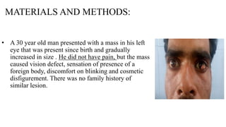
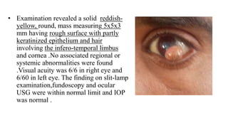
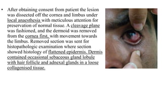
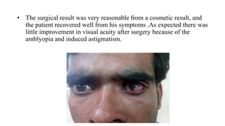
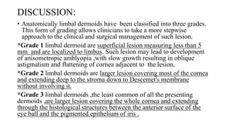
SlideShare a Scribd company logo
THE HAIRY EYEBALL-
LIMBAL DERMOID
DR.PAWAN N. JARWAL,
DR.KINJAL H. DESAI,
THIRD YEAR RESIDENTS
DEPARTMENT OF OPHTHALMOLOGY
S. S. G. HOSPITAL,VADODARA
INTRODUCTION:
• The estimated worldwide incidence of limbal dermoid is 1 per 10,000 to 3
per 10,000.
• Limbal dermoid are benign congenital tumours that contain choristomatous
tissue (tissue not found normally at that site).
• They appear most frequently at the inferior temporal quadrant of corneal
limbus and remain localised mostly to the superficial layer of the cornea and
sclera.
• They may contain a varity of histologically aberrant tissues, including
epidermal appendages, connective tissue,skin,fat,sweat gland,lacrimal gland
,muscle ,teeth,cartilage,bone,vascular structure and neurologic tissue
including the brain. Malignant degeneration is extremely rare.
• Limbal dermoid are thought to arise from an early embryological
anomaly (occurring at 5-10 weeks gestation)resulting in metaplastic
transformation of mesoblast between the rim of optic nerve and
surface ectoderm .
• Limbal dermoid are present at birth but may not be recognized until
the first or second decade of life . They may appear to enlarge as the
body mature.
MATERIALS AND METHODS:
• A 30 year old man presented with a mass in his left
eye that was present since birth and gradually
increased in size . He did not have pain, but the mass
caused vision defect, sensation of presence of a
foreign body, discomfort on blinking and cosmetic
disfigurement. There was no family history of
similar lesion.
• Examination revealed a solid reddish-
yellow, round, mass measuring 5x5x3
mm having rough surface with partly
keratinized epithelium and hair
involving the infero-temporal limbus
and cornea .No associated regional or
systemic abnormalities were found
.Visual acuity was 6/6 in right eye and
6/60 in left eye. The finding on slit-lamp
examination,fundoscopy and ocular
USG were within normal limit and IOP
was normal .
• After obtaining consent from patient the lesion
was dissected off the cornea and limbus under
local anaesthesia with meticulous attention for
preservation of normal tissue. A cleavage plane
was fashioned, and the dermoid was removed
from the cornea first, with movement towards
the limbus. Removed section was sent for
histopathologic examination where section
showed histology of flattened epidermis. Dermis
contained occasional sebaceous gland lobule
with hair follicle and adnexal glands in a loose
collagenised tissue.
• The surgical result was very reasonable from a cosmetic result, and
the patient recovered well from his symptoms .As expected there was
little improvement in visual acuity after surgery because of the
amblyopia and induced astigmatism.
DISCUSSION:
• Anatomically limbal dermoids have been classified into three grades.
This form of grading allows clinicians to take a more stepwise
approach to the clinical and surgical management of such lesion.
*Grade 1 limbal dermoid are superficial lesion measuring less than 5
mm and are localized to limbus. Such lesion may lead to development
of anisometropic amblyopia ,with slow growth resulting in oblique
astigmatism and flattening of cornea adjacent to the lesion.
*Grade 2 limbal dermoids are larger lesion covering most of the cornea
and extending deep to the stroma down to Descemet's membrane
without involving it.
*Grade 3 limbal dermoids ,the least common of all the presenting
dermoids ,are larger lesion covering the whole cornea and extending
through the histological structures between the anterior surface of the
eye ball and the pigmented epithelium of iris .
• Visual morbidity in limbal dermoid mainly results from
encroachment of the lesion into the visual axis, development of
astigmatism or formation of lipid infiltration of the cornea, which
obstructs the visual axis . Large Limbal dermoid can be cosmetically
disfiguring. In some cases staphyloma formation adjacent to dermoid
has been reported and may be associated with spontaneous
perforation of cornea or sclera.
• A varity of surgical techniques has been described in the literature ,
ranging from simple excision to lamellar and/or penetrating keratoplasty
with relaxing corneal incision, depending on the grade of lesion.
Depth,size,and site of such lesion are critical factor .Other techniques
include corneal-limbal scleral donor graft transplantation and surgical
resection followed by reconstructive multi-layered amniotic membrane
transplantation.
Conclusion:
• Treatment of limbal dermoid may consist of periodic removal of
irritating cilia, topical lubrication to prevent foreign body sensation, or
excision of the lesion if it causing significant cosmetic disfigurement
or interfering with vision.
• Attempts at complete removal are unnecessary because lesion may
extend into the deeper structure of the eye and risk of perforation
increase if attempts are made to remove lesion completely. If a deep
excision is necessary, then a lamellar keratoplasty can be performed to
reinforce the site of excision.
THANK YOU

More Related Content

What's hot

Dry eye
Dry eyeDry eye
Dry eye
Sheim Elteb
 
Keratoconus
KeratoconusKeratoconus
Keratoconus
Sarah Khan
 
Keratoconus
KeratoconusKeratoconus
Keratoconus
drkvasantha
 
Intraocular tumors
Intraocular tumorsIntraocular tumors
Intraocular tumors
Pratik Lakhmawar
 
Macular Pucker
Macular PuckerMacular Pucker
Macular Pucker
Amit Srivastava
 
SYSTEMIC DISORDERS WITH CORNEAL CHANGES by Dr. Iddi.pptx
SYSTEMIC DISORDERS WITH CORNEAL CHANGES by Dr. Iddi.pptxSYSTEMIC DISORDERS WITH CORNEAL CHANGES by Dr. Iddi.pptx
SYSTEMIC DISORDERS WITH CORNEAL CHANGES by Dr. Iddi.pptx
Iddi Ndyabawe
 
Lamellar Keratoplasty in ophthalmology
Lamellar Keratoplasty in ophthalmologyLamellar Keratoplasty in ophthalmology
Lamellar Keratoplasty in ophthalmology
pratik mohod
 
Keratoconus...
Keratoconus...Keratoconus...
Keratoconus...
Reema Dandavate
 
Limbal Stem Cell Deficiency & its management
Limbal Stem Cell Deficiency & its  managementLimbal Stem Cell Deficiency & its  management
Limbal Stem Cell Deficiency & its management
Karan Bhatia
 
Ferrara final the vitreoretinal interface a new interest for an old story
Ferrara    final the vitreoretinal interface a new interest for an old storyFerrara    final the vitreoretinal interface a new interest for an old story
Ferrara final the vitreoretinal interface a new interest for an old story
Thessaloniki International Vitreo-Retinal Summer School
 
Pseudoexfoliative syndrome and pigment dispersion syndrome and glaucoma
Pseudoexfoliative syndrome and pigment dispersion syndrome and glaucomaPseudoexfoliative syndrome and pigment dispersion syndrome and glaucoma
Pseudoexfoliative syndrome and pigment dispersion syndrome and glaucoma
Bipin Bista
 
Ptosis
PtosisPtosis
Ptosis
Amr Mounir
 
Keratoconus 2
Keratoconus 2Keratoconus 2
Keratoconus 2
Atif Rahman
 
Femtolasik... indications and limitations
Femtolasik... indications and limitationsFemtolasik... indications and limitations
Femtolasik... indications and limitations
Amr Mounir
 
Pigment dispersion syndrome
Pigment dispersion syndromePigment dispersion syndrome
Pigment dispersion syndrome
aditisingh77985
 
Vitreoretinal interface disorders
Vitreoretinal interface disordersVitreoretinal interface disorders
Vitreoretinal interface disorders
Eranda Wannigama
 
Macular hole
Macular hole Macular hole
Macular hole
Kumar Vaibhav
 
Proptosis in ophthalmology
Proptosis  in ophthalmologyProptosis  in ophthalmology
Proptosis in ophthalmology
Dr.Juleena Kunhimohammed
 
Corneal ulcer bact & fungal n
Corneal ulcer   bact & fungal nCorneal ulcer   bact & fungal n
Corneal ulcer bact & fungal n
anupama manoharan
 
Epiretinal membrane
Epiretinal membraneEpiretinal membrane
Epiretinal membrane
Pushkal Katara
 

What's hot (20)

Dry eye
Dry eyeDry eye
Dry eye
 
Keratoconus
KeratoconusKeratoconus
Keratoconus
 
Keratoconus
KeratoconusKeratoconus
Keratoconus
 
Intraocular tumors
Intraocular tumorsIntraocular tumors
Intraocular tumors
 
Macular Pucker
Macular PuckerMacular Pucker
Macular Pucker
 
SYSTEMIC DISORDERS WITH CORNEAL CHANGES by Dr. Iddi.pptx
SYSTEMIC DISORDERS WITH CORNEAL CHANGES by Dr. Iddi.pptxSYSTEMIC DISORDERS WITH CORNEAL CHANGES by Dr. Iddi.pptx
SYSTEMIC DISORDERS WITH CORNEAL CHANGES by Dr. Iddi.pptx
 
Lamellar Keratoplasty in ophthalmology
Lamellar Keratoplasty in ophthalmologyLamellar Keratoplasty in ophthalmology
Lamellar Keratoplasty in ophthalmology
 
Keratoconus...
Keratoconus...Keratoconus...
Keratoconus...
 
Limbal Stem Cell Deficiency & its management
Limbal Stem Cell Deficiency & its  managementLimbal Stem Cell Deficiency & its  management
Limbal Stem Cell Deficiency & its management
 
Ferrara final the vitreoretinal interface a new interest for an old story
Ferrara    final the vitreoretinal interface a new interest for an old storyFerrara    final the vitreoretinal interface a new interest for an old story
Ferrara final the vitreoretinal interface a new interest for an old story
 
Pseudoexfoliative syndrome and pigment dispersion syndrome and glaucoma
Pseudoexfoliative syndrome and pigment dispersion syndrome and glaucomaPseudoexfoliative syndrome and pigment dispersion syndrome and glaucoma
Pseudoexfoliative syndrome and pigment dispersion syndrome and glaucoma
 
Ptosis
PtosisPtosis
Ptosis
 
Keratoconus 2
Keratoconus 2Keratoconus 2
Keratoconus 2
 
Femtolasik... indications and limitations
Femtolasik... indications and limitationsFemtolasik... indications and limitations
Femtolasik... indications and limitations
 
Pigment dispersion syndrome
Pigment dispersion syndromePigment dispersion syndrome
Pigment dispersion syndrome
 
Vitreoretinal interface disorders
Vitreoretinal interface disordersVitreoretinal interface disorders
Vitreoretinal interface disorders
 
Macular hole
Macular hole Macular hole
Macular hole
 
Proptosis in ophthalmology
Proptosis  in ophthalmologyProptosis  in ophthalmology
Proptosis in ophthalmology
 
Corneal ulcer bact & fungal n
Corneal ulcer   bact & fungal nCorneal ulcer   bact & fungal n
Corneal ulcer bact & fungal n
 
Epiretinal membrane
Epiretinal membraneEpiretinal membrane
Epiretinal membrane
 

Similar to LIMBAL DERMOID -HAIRY EYEBALL

Common Cases: Lids, Conjunctiva and Sclera
Common Cases: Lids, Conjunctiva and ScleraCommon Cases: Lids, Conjunctiva and Sclera
Common Cases: Lids, Conjunctiva and Sclera
Riyad Banayot
 
Ophthalmology 5th year, 2nd lecture (Dr. Tara)
Ophthalmology 5th year, 2nd lecture (Dr. Tara)Ophthalmology 5th year, 2nd lecture (Dr. Tara)
Ophthalmology 5th year, 2nd lecture (Dr. Tara)
College of Medicine, Sulaymaniyah
 
Surgical excision of congenital Dermoid cysts in the orbit
Surgical excision of congenital Dermoid cysts in the orbitSurgical excision of congenital Dermoid cysts in the orbit
Surgical excision of congenital Dermoid cysts in the orbit
iosrjce
 
Pappilomatous Squamous Cell Carcinoma At The Limbus
Pappilomatous Squamous Cell Carcinoma At The LimbusPappilomatous Squamous Cell Carcinoma At The Limbus
Pappilomatous Squamous Cell Carcinoma At The Limbus
Dr. Jagannath Boramani
 
Lecture on Common Tumours of Lids For 4th Year MBBS Undergraduate Students B...
Lecture on Common Tumours  of Lids For 4th Year MBBS Undergraduate Students B...Lecture on Common Tumours  of Lids For 4th Year MBBS Undergraduate Students B...
Lecture on Common Tumours of Lids For 4th Year MBBS Undergraduate Students B...
DrHussainAhmadKhaqan
 
keratoplasty.pptx
keratoplasty.pptxkeratoplasty.pptx
keratoplasty.pptx
dratulkranand
 
Keratoplasty
KeratoplastyKeratoplasty
Keratoplasty
Nitish Narang
 
Retinoblastoma, general veiw.. by DR.ZEINAB MEDANI
Retinoblastoma, general veiw.. by DR.ZEINAB MEDANIRetinoblastoma, general veiw.. by DR.ZEINAB MEDANI
Retinoblastoma, general veiw.. by DR.ZEINAB MEDANI
Omdurman Islamic University
 
Presentation
PresentationPresentation
Presentation
Mohammed Sayed
 
Ocular surface squamous neoplasia
Ocular surface squamous neoplasiaOcular surface squamous neoplasia
Ocular surface squamous neoplasia
Rashed-Ul-Hasan Rasu
 
Depositions and Degenerations of Conjuctiva and Cornea.docx
Depositions and Degenerations of Conjuctiva and Cornea.docxDepositions and Degenerations of Conjuctiva and Cornea.docx
Depositions and Degenerations of Conjuctiva and Cornea.docx
Iddi Ndyabawe
 
Ocular surface squamous neoplasia(ossn)
Ocular surface squamous neoplasia(ossn)Ocular surface squamous neoplasia(ossn)
Ocular surface squamous neoplasia(ossn)
SSSIHMS-PG
 
Keratoplasty
KeratoplastyKeratoplasty
Keratoplasty
Pravda Chaturvedi
 
Scleral buckling for rhegmatogenous retinal detachment
Scleral buckling for rhegmatogenous retinal detachmentScleral buckling for rhegmatogenous retinal detachment
Scleral buckling for rhegmatogenous retinal detachment
reboca smith
 
Ocular Surface Squamos Neoplasia.docx
Ocular Surface Squamos Neoplasia.docxOcular Surface Squamos Neoplasia.docx
Ocular Surface Squamos Neoplasia.docx
Iddi Ndyabawe
 
Urrets-Zavalia Syndrome.ophthalmololg pptx
Urrets-Zavalia Syndrome.ophthalmololg pptxUrrets-Zavalia Syndrome.ophthalmololg pptx
Urrets-Zavalia Syndrome.ophthalmololg pptx
fajrimohammed
 
Eyelid disorders
Eyelid disordersEyelid disorders
Eyelid disorders
SOUMYA SUBRAMANI
 
INDICATIONS OF KERATOPLASTY (Bindu K M)
INDICATIONS OF KERATOPLASTY (Bindu K M)INDICATIONS OF KERATOPLASTY (Bindu K M)
INDICATIONS OF KERATOPLASTY (Bindu K M)
Bindu K M
 
Short case Cornea
Short case CorneaShort case Cornea
Short case Cornea
Anisur Rahman
 
Radiation of eye AND ORBIT
Radiation of eye AND ORBITRadiation of eye AND ORBIT
Radiation of eye AND ORBIT
Kiran Ramakrishna
 

Similar to LIMBAL DERMOID -HAIRY EYEBALL (20)

Common Cases: Lids, Conjunctiva and Sclera
Common Cases: Lids, Conjunctiva and ScleraCommon Cases: Lids, Conjunctiva and Sclera
Common Cases: Lids, Conjunctiva and Sclera
 
Ophthalmology 5th year, 2nd lecture (Dr. Tara)
Ophthalmology 5th year, 2nd lecture (Dr. Tara)Ophthalmology 5th year, 2nd lecture (Dr. Tara)
Ophthalmology 5th year, 2nd lecture (Dr. Tara)
 
Surgical excision of congenital Dermoid cysts in the orbit
Surgical excision of congenital Dermoid cysts in the orbitSurgical excision of congenital Dermoid cysts in the orbit
Surgical excision of congenital Dermoid cysts in the orbit
 
Pappilomatous Squamous Cell Carcinoma At The Limbus
Pappilomatous Squamous Cell Carcinoma At The LimbusPappilomatous Squamous Cell Carcinoma At The Limbus
Pappilomatous Squamous Cell Carcinoma At The Limbus
 
Lecture on Common Tumours of Lids For 4th Year MBBS Undergraduate Students B...
Lecture on Common Tumours  of Lids For 4th Year MBBS Undergraduate Students B...Lecture on Common Tumours  of Lids For 4th Year MBBS Undergraduate Students B...
Lecture on Common Tumours of Lids For 4th Year MBBS Undergraduate Students B...
 
keratoplasty.pptx
keratoplasty.pptxkeratoplasty.pptx
keratoplasty.pptx
 
Keratoplasty
KeratoplastyKeratoplasty
Keratoplasty
 
Retinoblastoma, general veiw.. by DR.ZEINAB MEDANI
Retinoblastoma, general veiw.. by DR.ZEINAB MEDANIRetinoblastoma, general veiw.. by DR.ZEINAB MEDANI
Retinoblastoma, general veiw.. by DR.ZEINAB MEDANI
 
Presentation
PresentationPresentation
Presentation
 
Ocular surface squamous neoplasia
Ocular surface squamous neoplasiaOcular surface squamous neoplasia
Ocular surface squamous neoplasia
 
Depositions and Degenerations of Conjuctiva and Cornea.docx
Depositions and Degenerations of Conjuctiva and Cornea.docxDepositions and Degenerations of Conjuctiva and Cornea.docx
Depositions and Degenerations of Conjuctiva and Cornea.docx
 
Ocular surface squamous neoplasia(ossn)
Ocular surface squamous neoplasia(ossn)Ocular surface squamous neoplasia(ossn)
Ocular surface squamous neoplasia(ossn)
 
Keratoplasty
KeratoplastyKeratoplasty
Keratoplasty
 
Scleral buckling for rhegmatogenous retinal detachment
Scleral buckling for rhegmatogenous retinal detachmentScleral buckling for rhegmatogenous retinal detachment
Scleral buckling for rhegmatogenous retinal detachment
 
Ocular Surface Squamos Neoplasia.docx
Ocular Surface Squamos Neoplasia.docxOcular Surface Squamos Neoplasia.docx
Ocular Surface Squamos Neoplasia.docx
 
Urrets-Zavalia Syndrome.ophthalmololg pptx
Urrets-Zavalia Syndrome.ophthalmololg pptxUrrets-Zavalia Syndrome.ophthalmololg pptx
Urrets-Zavalia Syndrome.ophthalmololg pptx
 
Eyelid disorders
Eyelid disordersEyelid disorders
Eyelid disorders
 
INDICATIONS OF KERATOPLASTY (Bindu K M)
INDICATIONS OF KERATOPLASTY (Bindu K M)INDICATIONS OF KERATOPLASTY (Bindu K M)
INDICATIONS OF KERATOPLASTY (Bindu K M)
 
Short case Cornea
Short case CorneaShort case Cornea
Short case Cornea
 
Radiation of eye AND ORBIT
Radiation of eye AND ORBITRadiation of eye AND ORBIT
Radiation of eye AND ORBIT
 

Recently uploaded

Physiology of Chemical Sensation of smell.pdf
Physiology of Chemical Sensation of smell.pdfPhysiology of Chemical Sensation of smell.pdf
Physiology of Chemical Sensation of smell.pdf
MedicoseAcademics
 
Triangles of Neck and Clinical Correlation by Dr. RIG.pptx
Triangles of Neck and Clinical Correlation by Dr. RIG.pptxTriangles of Neck and Clinical Correlation by Dr. RIG.pptx
Triangles of Neck and Clinical Correlation by Dr. RIG.pptx
Dr. Rabia Inam Gandapore
 
How STIs Influence the Development of Pelvic Inflammatory Disease.pptx
How STIs Influence the Development of Pelvic Inflammatory Disease.pptxHow STIs Influence the Development of Pelvic Inflammatory Disease.pptx
How STIs Influence the Development of Pelvic Inflammatory Disease.pptx
FFragrant
 
Physiology of Special Chemical Sensation of Taste
Physiology of Special Chemical Sensation of TastePhysiology of Special Chemical Sensation of Taste
Physiology of Special Chemical Sensation of Taste
MedicoseAcademics
 
Effective-Soaps-for-Fungal-Skin-Infections.pptx
Effective-Soaps-for-Fungal-Skin-Infections.pptxEffective-Soaps-for-Fungal-Skin-Infections.pptx
Effective-Soaps-for-Fungal-Skin-Infections.pptx
SwisschemDerma
 
Ophthalmology Clinical Tests for OSCE exam
Ophthalmology Clinical Tests for OSCE examOphthalmology Clinical Tests for OSCE exam
Ophthalmology Clinical Tests for OSCE exam
KafrELShiekh University
 
Light House Retreats: Plant Medicine Retreat Europe
Light House Retreats: Plant Medicine Retreat EuropeLight House Retreats: Plant Medicine Retreat Europe
Light House Retreats: Plant Medicine Retreat Europe
Lighthouse Retreat
 
Local Advanced Lung Cancer: Artificial Intelligence, Synergetics, Complex Sys...
Local Advanced Lung Cancer: Artificial Intelligence, Synergetics, Complex Sys...Local Advanced Lung Cancer: Artificial Intelligence, Synergetics, Complex Sys...
Local Advanced Lung Cancer: Artificial Intelligence, Synergetics, Complex Sys...
Oleg Kshivets
 
Ocular injury ppt Upendra pal optometrist upums saifai etawah
Ocular injury  ppt  Upendra pal  optometrist upums saifai etawahOcular injury  ppt  Upendra pal  optometrist upums saifai etawah
Ocular injury ppt Upendra pal optometrist upums saifai etawah
pal078100
 
micro teaching on communication m.sc nursing.pdf
micro teaching on communication m.sc nursing.pdfmicro teaching on communication m.sc nursing.pdf
micro teaching on communication m.sc nursing.pdf
Anurag Sharma
 
Top-Vitamin-Supplement-Brands-in-India.pptx
Top-Vitamin-Supplement-Brands-in-India.pptxTop-Vitamin-Supplement-Brands-in-India.pptx
Top-Vitamin-Supplement-Brands-in-India.pptx
SwisschemDerma
 
Ozempic: Preoperative Management of Patients on GLP-1 Receptor Agonists
Ozempic: Preoperative Management of Patients on GLP-1 Receptor Agonists  Ozempic: Preoperative Management of Patients on GLP-1 Receptor Agonists
Ozempic: Preoperative Management of Patients on GLP-1 Receptor Agonists
Saeid Safari
 
Superficial & Deep Fascia of the NECK.pptx
Superficial & Deep Fascia of the NECK.pptxSuperficial & Deep Fascia of the NECK.pptx
Superficial & Deep Fascia of the NECK.pptx
Dr. Rabia Inam Gandapore
 
Non-respiratory Functions of the Lungs.pdf
Non-respiratory Functions of the Lungs.pdfNon-respiratory Functions of the Lungs.pdf
Non-respiratory Functions of the Lungs.pdf
MedicoseAcademics
 
Hemodialysis: Chapter 3, Dialysis Water Unit - Dr.Gawad
Hemodialysis: Chapter 3, Dialysis Water Unit - Dr.GawadHemodialysis: Chapter 3, Dialysis Water Unit - Dr.Gawad
Hemodialysis: Chapter 3, Dialysis Water Unit - Dr.Gawad
NephroTube - Dr.Gawad
 
KDIGO 2024 guidelines for diabetologists
KDIGO 2024 guidelines for diabetologistsKDIGO 2024 guidelines for diabetologists
KDIGO 2024 guidelines for diabetologists
د.محمود نجيب
 
Pharma Pcd Franchise in Jharkhand - Yodley Lifesciences
Pharma Pcd Franchise in Jharkhand - Yodley LifesciencesPharma Pcd Franchise in Jharkhand - Yodley Lifesciences
Pharma Pcd Franchise in Jharkhand - Yodley Lifesciences
Yodley Lifesciences
 
NVBDCP.pptx Nation vector borne disease control program
NVBDCP.pptx Nation vector borne disease control programNVBDCP.pptx Nation vector borne disease control program
NVBDCP.pptx Nation vector borne disease control program
Sapna Thakur
 
263778731218 Abortion Clinic /Pills In Harare ,
263778731218 Abortion Clinic /Pills In Harare ,263778731218 Abortion Clinic /Pills In Harare ,
263778731218 Abortion Clinic /Pills In Harare ,
sisternakatoto
 
Vision-1.pptx, Eye structure, basics of optics
Vision-1.pptx, Eye structure, basics of opticsVision-1.pptx, Eye structure, basics of optics
Vision-1.pptx, Eye structure, basics of optics
Sai Sailesh Kumar Goothy
 

Recently uploaded (20)

Physiology of Chemical Sensation of smell.pdf
Physiology of Chemical Sensation of smell.pdfPhysiology of Chemical Sensation of smell.pdf
Physiology of Chemical Sensation of smell.pdf
 
Triangles of Neck and Clinical Correlation by Dr. RIG.pptx
Triangles of Neck and Clinical Correlation by Dr. RIG.pptxTriangles of Neck and Clinical Correlation by Dr. RIG.pptx
Triangles of Neck and Clinical Correlation by Dr. RIG.pptx
 
How STIs Influence the Development of Pelvic Inflammatory Disease.pptx
How STIs Influence the Development of Pelvic Inflammatory Disease.pptxHow STIs Influence the Development of Pelvic Inflammatory Disease.pptx
How STIs Influence the Development of Pelvic Inflammatory Disease.pptx
 
Physiology of Special Chemical Sensation of Taste
Physiology of Special Chemical Sensation of TastePhysiology of Special Chemical Sensation of Taste
Physiology of Special Chemical Sensation of Taste
 
Effective-Soaps-for-Fungal-Skin-Infections.pptx
Effective-Soaps-for-Fungal-Skin-Infections.pptxEffective-Soaps-for-Fungal-Skin-Infections.pptx
Effective-Soaps-for-Fungal-Skin-Infections.pptx
 
Ophthalmology Clinical Tests for OSCE exam
Ophthalmology Clinical Tests for OSCE examOphthalmology Clinical Tests for OSCE exam
Ophthalmology Clinical Tests for OSCE exam
 
Light House Retreats: Plant Medicine Retreat Europe
Light House Retreats: Plant Medicine Retreat EuropeLight House Retreats: Plant Medicine Retreat Europe
Light House Retreats: Plant Medicine Retreat Europe
 
Local Advanced Lung Cancer: Artificial Intelligence, Synergetics, Complex Sys...
Local Advanced Lung Cancer: Artificial Intelligence, Synergetics, Complex Sys...Local Advanced Lung Cancer: Artificial Intelligence, Synergetics, Complex Sys...
Local Advanced Lung Cancer: Artificial Intelligence, Synergetics, Complex Sys...
 
Ocular injury ppt Upendra pal optometrist upums saifai etawah
Ocular injury  ppt  Upendra pal  optometrist upums saifai etawahOcular injury  ppt  Upendra pal  optometrist upums saifai etawah
Ocular injury ppt Upendra pal optometrist upums saifai etawah
 
micro teaching on communication m.sc nursing.pdf
micro teaching on communication m.sc nursing.pdfmicro teaching on communication m.sc nursing.pdf
micro teaching on communication m.sc nursing.pdf
 
Top-Vitamin-Supplement-Brands-in-India.pptx
Top-Vitamin-Supplement-Brands-in-India.pptxTop-Vitamin-Supplement-Brands-in-India.pptx
Top-Vitamin-Supplement-Brands-in-India.pptx
 
Ozempic: Preoperative Management of Patients on GLP-1 Receptor Agonists
Ozempic: Preoperative Management of Patients on GLP-1 Receptor Agonists  Ozempic: Preoperative Management of Patients on GLP-1 Receptor Agonists
Ozempic: Preoperative Management of Patients on GLP-1 Receptor Agonists
 
Superficial & Deep Fascia of the NECK.pptx
Superficial & Deep Fascia of the NECK.pptxSuperficial & Deep Fascia of the NECK.pptx
Superficial & Deep Fascia of the NECK.pptx
 
Non-respiratory Functions of the Lungs.pdf
Non-respiratory Functions of the Lungs.pdfNon-respiratory Functions of the Lungs.pdf
Non-respiratory Functions of the Lungs.pdf
 
Hemodialysis: Chapter 3, Dialysis Water Unit - Dr.Gawad
Hemodialysis: Chapter 3, Dialysis Water Unit - Dr.GawadHemodialysis: Chapter 3, Dialysis Water Unit - Dr.Gawad
Hemodialysis: Chapter 3, Dialysis Water Unit - Dr.Gawad
 
KDIGO 2024 guidelines for diabetologists
KDIGO 2024 guidelines for diabetologistsKDIGO 2024 guidelines for diabetologists
KDIGO 2024 guidelines for diabetologists
 
Pharma Pcd Franchise in Jharkhand - Yodley Lifesciences
Pharma Pcd Franchise in Jharkhand - Yodley LifesciencesPharma Pcd Franchise in Jharkhand - Yodley Lifesciences
Pharma Pcd Franchise in Jharkhand - Yodley Lifesciences
 
NVBDCP.pptx Nation vector borne disease control program
NVBDCP.pptx Nation vector borne disease control programNVBDCP.pptx Nation vector borne disease control program
NVBDCP.pptx Nation vector borne disease control program
 
263778731218 Abortion Clinic /Pills In Harare ,
263778731218 Abortion Clinic /Pills In Harare ,263778731218 Abortion Clinic /Pills In Harare ,
263778731218 Abortion Clinic /Pills In Harare ,
 
Vision-1.pptx, Eye structure, basics of optics
Vision-1.pptx, Eye structure, basics of opticsVision-1.pptx, Eye structure, basics of optics
Vision-1.pptx, Eye structure, basics of optics
 

LIMBAL DERMOID -HAIRY EYEBALL

  • 1. THE HAIRY EYEBALL- LIMBAL DERMOID DR.PAWAN N. JARWAL, DR.KINJAL H. DESAI, THIRD YEAR RESIDENTS DEPARTMENT OF OPHTHALMOLOGY S. S. G. HOSPITAL,VADODARA
  • 2. INTRODUCTION: • The estimated worldwide incidence of limbal dermoid is 1 per 10,000 to 3 per 10,000. • Limbal dermoid are benign congenital tumours that contain choristomatous tissue (tissue not found normally at that site). • They appear most frequently at the inferior temporal quadrant of corneal limbus and remain localised mostly to the superficial layer of the cornea and sclera. • They may contain a varity of histologically aberrant tissues, including epidermal appendages, connective tissue,skin,fat,sweat gland,lacrimal gland ,muscle ,teeth,cartilage,bone,vascular structure and neurologic tissue including the brain. Malignant degeneration is extremely rare.
  • 3. • Limbal dermoid are thought to arise from an early embryological anomaly (occurring at 5-10 weeks gestation)resulting in metaplastic transformation of mesoblast between the rim of optic nerve and surface ectoderm . • Limbal dermoid are present at birth but may not be recognized until the first or second decade of life . They may appear to enlarge as the body mature.
  • 4. MATERIALS AND METHODS: • A 30 year old man presented with a mass in his left eye that was present since birth and gradually increased in size . He did not have pain, but the mass caused vision defect, sensation of presence of a foreign body, discomfort on blinking and cosmetic disfigurement. There was no family history of similar lesion.
  • 5. • Examination revealed a solid reddish- yellow, round, mass measuring 5x5x3 mm having rough surface with partly keratinized epithelium and hair involving the infero-temporal limbus and cornea .No associated regional or systemic abnormalities were found .Visual acuity was 6/6 in right eye and 6/60 in left eye. The finding on slit-lamp examination,fundoscopy and ocular USG were within normal limit and IOP was normal .
  • 6. • After obtaining consent from patient the lesion was dissected off the cornea and limbus under local anaesthesia with meticulous attention for preservation of normal tissue. A cleavage plane was fashioned, and the dermoid was removed from the cornea first, with movement towards the limbus. Removed section was sent for histopathologic examination where section showed histology of flattened epidermis. Dermis contained occasional sebaceous gland lobule with hair follicle and adnexal glands in a loose collagenised tissue.
  • 7. • The surgical result was very reasonable from a cosmetic result, and the patient recovered well from his symptoms .As expected there was little improvement in visual acuity after surgery because of the amblyopia and induced astigmatism.
  • 8. DISCUSSION: • Anatomically limbal dermoids have been classified into three grades. This form of grading allows clinicians to take a more stepwise approach to the clinical and surgical management of such lesion. *Grade 1 limbal dermoid are superficial lesion measuring less than 5 mm and are localized to limbus. Such lesion may lead to development of anisometropic amblyopia ,with slow growth resulting in oblique astigmatism and flattening of cornea adjacent to the lesion. *Grade 2 limbal dermoids are larger lesion covering most of the cornea and extending deep to the stroma down to Descemet's membrane without involving it. *Grade 3 limbal dermoids ,the least common of all the presenting dermoids ,are larger lesion covering the whole cornea and extending through the histological structures between the anterior surface of the eye ball and the pigmented epithelium of iris .
  • 9. • Visual morbidity in limbal dermoid mainly results from encroachment of the lesion into the visual axis, development of astigmatism or formation of lipid infiltration of the cornea, which obstructs the visual axis . Large Limbal dermoid can be cosmetically disfiguring. In some cases staphyloma formation adjacent to dermoid has been reported and may be associated with spontaneous perforation of cornea or sclera. • A varity of surgical techniques has been described in the literature , ranging from simple excision to lamellar and/or penetrating keratoplasty with relaxing corneal incision, depending on the grade of lesion. Depth,size,and site of such lesion are critical factor .Other techniques include corneal-limbal scleral donor graft transplantation and surgical resection followed by reconstructive multi-layered amniotic membrane transplantation.
  • 10. Conclusion: • Treatment of limbal dermoid may consist of periodic removal of irritating cilia, topical lubrication to prevent foreign body sensation, or excision of the lesion if it causing significant cosmetic disfigurement or interfering with vision. • Attempts at complete removal are unnecessary because lesion may extend into the deeper structure of the eye and risk of perforation increase if attempts are made to remove lesion completely. If a deep excision is necessary, then a lamellar keratoplasty can be performed to reinforce the site of excision.