Noncompaction
Cardiomyopathy
Dr. Sarvesh Prajapati
Bombay Hospital, Mumbai
Introduction
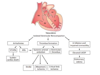
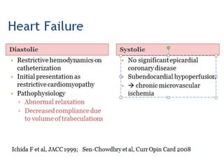
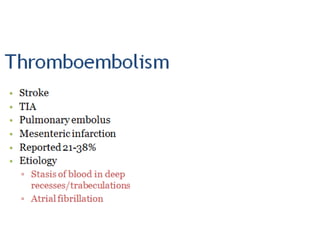
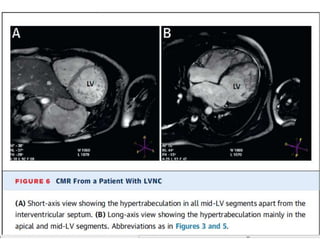
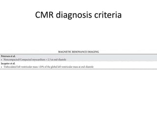
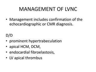
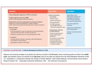
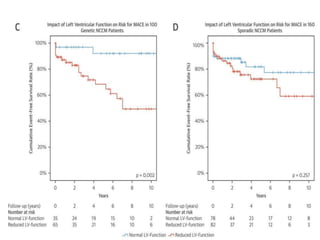
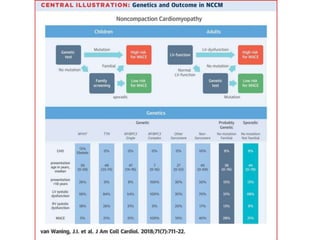
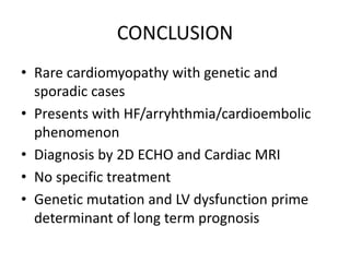
• Left ventricular noncompaction (LVNC) is defined by 3
markers:
– prominent left ventricular (LV) trabeculae,
– deep intertrabecular recesses,
– the thin compacted layer
• The spectrum of morphologic variability is extreme
• LVNC can be isolated or associated with
cardiomyopathies, congenital heart diseases, and
complex syndromes involving the heart.
• Hypertrabeculation Syndrome
• Persistent myocardial sinusoids
• Spongy myocardium
History
• Dusek - first described the postnatal persistence
of spongy myocardium in 1975 pathologically,
• Engberding and Bender made the first clinical
recognition with 2D ECHO in 1984
• Three decades later, with only morphologic
assessment available and no definitive genetic
pathway, isolated left ventricular
noncompaction (LVNC) remains a diagnostic
and management challenge.
Classification
• The AHA - genetic cardiomyopathy,
• The ESC - unclassified cardiomyopathy.
• The LVNC trait may be familial (inherited) or
nonfamilial (sporadic)
• Nonfamilial forms are diagnosed when LVNC
is proven absent in relatives.
• Sporadic LVNC can be acquired, as in highly-
trained athletes, sickle cell anemia patients,
and pregnancy.
• The genetic bases of familial LVNC are still a
matter of research.
hypothesis
Embryogenic
Nonembryogenic
NONEMBRYOGENIC HYPOTHESES
• May occur in adult life, leading to acquired
LVNC.
• Athelets - compacted layer well preservesd
• pregnant women – 25% because of preload
• sickle cell anemia - increased cardiac pre-load
LVNC: PHYSIOLOGIC OR PATHOLOGIC
REMODELING OF THE MYOCARDIUM?
EMBRYOGENIC HYPOTHESIS
• Studies in experimental models suggest that the
process of cardiac trabeculation begins after the
cardiac looping stage.
• Emerging evidence suggests that the myocytes
forming the trabeculae arise from a different
clonal origin in the heart wall.
• Notch signaling in the endocardium is also critical
for cardiac trabecular formation
• The trabecular portion of the myocardial wall is tiny
and thinner in the LV than in the right ventricle, and
the compacted wall is more prominent in the thicker LV
wall.
• Hypothesis 1 states - myocyte organization fails to
evolve from the embryonic spongiform condition to
the compacted,mature state. Although both ventricles
may be involved, the LV is generally affected.
• Hypothesis2 states - that LVNC occurs as a result of
inhibiting the regression of embryonic structures.
Early Embryology, <5 weeks
Neuregulin growth factors
3 weeks
↓N-Cadherin
During the early developmental stages, the heart tube is still without epicardial overlay.
As an adaptation to improve nourishment of the rapidly growing heart, the
Embryology, 5-8 weeks
• Compaction begins in the human embryo by 8
to 12 weeks and by the fourth month of
gestation the compacted myocardium
composes the majority of the ventricular
volume.
Genetics
• Multiple genetic proposals for the phenotypic
development of noncompaction.
• It is established that a thorough, three-
generational family history should be obtained
for evaluation of genetic influence, which may
also impact the screening of additional family
members
• Genes Identified. The identified genes are as
follows: 
– Fbkp1a/Notch pathway. 
– G4.5 gene/TAZ protein. 
– 14-3-3 deletion. 
– ZASP protein. 
– TNNT2 protein. 
– MYH7 protein. 
– TPM1 protein. 
– MYBPC3 protein. 
– ACTC1 protein.
• The Heart Rhythm Society states that genetic
testing is recommended (Class I) for relatives
and appropriate family members when a
mutation-specific gene has been identified in
the index case
ANATOMY AND PATHOLOGY
• LVNC describes a macroscopic mismatch
between the noncompacted trabeculae and
the compacted myocyte layers.
• Myocytes in the trabeculae do not show
histologic differences from those forming the
compacted layer, which explains why LVNC
histology (i.e., endomyocardial biopsy) does
not specifically contribute to the diagnosis.
Epidemiology of isolated
noncompaction
• Children  Adults, elderly
• 0.05% (Ritter M et al, Mayo Clin Proc
1997)
• 0.014%(Oechslin EN et al, JACC 2000)
• Male>Female
• 70% sporadic and 30% familial
• Neuromuscular disorders
• Metabolic disease
• Genetic syndromes
• ▫ Barth syndrome
 X-linked, dilated CMP, neutropenia, skeletal myopathy,
mitochondrial abnormalities, lactic acidosis
 G4.5 gene in Xq28: encodes tafazzins proteins:
acyltransferase functions in mitochondria, expressed in
heart/muscle cells
• ▫ Charcot-Marie-Tooth
• ▫ Nail-patella
Clinical features
ECG
• Left or right axis deviation
• PR prolongation
• Left ventricular hypertrophy
• LBBB, RBBB, IVCD
• Repolarization abnormalities
• In pediatric population:
• ▫ Sinus bradycardia
• ▫ WPW
2D ECHO
• Two-dimensional grayscale echocardiography is
the most common and useful tool for LVNC
diagnosis, showing
broad trabeculae
deep intertrabecular recesses
in the LV myocardium, typically located in the LV
apex and the midinferior and lateral walls.
• basal and midinterventricular septum – Free of
trabecule
• atypical viewsto image the more apical segments of the
LV and detect the prominent trabeculae.
• After careful evaluation of all criteria, the most
important echocardiographic criterion remains
Noncompacted/compacted ratio >2.0 in end-systole.
 Problem is interobserver and intraobserver variability
Other findings
• Systolic dysfunction (impaired coronary flow reserve and
microcirculatory defects, together with a primary myocardial
Disease)
• significantly higher longitudinal systolic strain rate and strain
in the basal segments than in the apex.
• Diastolic dysfunction
CONTRAST ECHOCARDIOGRAPHY
• because of imaging quality limitations,
especially in the more apical region of the
heart.
CARDIAC MAGNETIC RESONANCE
• Accurately describe and diagnose LVNC and
distinguishtrue LVNC from the prominent
hypertrabeculationthat can be seen in normal
hearts and individuals.
• A noncompacted/ compacted ratio >2.3 on CMR
is considered the cutoff for LVNC diagnosis.
• Compact papillary muscle should be
distinguished from prominent trabeculation
• The typical 2-layered structure of the LV wall can be
better measured in CMR, where the thinner,
compacted layer can be precisely measured in affected
ventricular segments.
• Functional data (hypokinesis of the noncompacted
segments vs. normal kinesis of unaffected segments)
may further strengthen the diagnostic hypothesis.
• High-intensity endocardial T2 signals, subendocardial
perfusion defects, and delayed enhancement of the
subendocardial layer
CMR diagnosis criteria
MANAGEMENT OF LVNC
• Management includes confirmation of the
echocardiographic or CMR diagnosis.
D/D
• prominent hypertrabeculation
• apical HCM, DCM,
• endocardial fibroelastosis,
• LV apical thrombus
• Clinical management of LVNC depends on the
presence or absence of cardiac dysfunction or
arrhythmias.
• Familial LVNC should be diagnosed by
echocardiographic screening of family members.
• Genetic testing - it may be helpful for confirming
diagnoses in family members and/or determining
potential development in family members to aid
in the timing of screening.
• Currently, there are no specific treatments for
LVNC.
• Patient should be treated according to their
clinical needs and corresponding guidelines.
• OAC – for primary prevention or secondary
prevention
• Complications with LVNC include
– heart failure,
– arrhythmias including sudden cardiac death,
– systemic embolic events
Arrythmia
• Atrial tachycardia and fibrillation are common.
• Ventricular tachyarrhythmias in 47% of symptomatic
patients.
• SCD - 13% to 18% of (mostly adult) patients with LVNC.
• LVNC has been considered a reason to restrict athletic
participation.
• ICD therapy may be effective in patients with LVNC.
• OAC
Heart failure
• Heart failure (27%)
• GDMT for Heart failure
• CRT improves NYHA functional class in
patients with LVNC and may hence be
considered in patients with an LVEF 35% and
signs of ventricular dyssynchrony.
Systemic embolic events
• The event rate of stroke in patients with LVNC is 1-2%
per year or a total risk thromboembolism of 21–38%.
• Oral anticoagulation should be used when a definite
left ventricular clot has been identified on imaging
or the patient has documented atrial fibrillation.
• CHADS2/CHADS2-Vasc scores as guidance
• It is unknown whether or not the small
compacted layer and the deep recesses of the
heart in patients with LVNC increases the risk
of complications, such as ventricular
perforation in interventional occasions or
implantation of devices.
Prognosis
Controversies still unanswered
• Myocardial embryogenesis,
• Primary genetic or unclassified
cardiomyopathy
• presence of different diagnostic criteria, with
their limitations
• Lack of robust data,
CONCLUSION
• Rare cardiomyopathy with genetic and
sporadic cases
• Presents with HF/arryhthmia/cardioembolic
phenomenon
• Diagnosis by 2D ECHO and Cardiac MRI
• No specific treatment
• Genetic mutation and LV dysfunction prime
determinant of long term prognosis
Thank You

Left ventricular noncompaction

  • 1.
  • 2.
    Introduction • Left ventricularnoncompaction (LVNC) is defined by 3 markers: – prominent left ventricular (LV) trabeculae, – deep intertrabecular recesses, – the thin compacted layer • The spectrum of morphologic variability is extreme • LVNC can be isolated or associated with cardiomyopathies, congenital heart diseases, and complex syndromes involving the heart.
  • 4.
    • Hypertrabeculation Syndrome •Persistent myocardial sinusoids • Spongy myocardium
  • 5.
    History • Dusek -first described the postnatal persistence of spongy myocardium in 1975 pathologically, • Engberding and Bender made the first clinical recognition with 2D ECHO in 1984 • Three decades later, with only morphologic assessment available and no definitive genetic pathway, isolated left ventricular noncompaction (LVNC) remains a diagnostic and management challenge.
  • 6.
    Classification • The AHA- genetic cardiomyopathy, • The ESC - unclassified cardiomyopathy. • The LVNC trait may be familial (inherited) or nonfamilial (sporadic)
  • 7.
    • Nonfamilial formsare diagnosed when LVNC is proven absent in relatives. • Sporadic LVNC can be acquired, as in highly- trained athletes, sickle cell anemia patients, and pregnancy. • The genetic bases of familial LVNC are still a matter of research.
  • 8.
  • 9.
    NONEMBRYOGENIC HYPOTHESES • Mayoccur in adult life, leading to acquired LVNC. • Athelets - compacted layer well preservesd • pregnant women – 25% because of preload • sickle cell anemia - increased cardiac pre-load
  • 10.
    LVNC: PHYSIOLOGIC ORPATHOLOGIC REMODELING OF THE MYOCARDIUM?
  • 11.
    EMBRYOGENIC HYPOTHESIS • Studiesin experimental models suggest that the process of cardiac trabeculation begins after the cardiac looping stage. • Emerging evidence suggests that the myocytes forming the trabeculae arise from a different clonal origin in the heart wall. • Notch signaling in the endocardium is also critical for cardiac trabecular formation
  • 12.
    • The trabecularportion of the myocardial wall is tiny and thinner in the LV than in the right ventricle, and the compacted wall is more prominent in the thicker LV wall. • Hypothesis 1 states - myocyte organization fails to evolve from the embryonic spongiform condition to the compacted,mature state. Although both ventricles may be involved, the LV is generally affected. • Hypothesis2 states - that LVNC occurs as a result of inhibiting the regression of embryonic structures.
  • 13.
    Early Embryology, <5weeks Neuregulin growth factors 3 weeks ↓N-Cadherin During the early developmental stages, the heart tube is still without epicardial overlay. As an adaptation to improve nourishment of the rapidly growing heart, the
  • 14.
  • 15.
    • Compaction beginsin the human embryo by 8 to 12 weeks and by the fourth month of gestation the compacted myocardium composes the majority of the ventricular volume.
  • 17.
    Genetics • Multiple geneticproposals for the phenotypic development of noncompaction. • It is established that a thorough, three- generational family history should be obtained for evaluation of genetic influence, which may also impact the screening of additional family members
  • 18.
    • Genes Identified.The identified genes are as follows:  – Fbkp1a/Notch pathway.  – G4.5 gene/TAZ protein.  – 14-3-3 deletion.  – ZASP protein.  – TNNT2 protein.  – MYH7 protein.  – TPM1 protein.  – MYBPC3 protein.  – ACTC1 protein.
  • 19.
    • The HeartRhythm Society states that genetic testing is recommended (Class I) for relatives and appropriate family members when a mutation-specific gene has been identified in the index case
  • 20.
    ANATOMY AND PATHOLOGY •LVNC describes a macroscopic mismatch between the noncompacted trabeculae and the compacted myocyte layers. • Myocytes in the trabeculae do not show histologic differences from those forming the compacted layer, which explains why LVNC histology (i.e., endomyocardial biopsy) does not specifically contribute to the diagnosis.
  • 22.
    Epidemiology of isolated noncompaction •Children  Adults, elderly • 0.05% (Ritter M et al, Mayo Clin Proc 1997) • 0.014%(Oechslin EN et al, JACC 2000) • Male>Female • 70% sporadic and 30% familial
  • 23.
    • Neuromuscular disorders •Metabolic disease • Genetic syndromes • ▫ Barth syndrome  X-linked, dilated CMP, neutropenia, skeletal myopathy, mitochondrial abnormalities, lactic acidosis  G4.5 gene in Xq28: encodes tafazzins proteins: acyltransferase functions in mitochondria, expressed in heart/muscle cells • ▫ Charcot-Marie-Tooth • ▫ Nail-patella
  • 28.
  • 32.
    ECG • Left orright axis deviation • PR prolongation • Left ventricular hypertrophy • LBBB, RBBB, IVCD • Repolarization abnormalities • In pediatric population: • ▫ Sinus bradycardia • ▫ WPW
  • 33.
    2D ECHO • Two-dimensionalgrayscale echocardiography is the most common and useful tool for LVNC diagnosis, showing broad trabeculae deep intertrabecular recesses in the LV myocardium, typically located in the LV apex and the midinferior and lateral walls. • basal and midinterventricular septum – Free of trabecule
  • 34.
    • atypical viewstoimage the more apical segments of the LV and detect the prominent trabeculae. • After careful evaluation of all criteria, the most important echocardiographic criterion remains Noncompacted/compacted ratio >2.0 in end-systole.  Problem is interobserver and intraobserver variability
  • 37.
    Other findings • Systolicdysfunction (impaired coronary flow reserve and microcirculatory defects, together with a primary myocardial Disease) • significantly higher longitudinal systolic strain rate and strain in the basal segments than in the apex. • Diastolic dysfunction
  • 39.
    CONTRAST ECHOCARDIOGRAPHY • becauseof imaging quality limitations, especially in the more apical region of the heart.
  • 41.
    CARDIAC MAGNETIC RESONANCE •Accurately describe and diagnose LVNC and distinguishtrue LVNC from the prominent hypertrabeculationthat can be seen in normal hearts and individuals. • A noncompacted/ compacted ratio >2.3 on CMR is considered the cutoff for LVNC diagnosis. • Compact papillary muscle should be distinguished from prominent trabeculation
  • 42.
    • The typical2-layered structure of the LV wall can be better measured in CMR, where the thinner, compacted layer can be precisely measured in affected ventricular segments. • Functional data (hypokinesis of the noncompacted segments vs. normal kinesis of unaffected segments) may further strengthen the diagnostic hypothesis. • High-intensity endocardial T2 signals, subendocardial perfusion defects, and delayed enhancement of the subendocardial layer
  • 45.
  • 46.
    MANAGEMENT OF LVNC •Management includes confirmation of the echocardiographic or CMR diagnosis. D/D • prominent hypertrabeculation • apical HCM, DCM, • endocardial fibroelastosis, • LV apical thrombus
  • 48.
    • Clinical managementof LVNC depends on the presence or absence of cardiac dysfunction or arrhythmias. • Familial LVNC should be diagnosed by echocardiographic screening of family members. • Genetic testing - it may be helpful for confirming diagnoses in family members and/or determining potential development in family members to aid in the timing of screening.
  • 49.
    • Currently, thereare no specific treatments for LVNC. • Patient should be treated according to their clinical needs and corresponding guidelines. • OAC – for primary prevention or secondary prevention
  • 50.
    • Complications withLVNC include – heart failure, – arrhythmias including sudden cardiac death, – systemic embolic events
  • 51.
    Arrythmia • Atrial tachycardiaand fibrillation are common. • Ventricular tachyarrhythmias in 47% of symptomatic patients. • SCD - 13% to 18% of (mostly adult) patients with LVNC. • LVNC has been considered a reason to restrict athletic participation. • ICD therapy may be effective in patients with LVNC. • OAC
  • 52.
    Heart failure • Heartfailure (27%) • GDMT for Heart failure • CRT improves NYHA functional class in patients with LVNC and may hence be considered in patients with an LVEF 35% and signs of ventricular dyssynchrony.
  • 53.
    Systemic embolic events •The event rate of stroke in patients with LVNC is 1-2% per year or a total risk thromboembolism of 21–38%. • Oral anticoagulation should be used when a definite left ventricular clot has been identified on imaging or the patient has documented atrial fibrillation. • CHADS2/CHADS2-Vasc scores as guidance
  • 54.
    • It isunknown whether or not the small compacted layer and the deep recesses of the heart in patients with LVNC increases the risk of complications, such as ventricular perforation in interventional occasions or implantation of devices.
  • 55.
  • 58.
    Controversies still unanswered •Myocardial embryogenesis, • Primary genetic or unclassified cardiomyopathy • presence of different diagnostic criteria, with their limitations • Lack of robust data,
  • 59.
    CONCLUSION • Rare cardiomyopathywith genetic and sporadic cases • Presents with HF/arryhthmia/cardioembolic phenomenon • Diagnosis by 2D ECHO and Cardiac MRI • No specific treatment • Genetic mutation and LV dysfunction prime determinant of long term prognosis
  • 60.