2
Nervous System
• TheNervous System:
• Made of millions of nerve cells that communicate with one another to
control the body and maintain homeostasis
• The nerve cells detect happening both inside and outside the body,
interpret these happenings, and cause a response
• Monitors and controls almost every organ system through a series of
positive and negative feedback loops
3.
3
Nervous System
• Functionof the Nervous Tissue:
• Maintains homeostasis: steady internal physical and chemical conditions
• Receives sensory input: such as sight, hearing smell, touch, pain, body
position and temperature, blood pH, blood gases, and blood pressure
• Integrates information: processing of sensory input and initiating
responses
4.
4
Nervous System
• Functionof the Nervous Tissue:
• Controls muscles and glands: controls the contraction of skeletal and
smooth muscles, controls secretions form many glands such as sweat
glands, slavery glands, and glands of the digestive system
• Establishes and maintains mental activity: controls thinking
consciousness, memory, and emotions
5.
5
Nervous System
• Dividedinto tow major sections:
• Central Nervous system (CNS)
• Peripheral Nervous system (PNS)
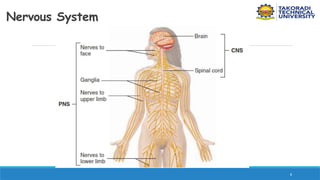
• The Central Nervous System (CNS):
• Made of the brain located with the skull , and spinal cord located
within the vertebral canal formed by the vertebrae
6.
6
Nervous System
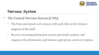
• TheCentral Nervous System (CNS):
• The brain and spinal cord connect with each other at the foramen
magnum of the skull
• Receives incoming information (action potential), analyses and
organizes the information, and initiates appropriate action or response
7.
7
Nervous System
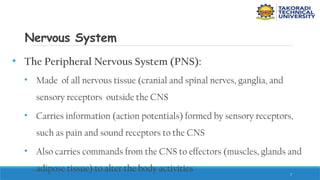
• ThePeripheral Nervous System (PNS):
• Made of all nervous tissue (cranial and spinal nerves, ganglia, and
sensory receptors outside the CNS
• Carries information (action potentials) formed by sensory receptors,
such as pain and sound receptors to the CNS
• Also carries commands from the CNS to effectors (muscles, glands and
adipose tissue) to alter the body activities
9
Nervous System
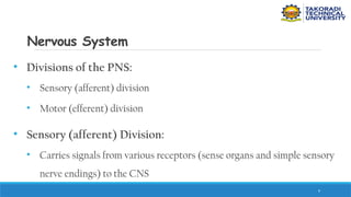
• Divisionsof the PNS:
• Sensory (afferent) division
• Motor (efferent) division
• Sensory (afferent) Division:
• Carries signals from various receptors (sense organs and simple sensory
nerve endings) to the CNS
10.
10
Nervous System
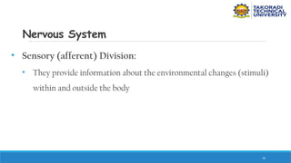
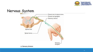
• Sensory(afferent) Division:
• They provide information about the environmental changes (stimuli)
within and outside the body
12
Nervous System
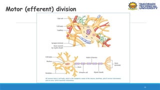
• Motor(efferent) Division:
• Carries signal s from the CNS to structures (i.e., muscles and gland)
that carry out the body’s responses
• Cells and organs responding to command from the CNS are called
effectors
• It is divided into somatic (voluntary) division and autonomic
(involuntary) division
14
Nervous System
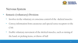
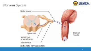
• Somatic(voluntary) Division:
• Involves in the voluntary or conscious control of the skeletal muscles
• Convey information from cutaneous and special sense receptors to the
CNS
• Enable voluntary movement of the skeletal muscles, such as raising of
the hand, or picking items, or throw of ball
15.
15
Nervous System
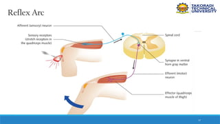
• Somatic(voluntary) Division:
• The skeletal muscles may also contract involuntarily through reflex arc
18
Nervous System
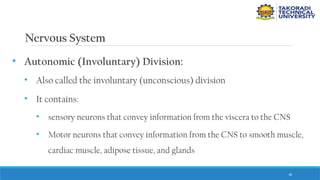
• Autonomic(Involuntary) Division:
• Also called the involuntary (unconscious) division
• It contains:
• sensory neurons that convey information from the viscera to the CNS
• Motor neurons that convey information from the CNS to smooth muscle,
cardiac muscle, adipose tissue, and glands
19.
19
Nervous System
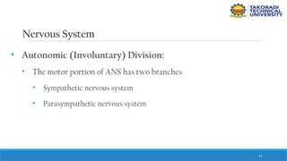
• Autonomic(Involuntary) Division:
• The motor portion of ANS has two branches
• Sympathetic nervous system
• Parasympathetic nervous system
20.
20
Nervous System
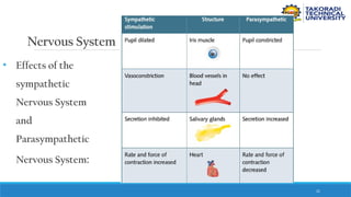
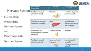
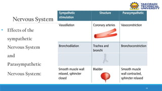
• SympatheticNervous System:
• Reacts to changes in the environment by stimulating activity and
therefore using energy (e.g.. Increases of the heartbeat)
• Parasympathetic Nervous System:
• Opposes the actions of the sympathetic nervous system by inhibiting
activity and therefore conserve energy (e.g., decrease in heartbeat)
24
Nervous System
• Thenervous system is made of organs comprised mainly of nerve
tissue supported and protected by connective tissues
• The nerves tissue is made of
• Neurons
• neuroglia
25.
25
Nervous System
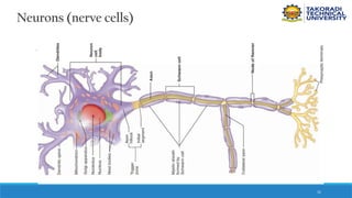
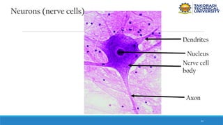
• Neurons(nerve cells):
• The structural and functional units of the nervous system
• Delicate, and specialized to produce and transmit action potentials
• Nerve Cell body: portion of the neuron containing a large spherical
nucleus and cytoplasmic organelles
• Dendrites and Axons extend from the nerve cell body
26.
26
Nervous System
• Dendrites:
•short and highly branched, tapering processes; create impulses
(electrical signals) when stimulated by other neurons and sensory
receptors
• Dendrites carry impulses toward the nerve cell body or axon
27.
27
Nervous System
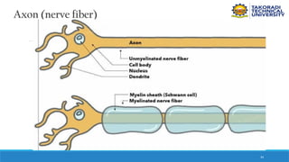
• Axon(nerve fiber):
• A long, thin process of the neuron
• May have one or more side branches called axon collaterals
• Collaterals enable the neuron to make contact with more neurons or
effectors
• Forms a number of short, fine branches (the terminal arborization) at
the distal tip
28.
28
Nervous System
• Axon(nerve fiber):
• A long, thin process of the neuron
• May have one or more side branches called axon collaterals
• Collaterals enable the neuron to make contact with more neurons or
effectors
• Forms a number of short, fine branches at the distal tip
29.
29
Nervous System
• Axon(nerve fiber):
• Terminal boutons (enlarged tips of the arborization) forms junctions
(synapses) with other neurons, muscles, adipose tissue, or glands
• Carries action potentials away from the nerve cell body or dendrites
• Myelinated axons are enclosed in insulating myelin sheath
• Myelin sheath increase the speed of action potential transmission
30.
30
Nervous System
• Axon(nerve fiber):
• Myelin sheath gaps: tiny space between adjacent myelin-forming cells
where an axon is exposed
• Axons without myelin sheath are called unmyelinated axons; they have
a slower speed of action potential transmission
35
Axon (nerve fiber)
Crosssection of a nerve as seen under the
microscope: two fascicles (subdivisions)
are shown
P – perineurium;
EP- epineurium;
Ax- axons; MS- myelin sheath;
En- endoneurium
Gartner LP, Hiatt JL., Color Atlas of Histology 3rd
ed. Philadelphia: Lippincott
Williams & Wilkins, 2000.
36.
36
Nervous System
• Typesof Neurons:
• Neurons are classified according to anatomy or function
• Anatomy classification:
• Multipolar Neurons
• Bipolar Neurons
• Pseudo-uipolar Neurons
37.
37
Nervous System
• MultipolarNeurons
• Have several dendrites and a single axon extending from the nerve cell
body
• Neurons with nerve cell bodies located in the brain and spinal cord are
multipolar neurons
• The most abundant neurons in the nervous system
38.
38
Nervous System
• MultipolarNeurons
• E.g., include Pyramidal cells of the cerebral cortex and Purkinje cells
and neurons of the cerebellar cortex
• Multipolar neurons are subclassified into
• Golgi type I: neurons, when the axon extends beyond the limits of the
dendritic tree
39.
39
Nervous System
• MultipolarNeurons
• Multipolar neurons are subclassified into
• Golgi type II: neurons, when an axon terminates in the immediate area
of the cell body and does not extend beyond the limits of the dendritic
tress (e.g., Small stellate cells of the cerebral cortex )
41
Nervous System
• Bipolarneurons:
• Have only two processes: a dendrite and an axon extending from
opposite ends of the nerve cell body
• Occurs in the sensory portions of the eyes, ears, and nose
42.
42
Nervous System
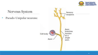
• Pseudo-Unipolarneurons:
• Have a single process extending from the nerve cell body
• The process quickly divides into two branches in opposite directions,
with both branches functioning as a single axon
• Localized in the sensory ganglia of cranial and spinal nerves
45
Nervous System
• SensoryNeurons:
• Carries action potential from the peripheral part of the body to the
CNS
• Their dendrites are specialized to detect changes directly (associated
with sensory receptors)
• Nerves cell bodies are located external to the CNS in ganglia
46.
46
Nervous System
• SensoryNeurons:
• Predominantly Pseudo-unipolar neurons, though, bipolar neurons are
found in special sense organs
47.
47
Nervous System
• Interneurons:
•Located completely inside the CNS and synapse with other neurons
• Responsible for the processing and interpretation of action potentials
by the CNS
• Receive action potentials from sensory neurons and transmit them
from place to place within the CNS
48.
48
Nervous System
• MotorNeurons:
• Carry action potentials from the CNS to effectors to produce an action
• Nerve cell bodies and dendrites are located with the CNS, the axon is
located in cranial and spinal nerves
• Are multipolar neurons
49.
49
Nervous System
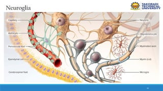
• Neuroglia:
•Non-neuronal cells in the nervous system
• Provide support and protection for neurons
• Schwann cells:
• Form the myelin sheath around the PNS myelinated axon
50.
50
Nervous System
• Schwanncells:
• It wraps its plasma membrane tightly around an axon many times so
that the nucleus and most of the cytoplasm become squeezed into the
outer layer
• Inner layers (formed by layers of plasma membrane) constitute the
myelin sheath
51.
51
Nervous System
• Schwanncells:
• Wrap themselves around small segments of a single axon
• The cytoplasm and nucleus form the outermost layer called
neurilemma (neurolemma)
• The outermost layer form the neurilemma (essential for axon
regeneration after injury)
52.
52
Nervous System
• Satellitecells:
• Found in ganglia (clusters of nerve cell bodies located within the PNS
• Insulate the nerve cell bodies within the ganglion, and regulate the
chemical environment of the ganglion for optimal function
53.
53
Nervous System
• Oligodendrocytes:
•Form the myelin sheath of myelinated axons within the CNS
• Lack neurilemma, hence the inability of axons within the brain and
spinal cord to regenerate after injury
54.
54
Nervous System
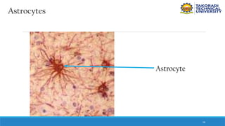
• Astrocytes:
•Primarily support cells for neurons in the CNS
• Stimulate the growth of neurons and influence synaptic transmission
• Joins with the epithelium of blood vessels to form the blood-brain
barrier (BBB)
57
Nervous System
• Microglialcells:
• Small phagocytic cells wandering through the CNS tissue
• Search out and destroy tissue debris, infectious microorganisms and
foreign matter
58.
58
Nervous System
• EpendymalCells:
• Form the epithelial lining of cavities in the brain and spinal cord and
aid in the production of cerebrospinal fluid (fluid within the CNS)
60
Nervous System
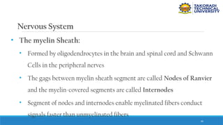
• Themyelin Sheath:
• Formed by oligodendrocytes in the brain and spinal cord and Schwann
Cells in the peripheral nerves
• The gags between myelin sheath segment are called Nodes of Ranvier
and the myelin-covered segments are called Internodes
• Segment of nodes and internodes enable myelinated fibers conduct
signals faster than unmyelinated fibers
61.
61
Nervous System
• Themyelin Sheath:
• Unmyelinated fibers are responsible for digestive secretions
• Nerve Tissues :
• White matter
• Gray matter
62.
62
Nervous System
• WhiteMatter:
• Made of bundles of nerves fibers called tracts, which travel up and
down the spinal cord between one region of the brain and another, and
between the brain and cord
• Most of the fibers are myelinated
• The myelin gives the white matter a glistening, pearly white colour
63.
63
Nervous System
• WhiteMatter:
• There are no nerves in the brain or spinal cord
• The body’s bundles of nerve fibers found in the PNS are called nerves
and those found in the CNS are called tracts
• Is more like the cable of the CNS, bundle of nerve fibers carrying
signals from place to place
64.
64
Nervous System
• GrayMatter:
• Neurosomas, dendrites, and synapses are located in the gray matter
• Has duller colour in fresh nervous tissue due to lack of myelin
• It is the information processing part of the CNS
• Forms the inner core of the spinal cord
65.
65
Synaptic Terminals andSynapses
• Synaptic terminal is specialized for the transmission of a
chemical message in response to an action potential
• Synapse is the junction between the presynaptic terminal of an
axon and a postsynaptic membrane receptor surface, generally a
dendrite
66.
66
Synaptic Terminals andSynapses
• Synaptic terminal is specialized for the transmission of a
chemical message in response to an action potential
• Synapse is the junction between the presynaptic terminal of an
axon and a postsynaptic membrane receptor surface, generally a
dendrite
67.
67
Synaptic Terminals andSynapses
• Presynaptic:
• Refers to the transmitting side (usually axonal)
• Postsynaptic:
• The receiving side (usually dendritic or somatic , sometimes axonal)
• Synaptic cleft:
• separates the pre- and postsynaptic membrane
68.
68
Synaptic Terminals andSynapses
• The inner surface of synaptic membranes are coated by a dense
material
• Presynaptic terminals contain a large number of membrane-bound
synaptic vesicles containing neurotransmitter and mitochondria
• synapses are classified by their location on the postsynaptic
neuron
69.
69
Synaptic Terminals andSynapses
• Classification of synapses:
• Axospinous synapses: axon terminals facing a dendritic spine
• Axodendritic synapses: axon terminals on the shaft of a dendrite
• Axosomatic synapses: axon terminals on the soma of a neuron
• Axoaxonic synapses: axon terminals ending on axon terminals
70.
70
Synaptic Terminals andSynapses
• Classification of synapses:
• Axospinous synapses: axon terminals facing a dendritic spine
• Axodendritic synapses: axon terminals on the shaft of a dendrite
• Axosomatic synapses: axon terminals on the soma of a neuron
• Axoaxonic synapses: axon terminals ending on axon terminals


![CTEV [ clubfoot] DR ARUN LAL ,DR MOHAMED ASHRAF travancore medical college k...](https://cdn.slidesharecdn.com/ss_thumbnails/ctevclubfootdrarunlaldrmohamedashraftravancoremedicalcollegekollamkeralaindia-260208063247-18fc466c-thumbnail.jpg?width=640&height=640&fit=bounds)


![ONFH[AVN HIP] -TRIPLE REGIME -A NOVAL SURGICAL CONCEPT .pptx](https://cdn.slidesharecdn.com/ss_thumbnails/onfhavnhip2026koaconcalicutdrgokuldevdrmashraf-260210064517-213ec005-thumbnail.jpg?width=640&height=640&fit=bounds)


