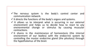
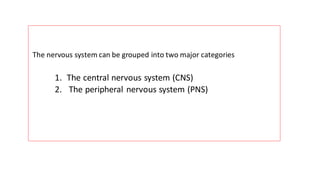
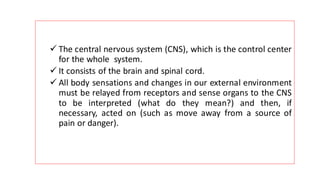
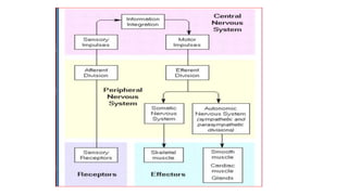
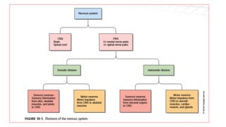
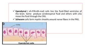
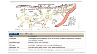
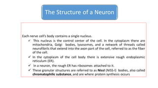
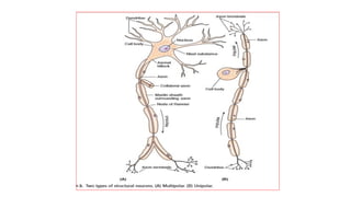
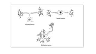
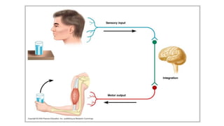
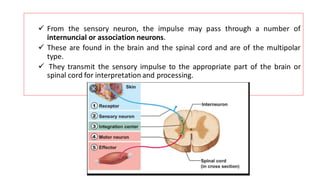
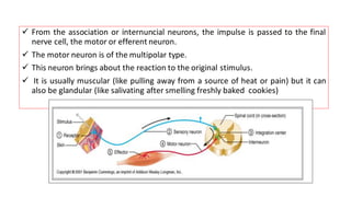
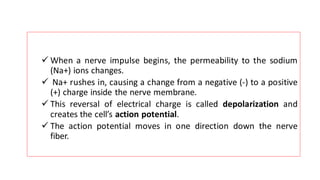
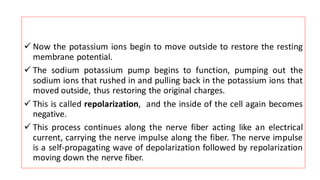
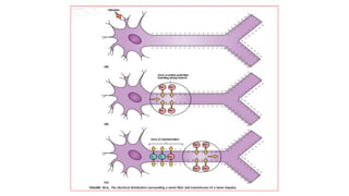
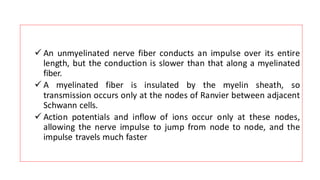
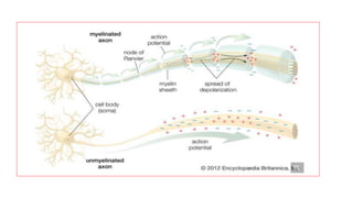
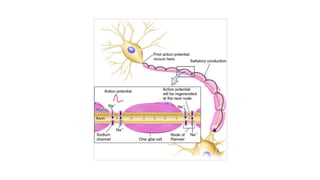
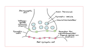
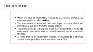
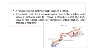
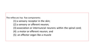
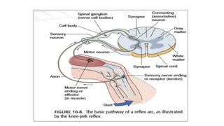
Scientificity and feasibility study of non-invasive central arterial pressure...
Nervous systems central nervous system and
1.
2. ✓ The nervous system is the body’s control center and
communication network.
✓ It directs the functions of the body’s organs and systems.
✓ It allows us to interpret what is occurring in our external
environment and helps us to decide how to react to any
environmental change or stimulus by causing muscular
contractions.
✓ It shares in the maintenance of homeostasis (the internal
environment of our bodies) with the endocrine system by
controlling the master endocrine gland (the pituitary) through
the hypothalamus of the brain
3. The nervous system can be grouped into two major categories
1. The central nervous system (CNS)
2. The peripheral nervous system (PNS)
4. ✓ The central nervous system (CNS), which is the control center
for the whole system.
✓ It consists of the brain and spinal cord.
✓ All body sensations and changes in our external environment
must be relayed from receptors and sense organs to the CNS
to be interpreted (what do they mean?) and then, if
necessary, acted on (such as move away from a source of
pain or danger).
5. ✓ The peripheral nervous system (PNS), which is subdivided into several
smaller units. This second category consists of all the nerves that
connect the brain and spinal cord with sensory receptors, muscles, and
glands.
✓ The PNS can be divided into two subcategories:
▪ The afferent peripheral system, which consists of afferent or sensory neurons that
convey information from receptors in the periphery of the body to the brain and
spinal cord,
▪ And the efferent peripheral system, which consists of efferent or motor neurons
that convey information from the brain and spinal cord to muscles and glands.
6. The efferent peripheral system can be further subdivided into two subcategories.
✓ The first is the somatic nervous system, which conducts impulses from the
brain and spinal cord to skeletal muscle, thereby causing us to respond or
react to changes in our external environment.
✓ The second is the autonomic nervous system (ANS), which conducts
impulses from the brain and spinal cord to smooth muscle tissue (like the
smooth muscles of the intestine that push food through the digestive
tract), to cardiac muscle tissue of the heart, and to glands (like the
endocrine glands). The ANS is considered to be involuntary.
7. The organs affected by this system receive nerve fibers from two
divisions of the ANS:
✓ The sympathetic division, which stimulates or speeds up activity and
thus involves energy expenditure and uses norepinephrine (nor-ep-ih-
NEH-frin) as a neurotransmitter
✓ And the parasympathetic division, which stimulates or speeds up the
body’s vegetative activities such as digestion, urination, and defecation
and restores or slows down other activities. It uses acetylcholine (ah-seh-
till-KOH-leen) as a neurotransmitter at nerve endings.
8.
9.
10. ✓ Nervous tissue consists of groupings of nerve cells or neurons (NOO-
ronz) that transmit information called nerve impulses in the form of
electrochemical changes.
✓ A nerve is a bundle of nerve cells or fibers.
✓ Nervous tissue is also composed of cells that perform support and
protection.
✓ These cells are called neuroglia (noo-ROWGlee-ah) or glial (GLEE-al) cells
(neuroglia means nerve glue).
✓ Over 60% of all brain cells are neuroglia cells.
11. There are different kinds of neuroglia cells, and, unlike neurons, they do
not conduct impulses
✓ Astrocytes are star-shaped cells that wrap around nerve cells to
form a supporting network in the brain and spinal cord.
✓ They attach neurons to their blood vessels, thus helping regulate
nutrients and ions that are needed by the nerve cells
Neuroglia Cells
12. ✓ Oligodendroglia(all-ih-gohDEN-droh-GLEE-ah) look like small astrocytes.
✓ They also provide support by forming semirigid connective-like tissue rows between
neurons in the brain and spinal cord.
✓ They produce the fatty myelin ( MY-eh-lin) sheath on the neurons of the brain and spinal
cord of the CNS
13. ✓ Microglia (my-KROWG-lee-al) cells are small cells that
protect the CNS and whose role is to engulf and
destroy microbes like bacteria and cellular debris.
14. ✓ Ependymal ( eh-PIN-dih-mal) cells line the fluid-filled ventricles of
the brain. Some produce cerebrospinal fluid and others with cilia
move the fluid through the CNS.
✓ Schwann cells form myelin sheaths around nerve fibers in the PNS.
15.
16.
17. Each nerve cell’s body contains a single nucleus.
✓ This nucleus is the control center of the cell. In the cytoplasm there are
mitochondria, Golgi bodies, lysosomes, and a network of threads called
neurofibrils that extend into the axon part of the cell, referred to as the fiber
of the cell.
✓ In the cytoplasm of the cell body there is extensive rough endoplasmic
reticulum (ER).
✓ In a neuron, the rough ER has ribosomes attached to it.
✓ These granular structures are referred to as Nissl (NISS-l) bodies, also called
chromatophilic substance, and are where protein synthesis occurs
The Structure of a Neuron
18.
19. ✓ There are two kinds of nerve fibers on the nerve cell:
dendrites (DEN-drightz) and axons.
✓ Dendrites are short and branched, like the branches of
trees.
✓ These are the receptive areas of the neuron and a
multipolar neuron will have many dendrites.
20. ✓ A nerve cell, however, will have only one axon, which begins
as a slight enlargement of the cell body called the axonal
hillock.
✓ The axon is a long process or fiber that begins singly but may
branch and at its end has many fine extensions called axon
terminals that contact with dendrites of other neurons.
✓ Numerous mitochondria and neurofibrils are in the axon.
21.
22. ✓ The large peripheral axons are enclosed in fatty myelin sheaths produced by
the Schwann cells.
✓ These are a type of neuroglial cell that wrap tightly in layers around the axon,
producing fatty sheets of lipoprotein.
✓ The portions of the Schwann cell that contain most of the cytoplasm of the
cell and the nucleus remain outside of the myelin sheath and make up a
portion called the neurilemma.
✓ Narrow gaps in the sheath are the nodes of Ranvier.
23.
24.
25. Cells that conduct impulses from one part of the body to another are called neurons.
They may be classified by both function and structure. The structural classification
consistsof three types of cells.
1. Multipolar neurons are neurons that have several (multi) dendrites and one
axon. Most neurons in the brain and spinal cord are this type.
2. Bipolar neurons have one dendrite and one axon. They function as receptor
cells in special sense organs. Only two (bi) processes come off the cell body.
They are found in only three areas of the body: the retina of the eye, the
inner ear, and the olfactory area of the nose
Structural Classification of Neurons
26. 3. Unipolar neurons have only one process extending from the
cell body. This single process then branches into a central
branch that functions as an axon and a peripheral branch that
functions as a dendrite. Most sensory neurons are unipolar
neurons. The branch functioning as an axon enters the brain or
spinal cord; the branch functioning as a dendrite connects to a
peripheral part of the body.
27.
28.
29. ✓ Nerve cells pick up various changes in the environment (stimuli such
as changes in temperature or pressure) from receptors.
✓ Receptors are the peripheral nerve endings of sensory nerves that
respond to stimuli.
✓ There are many different types of receptors. Our skin has an
enormous number of such receptors. These receptors change the
energy of a stimulus, like heat, into nerve impulses.
Functional Classification of Neurons
30. ✓ The first nerve cell receiving this impulse directly from a
receptor is called a sensory or afferent neuron. These neurons
are of the unipolar type.
✓ The receptors are in contact with only one end of the sensory
neuron (the peripheral process in the skin), thus ensuring a
one-way transmission of the impulse. The central process of
the sensory neuron goes to the spinal cord.
31. ✓ From the sensory neuron, the impulse may pass through a number of
internuncial or association neurons.
✓ These are found in the brain and the spinal cord and are of the multipolar
type.
✓ They transmit the sensory impulse to the appropriate part of the brain or
spinal cord for interpretation and processing.
32. ✓ From the association or internuncial neurons, the impulse is passed to the final
nerve cell, the motor or efferent neuron.
✓ The motor neuron is of the multipolar type.
✓ This neuron brings about the reaction to the original stimulus.
✓ It is usually muscular (like pulling away from a source of heat or pain) but it can
also be glandular (like salivating after smelling freshly baked cookies)
33. ✓ Positively charged sodium (Na+) ions are in greater concentration outside the
cell than inside.
✓ There is a greater concentration of positively charged potassium (K+) ions inside
the cell than outside.
✓ This situation is maintained by the cell m embrane’s s odium-potassium pump.
✓ In addition to the potassium ion, the inside of the fiber has negatively charged
chloride (Cl-) ions and other negatively charged organic molecules. Thus, the
nerve fiber has an electrical distribution as well, such that the outside is
positively charged while the inside is negatively charged.
✓ This condition is known as the membrane or resting potential
Na+ and K+ ions tend to diffuse across the membrane but the cell maintains the resting potential
through the channels of the sodium-potassium pump that actively extrudes Na+ and accumulates
K+ ions
34.
35. ✓ When a nerve impulse begins, the permeability to the sodium
(Na+) ions changes.
✓ Na+ rushes in, causing a change from a negative (-) to a positive
(+) charge inside the nerve membrane.
✓ This reversal of electrical charge is called depolarization and
creates the cell’s action potential.
✓ The action potential moves in one direction down the nerve
fiber.
36. ✓ Now the potassium ions begin to move outside to restore the resting
membrane potential.
✓ The sodium potassium pump begins to function, pumping out the
sodium ions that rushed in and pulling back in the potassium ions that
moved outside, thus restoring the original charges.
✓ This is called repolarization, and the inside of the cell again becomes
negative.
✓ This process continues along the nerve fiber acting like an electrical
current, carrying the nerve impulse along the fiber. The nerve impulse
is a self-propagating wave of depolarization followed by repolarization
moving down the nerve fiber.
37.
38. ✓ An unmyelinated nerve fiber conducts an impulse over its entire
length, but the conduction is slower than that along a myelinated
fiber.
✓ A myelinated fiber is insulated by the myelin sheath, so
transmission occurs only at the nodes of Ranvier between adjacent
Schwann cells.
✓ Action potentials and inflow of ions occur only at these nodes,
allowing the nerve impulse to jump from node to node, and the
impulse travels much faster
39.
40.
41. ✓ Synapses (sin-AP-seez) are the areas where the terminal branches of an
axon (the axon terminals) are anchored close to, but not touching, the
ends of the dendrites of another neuron.
✓ These synapses are one-way junctions that ensure that the nerve impulse
travels in only one direction. This area is called a synaptic cleft.
✓ Other such areas of synapses are between axon endings and muscles or
between axon endings and glands.
THE SYNAPTIC TRANSMISSION
42. ✓ Transmission across synapses is brought about by the secretions of
very low concentrations of chemicals called neurotransmitters that
move across the gap.
✓ As the nerve impulse travels down the fiber, it causes vesicles in the
axon endings of a presynaptic neuron to release the chemical
neurotransmitter.
✓ Most of the synapses in our bodies use acetylcholine as the
neurotransmitter.
43.
44. ✓ The acetylcholine allows the impulse to travel across the synaptic cleft to the
postsynaptic neuron.
✓ However, it does not remain there because an enzyme in the cleft,
acetylcholinesterase, immediately begins to break down the acetylcholine after it
performs its function .
✓ The autonomic nervous system in addition uses adrenaline (also called epinephrine)
as a transmitting agent.
✓ Many kinds of neurotransmitters are found in the nervous system.
45.
46. ✓ Many kinds of neurotransmitters are found in the nervous system.
✓ Some neurons produce only one type; others produce two or three.
✓ The best known neurotransmitters are acetylcholine and norepinephrine.
✓ Some others are serotonin (sayr-oh-TOH-nin), dopamine (DOH-pah-meen),
and the endorphins (in-DOHR-fi nz).
47. ▪ When we have an involuntary reaction to an external stimulus, we
experience what is called a reflex.
▪ This is experienced when we prick our finger on a rose thorn and
immediately pull away from the source of pain.
▪ The reflex allowed us to respond much more quickly than if we had to
consciously think about what to do and interpret the information in
the CNS.
▪ A reflex then is an involuntary reaction or response to a stimulus
applied to our periphery and transmitted to the CNS.
THE REFLEX ARC
48. ▪ A reflex arc is the pathway that results in a reflex .
▪ It is a basic unit of the nervous system and is the smallest and
simplest pathway able to receive a stimulus, enter the CNS
(usually the spinal cord) for immediate interpretation, and
produce a response
49. The reflex arc has five components:
(1) a sensory receptor in the skin;
(2) a sensory or afferent neuron;
(3) association or internuncial neurons within the spinal cord;
(4) a motor or efferent neuron; and
(5) an effector organ like a muscle