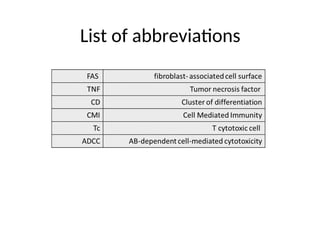
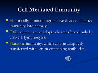
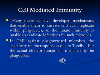
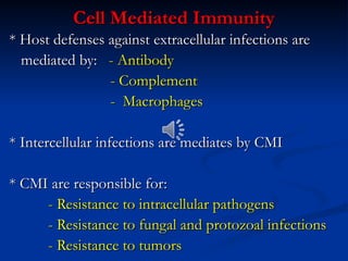
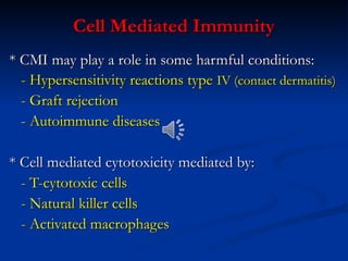
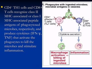
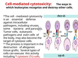
BY
Dr. Dhafer Abdullah Farhan
Ph. D Cancer research
Today
We are
here
List of abbreviations
Cell Mediated Immunity
 Historically, immunologists have divided adaptive
Historically, immunologists have divided adaptive
immunity into namely:
 CMI
CMI, which can be adoptively transferred only by
, which can be adoptively transferred only by
viable T lymphocytes.
 Humoral
Humoral immunity, which can be adoptively
immunity, which can be adoptively
transferred with serum containing antibodies.
Cell Mediated Immunity
 Many microbes have developed mechanisms
Many microbes have developed mechanisms
that enable them to survive and even replicate
within phagocytes, so the innate immunity is
unable to eradicate infections by such microbes.
 In CMI against phagocytosed microbes, the
In CMI against phagocytosed microbes, the
specificity of the response is due to T cells – but
the actual effector function is mediated by the
phagocytes.
phagocytes.
Cell Mediated Immunity
* Host defenses against extracellular infections are
mediated by:
mediated by: - Antibody
- Antibody
- Complement
- Complement
- Macrophages
- Macrophages
* Intercellular infections are mediates by CMI
* CMI are responsible for:
- Resistance to intracellular pathogens
- Resistance to fungal and protozoal infections
- Resistance to tumors
Cell Mediated Immunity
* CMI may play a role in some harmful conditions:
- Hypersensitivity reactions type
- Hypersensitivity reactions type IV (contact dermatitis)
IV (contact dermatitis)
- Graft rejection
- Graft rejection
- Autoimmune diseases
* Cell mediated cytotoxicity mediated by:
- T-cytotoxic cells
- T-cytotoxic cells
- Natural killer cells
- Activated macrophages
Characters Of CMI
Characters Of CMI
Cellular immune response is mediated by:
- Subpopulation of T-lymphocytes
- Macrophages and their products
 CD4
CD4+
+
TH1 cells and CD8+
TH1 cells and CD8+
T cells recognize class II
MHC-associated or class I
MHC-associated peptide
antigens of phagocytosed
microbes, respectively, and
produce cytokines (IFN-
produce cytokines (IFN-
,
,
TNF) that activate the
phagocytes to kill the
microbes and stimulate
inflammation.
inflammation.
*The cell - mediated cytotoxicity
is an essential defense
against intracellular
pathogens, including viruses,
some bacteria and parasites.
Tumor cells, eukaryotic
pathogens and even cells of
the body, may also become the
target of cytotoxic cells.
* The process is important in the
destruction of allogeneic
tissue grafts. Several types of
cells can execute this activity
including T-cytotoxic (Tc) cells,
Cell-mediated cytotoxicity: The ways in
which leukocytes recognize and destroy other cells.
Tc-cells recognize specific Ag (e.g. viral peptides on
infected cells) presented by MHC-I most Tc are CD8+ve,
but about 10% of MHC-restricted cytotoxic T-cells are CD4+ve
& recognize Ag presented on MHC-II.
NK-cells recognize cells which fail to express MHCI
molecules, they can also bind to Ab already attached to Ag an
a target cell, using their Fc-R (CD16), in a what is known as
AB-dependent cell-mediated cytotoxicity (ADCC). Several
viruses have evolved mechanisms to avoid recognition by Tc
cell. They reduce the expression of MHC molecules or even
produce proteins which divert MHC molecules out of the
endoplasmic reticulum. So NK-cells specifically recognize
cells which have lost their MHC - I molecules.
Tc-cells and NK-cells act in a complementary
way to protect the body.
Mechanisms of cell-mediated
cytotoxicity
Tc, NK- and K-cells use
variety of different
mechanisms to kill their
targets, including: * direct
cell-cell signaling via surface
molecules. *
indirect signaling via cytokines.
in addition many CD8 Tc and large
granular lymphocytes (NK and K-
cells) have granules which contain
proteins that damage target cells if
they are released directly against
the target cell plasma membrane.
•a cytotoxicity which may be signaled via Fas or a TNF-R on
a target cell.
•Tc cells signal to their targets using members of TNF-R
group of molecules. These include Fas (CD95) and the type-1
TNF-R (TNFR-1) which are widely distributed in the
body.Ligation of Fas-induces trimerization of the Fas-
molecule on the cell surface, which causes them to
associate with a transducing molecule which recrutes and
activates caspases 8 and 10.
• For the cytotoxic lymphocytes which lack granules the Fas-
pathway is thought to be the principle means of signaling to
the target.
** Most CD8 TC and NK-cells (and
macrophages) have vesicles containing
TNF and lymphotoxin which can be
released onto a target cells. TNF acts in a
very similar way to the Fas-ligand. It
causes trimerization of the TNFR-1 so that
the receptor associates with adaptor
proteins which recruite caspases. Both
TNFR-1 and Fas containing
intracytoplasmic domains (death-
domains) which are found on a number of
proteins involved in cell survival.
*** The specific granules of NK-cells and Tc contain several
proteins, including perforin and granzymes (granules
associated enzymes). After binding to its target, the Tc cells directs
its granules towards the membrane a joining the target. Then, in a
Ca-dependent phase, the granule contents are discharge into the
cleft between the two cells.
Perforin is a pore-forming proteins and in the presence of
Ca , the perforin bind to the target cell membrane and polymerize
to form transmembrane channels. Tc-protected from
autodestruction with this protein by a proteoglycan (chondroitin-
sulphate A) which may bind to and inactivate the perforin,
thus Tc-survives and can continue kill further targets.
Granzymes are collection of serine estrases (enzymes)
which are also released upon granule exocytosis and become
active after release. Some of these enzyme trigger apoptosis and
DNA-degradation.
Both perforin and granzymes act synergistically; the granzymes
enter the target cells via pore created by perforin.
To summarize the lymphoid
cytotoxic effectors; CD8 Tc use
both Fas- L and granule release to
kill their targets, CD4Tc-cells use
principally FasL, and NK-cells use
primarily their granules.
TNF may contribute to the
cytotoxic damage produced by
any of these cells.
*cytotoxicity may be triggered specifically to a target by
ADCC or may involve a range of non-specific toxic
mediators. Macrophage and neutrophile express Fc-
gR1&2 which allow them to engage their targets by
ADCC.`
** Macrophages and neutrophiles granules which contain
neutrophil defensins, lysosomal enzymes and cytostatic
proteins. If the phagocyte fails to internalize its target,
then these mediators may be released into the
extracellular environment and contribute to localized cell
damage ((frustrated phagocytosis)), the effect of these
mediators tends to produce necrosis and inflammation.
*** Activated macrophage secrete TNF which can induce
A number of non-lymphoid cells may be cytotoxic to other cells,
Eosinophils are characterized by their granules.
Eosinophils are only weakly phagocytic, they ingest some
bacteria following activation but are less efficient than
neutrophils at intracellular killing. Their major function
appears to be the secretion of various toxic granules.
Constituents. They are therefore effective for the
extracellular killing of microorganisms, particularly large
parasites such as schistosoma. The components of the
eosinophil granule include major basic protein (MBP),
eosinophil peroxidase (EPO) and eosinophil cationic
protein (ECP). Other molecules produced by eosinophils
are lysophospholipase and eosinophil-derived neurotoxin
(EDN), which is also a ribonuclease but with strong
neurotoxic activity. Thus, killing may involve contact-
dependent degranulation or may simply require
deposition of toxins within the local tissue.
THANK YOU

lect Cell mediate immunity.ppt3456789098765

  • 1.
    BY Dr. Dhafer AbdullahFarhan Ph. D Cancer research Today We are here
  • 2.
  • 3.
    Cell Mediated Immunity CellMediated Immunity  Historically, immunologists have divided adaptive Historically, immunologists have divided adaptive immunity into namely: immunity into namely:  CMI CMI, which can be adoptively transferred only by , which can be adoptively transferred only by viable T lymphocytes. viable T lymphocytes.  Humoral Humoral immunity, which can be adoptively immunity, which can be adoptively transferred with serum containing antibodies. transferred with serum containing antibodies.
  • 4.
    Cell Mediated Immunity CellMediated Immunity  Many microbes have developed mechanisms Many microbes have developed mechanisms that enable them to survive and even replicate that enable them to survive and even replicate within phagocytes, so the innate immunity is within phagocytes, so the innate immunity is unable to eradicate infections by such microbes. unable to eradicate infections by such microbes.  In CMI against phagocytosed microbes, the In CMI against phagocytosed microbes, the specificity of the response is due to T cells – but specificity of the response is due to T cells – but the actual effector function is mediated by the the actual effector function is mediated by the phagocytes. phagocytes.
  • 5.
    Cell Mediated Immunity CellMediated Immunity * Host defenses against extracellular infections are * Host defenses against extracellular infections are mediated by: mediated by: - Antibody - Antibody - Complement - Complement - Macrophages - Macrophages * Intercellular infections are mediates by CMI * Intercellular infections are mediates by CMI * CMI are responsible for: * CMI are responsible for: - Resistance to intracellular pathogens - Resistance to intracellular pathogens - Resistance to fungal and protozoal infections - Resistance to fungal and protozoal infections - Resistance to tumors - Resistance to tumors
  • 6.
    Cell Mediated Immunity CellMediated Immunity * CMI may play a role in some harmful conditions: * CMI may play a role in some harmful conditions: - Hypersensitivity reactions type - Hypersensitivity reactions type IV (contact dermatitis) IV (contact dermatitis) - Graft rejection - Graft rejection - Autoimmune diseases - Autoimmune diseases * Cell mediated cytotoxicity mediated by: * Cell mediated cytotoxicity mediated by: - T-cytotoxic cells - T-cytotoxic cells - Natural killer cells - Natural killer cells - Activated macrophages - Activated macrophages
  • 7.
    Characters Of CMI CharactersOf CMI Cellular immune response is mediated by: Cellular immune response is mediated by: - Subpopulation of T-lymphocytes - Subpopulation of T-lymphocytes - Macrophages and their products - Macrophages and their products
  • 8.
     CD4 CD4+ + TH1 cellsand CD8+ TH1 cells and CD8+ T cells recognize class II T cells recognize class II MHC-associated or class I MHC-associated or class I MHC-associated peptide MHC-associated peptide antigens of phagocytosed antigens of phagocytosed microbes, respectively, and microbes, respectively, and produce cytokines (IFN- produce cytokines (IFN- , , TNF) that activate the TNF) that activate the phagocytes to kill the phagocytes to kill the microbes and stimulate microbes and stimulate inflammation. inflammation.
  • 9.
    *The cell -mediated cytotoxicity is an essential defense against intracellular pathogens, including viruses, some bacteria and parasites. Tumor cells, eukaryotic pathogens and even cells of the body, may also become the target of cytotoxic cells. * The process is important in the destruction of allogeneic tissue grafts. Several types of cells can execute this activity including T-cytotoxic (Tc) cells, Cell-mediated cytotoxicity: The ways in which leukocytes recognize and destroy other cells.
  • 10.
    Tc-cells recognize specificAg (e.g. viral peptides on infected cells) presented by MHC-I most Tc are CD8+ve, but about 10% of MHC-restricted cytotoxic T-cells are CD4+ve & recognize Ag presented on MHC-II. NK-cells recognize cells which fail to express MHCI molecules, they can also bind to Ab already attached to Ag an a target cell, using their Fc-R (CD16), in a what is known as AB-dependent cell-mediated cytotoxicity (ADCC). Several viruses have evolved mechanisms to avoid recognition by Tc cell. They reduce the expression of MHC molecules or even produce proteins which divert MHC molecules out of the endoplasmic reticulum. So NK-cells specifically recognize cells which have lost their MHC - I molecules. Tc-cells and NK-cells act in a complementary way to protect the body.
  • 11.
    Mechanisms of cell-mediated cytotoxicity Tc,NK- and K-cells use variety of different mechanisms to kill their targets, including: * direct cell-cell signaling via surface molecules. * indirect signaling via cytokines. in addition many CD8 Tc and large granular lymphocytes (NK and K- cells) have granules which contain proteins that damage target cells if they are released directly against the target cell plasma membrane.
  • 12.
    •a cytotoxicity whichmay be signaled via Fas or a TNF-R on a target cell. •Tc cells signal to their targets using members of TNF-R group of molecules. These include Fas (CD95) and the type-1 TNF-R (TNFR-1) which are widely distributed in the body.Ligation of Fas-induces trimerization of the Fas- molecule on the cell surface, which causes them to associate with a transducing molecule which recrutes and activates caspases 8 and 10. • For the cytotoxic lymphocytes which lack granules the Fas- pathway is thought to be the principle means of signaling to the target.
  • 13.
    ** Most CD8TC and NK-cells (and macrophages) have vesicles containing TNF and lymphotoxin which can be released onto a target cells. TNF acts in a very similar way to the Fas-ligand. It causes trimerization of the TNFR-1 so that the receptor associates with adaptor proteins which recruite caspases. Both TNFR-1 and Fas containing intracytoplasmic domains (death- domains) which are found on a number of proteins involved in cell survival.
  • 14.
    *** The specificgranules of NK-cells and Tc contain several proteins, including perforin and granzymes (granules associated enzymes). After binding to its target, the Tc cells directs its granules towards the membrane a joining the target. Then, in a Ca-dependent phase, the granule contents are discharge into the cleft between the two cells. Perforin is a pore-forming proteins and in the presence of Ca , the perforin bind to the target cell membrane and polymerize to form transmembrane channels. Tc-protected from autodestruction with this protein by a proteoglycan (chondroitin- sulphate A) which may bind to and inactivate the perforin, thus Tc-survives and can continue kill further targets. Granzymes are collection of serine estrases (enzymes) which are also released upon granule exocytosis and become active after release. Some of these enzyme trigger apoptosis and DNA-degradation. Both perforin and granzymes act synergistically; the granzymes enter the target cells via pore created by perforin.
  • 15.
    To summarize thelymphoid cytotoxic effectors; CD8 Tc use both Fas- L and granule release to kill their targets, CD4Tc-cells use principally FasL, and NK-cells use primarily their granules. TNF may contribute to the cytotoxic damage produced by any of these cells.
  • 16.
    *cytotoxicity may betriggered specifically to a target by ADCC or may involve a range of non-specific toxic mediators. Macrophage and neutrophile express Fc- gR1&2 which allow them to engage their targets by ADCC.` ** Macrophages and neutrophiles granules which contain neutrophil defensins, lysosomal enzymes and cytostatic proteins. If the phagocyte fails to internalize its target, then these mediators may be released into the extracellular environment and contribute to localized cell damage ((frustrated phagocytosis)), the effect of these mediators tends to produce necrosis and inflammation. *** Activated macrophage secrete TNF which can induce A number of non-lymphoid cells may be cytotoxic to other cells,
  • 17.
    Eosinophils are characterizedby their granules. Eosinophils are only weakly phagocytic, they ingest some bacteria following activation but are less efficient than neutrophils at intracellular killing. Their major function appears to be the secretion of various toxic granules. Constituents. They are therefore effective for the extracellular killing of microorganisms, particularly large parasites such as schistosoma. The components of the eosinophil granule include major basic protein (MBP), eosinophil peroxidase (EPO) and eosinophil cationic protein (ECP). Other molecules produced by eosinophils are lysophospholipase and eosinophil-derived neurotoxin (EDN), which is also a ribonuclease but with strong neurotoxic activity. Thus, killing may involve contact- dependent degranulation or may simply require deposition of toxins within the local tissue.
  • 18.