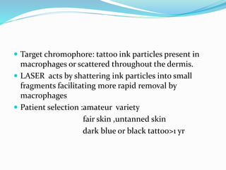
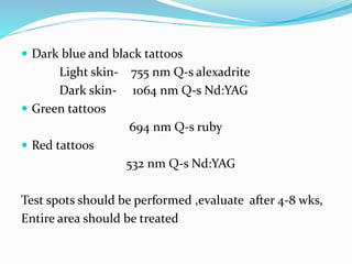
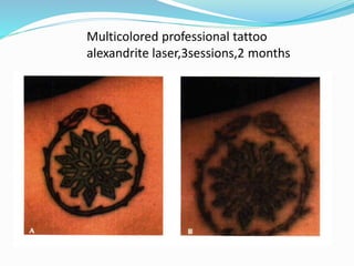
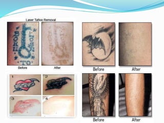
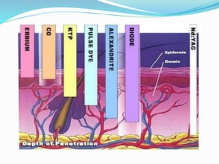
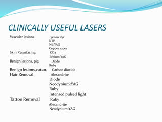
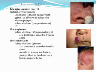
Dr. Hardik Dodia
HISTORY
 1960 -MAIMAN–Ruby rod(694 nm)
 1961-Nd:YAG
 1962-Argon
 1964-CO2
 Dr Leon Goldman-pioneered use of Ruby LASER on
cadaver and later on patients.
 1983-theory of selective photothermolysis by Anderson
and Parrish.
LASER
Light Amplification by Stimulate Emission of Radiation
Characteristics: 1)Monochromaticity-same wave length
2)Coherence-waves are in phase in time
and space
3)Collimation-waves remain parallel
 COMPONENTS OF LASER DEVICE:
1)Energy source
2)Lasing medium
3)Amplifier
TYPES OF LASER
LASING MEDIUM
 SOLID: 1) Ruby- 694nm Melanin ,Tatoo pigment
2)Nd:YAG- 1064nm Pigment
3)KTP- 532 OxyHB,Melanin
4)Er:YAG 2940 Water
5)Diode 800 OxyHB,Melanin
6)Alexandrite 755 Melanin ,Tatoo pigment
7)Copper vapor 578 OxyHB
 LIQUID:1)Yellow dye 585,595 OxyHB
2)Green dye 510 Melanin
 GAS: 1)Argon 488,514 OxyHB,Melanin
2)Helium:Neon 633
3)CO2 10600 water
4)Excimer UV breaks chemical bonds
LASER OUTPUT
 CONTINUOUS MODE- co2 LASER
 PULSD MODE- pulse duration in micro second
 QUALITY SWITCHING- pulse duration in
nanoseconds with extremely high peak power
 TISSUE OPTICS: FATE OF LASER LIGHT
1)Reflection-4to6%
2)Absorption-initial intensity
-chromophores
a)Endogenous-HB,melanin,water,collagen
b)Exogenous-tattoo ink,photosensitizer
3)Scattering-mainly due to collagen
more with shorter wave length
4)Transmission-residual light is transmitted
-more with large wave length as
there is less scattering
LASER OUTPUT
 Depends on lasing medium and electric supply
Energy: Joule( J )
Power is the rate at which energy is delivered
W=J / sec
Power density is the rate at which energy is delivered per
unit area ( W / cm2 )
FLUENCE :amount of energy delivered per cm2
J/cm2
THERMAL RELAXATION TIME:
 LIGHT TISSUE INTERACTION:
1)Photostimulation: wound healing
2)Photodynamic reactions: topical or systemic
administration of photo sensitizer dye and later
irradiation leads to oxidative reactions(cancer ,acne)
3)Photothermolytic and photomechanical reaction:
a) 37-60 C –tissue retraction
b) 60-90 C-protein denaturation and coagulation
c) 90-1oo C- carbonization & burning
d) >100 C- tissue is vaporized and ablated
TISSUE COOLING
AIM:
1)Preservation of epidermis.
2) Delivery of higher energy to target area.
3) Analgesia
1)Cold air convection-chilled air -30 c is directed onto
the area to be treated
2)Contact cooling-application of ice packs or chilled
water is passed in between transparent plates of
sapphire
3)Cryogen spray(dynamic cooling)-frozen gas is sprayed
onto the skin immediately before the laser pulse
ADVANTAGES
 Increased precision while cutting
 No contact
 Specificity
 Sterilization of operating site
 Less pain ,bleeding and quicker healing
 Lesser coagulation causes little surrounding damage
 Reduced hospitalization time
LASER HAZARDS
 EYE HAZARD- corneal and retinal burns,
corneal and lenticular opacities
cataract
visual loss
 SKIN HAZARDS-burns,purpura,vesicles
 FIRE HAZARDS
CO2 LASER - 10,600nm
 Infrared invisible LASER
 Target chromophore : water
 Penetration <1 mm
 CW /Superpulsed CO2
 USES:Focused Mode-skin tags, warts moles ,
as a knife
Defocused Mode-skin resurfacing,acne scars,
chickenpox scars,
rhinophyma
Pre op 3 months
Nd:YAG LASER -1064nm
 Infrared invisible LASER in which Nd is doped in a
crystal of YAG.
 Target chromophore: pigments
 Penetration -10-20mm
vessel- 3 mm
 USES: tattoo marks
pigmented skin lesions
vascular lesions
useful in hemorrhagic disorders
pre op at 3 months
Er:YAG - 2940nm
 Infrared invisible laser
 Target chromophore: water
 Tissue penetration less than co2 laser
 USES: skin resurfacing
 The 1540 erbium glass laser
pre op 1 month
PULSED DYE LASER(PDL)
 585nm -595nm
 Target chromophore-oxyhemoglobin
 VASCULAR LASER
 Uses: vascular lesions
hypertrophied , keloid scar
pigmented nevus
DIODE LASER 800nm
 Semiconductor laser.
 Target chromophore:
melanin,oxyhemoglobin
 Uses: hair removal
benign skin lesions
pigmented skin lesions
ALEXANDRITE LASER - 755nm
 Target chromophore-melanin ,tatoo pigments
 Uses: tattoo removal
pigmented skin lesions
hair removal
Solar lentigines(2 months)
ARGON LASER 488nm,514nm
 Bluish green coloured laser light
 Target chromophore:oxyhemoglobin,melanin
 Uses: vascular lesions
hair removal
RUBY LASER - 694nm
 Target chromophore: melanin , tattoo pigments
 Uses: tattoo marks
benign skin pigmented lesions-freckels ,nevi
lentigenes
hair removal
Long pulse ruby laser
EXCIMER LASER
 They are Ultraviolet LASERS( 193 ,248 ,308 ,355nm )
 The lasing medium uses a gas mixture of 0.1% Ar Fl
 or Kr Fl / Xe Cl /Xe Fl
 Used in LASIK
COPPER VAPOR LASER
 CVL use elemental copper to emit either green
light at 511 nm or yellow light at 578 nm.
 Target chromophore-oxyhemoglobin,melanin
 Uses: vascular lesions
 More scarring and pigmentary changes
Portwine stain-3months,2 session
KTP LASER 532 nm
 frequency- doubled YAG laser
 Target chromophore: melanin,oxyhemoglobin
 Uses: vascular lesions
varicose veins
Helium:Neon 633
 Laser with red colored aiming beam for accurate
placement of target spot, incorporated with laser
probes
IPL- 515-1200nm
 Emits polychromatic ,non coherent light beam by
flash lamp
 Follows principle of selective thermolysis
 Used along with filters
 USES: facial telangiectsia
port wine stain
superficial hemangiomas
NEWER ONE
 Ti-SAPPHIRE LASER: CUTTING PURPOSE
ANESTHESIA
Topical anesthetics - EMLA
LIDOCAINE-5%
TOPICAINE
TETRACAINE
Local injections and facial nerve blocks
Other drugs-anxiolytics and NSAIDs
According to need –regional block, G/A, conscious
sedation
SCARS
Currently-Pulsed dye laser(PDL-585nm)
selective photothermolysis
theories-
PATIENT SELECTION-light skinned individual(I-III)
relatively new(<1 yr)
red ,raised scar
Expected benefits: decrease in redness ,height,pruritus
improves pliability
Treatment protocol-hypertrophic scar
 RED-PDL alone or combined with steroids
or 5-FU every 4 weeks until desired results achieved
 NOT RED-occlusion, steroids or 5-FU every 4 weeks
until desired results achieved (5-FU: 50mg/ml)
 Steroids to be injected after laser
 multiple visits-4-6 wk interval
SIDE EFFECTS-burning, pain ,purpura
hyper pigmentations
Fitzpatrick skin type
 Type I White, never tans, always burns
 Type II White, sometimes tans, usually burns
 Type III White, tans at average rate, sometimes burns
 Type IV Moderate brown, tans with ease, rarely burns
 Type V Dark brown, tans very easily,
very rarely burns
 Type VI Black, tans very easily, does not burn
Pre op at 6 wks
Pre op 3 months
VASCULAR LESIONS
 SELECTIVE THERMOLYSIS:
It is the ability to target a specific
chromophore in the skin without damaging
surrounding structures through the selection of the
proper wavelength, pulse duration and fluence.
TARGET CHROMOPHORE: Oxyhemoglobin
PEAKS: 18nm, 542nm, 577nm
Pulse duration< thermal relaxation time of intended
target
Initial LASER:488nm &514 nm continuous argon LASER
HEMANGIOMA
 Indications:1)compromised airway
2)visual axis impairment
3)obstruction
4)auditory involvement
5)bleeding,ulceration,infection,facial
involvement
Superficial hemangioma-better outcome
Deep-poor
Multiple treatments
 SUPERFICIAL HEMANGIOMAS:
1) Pulsed dye LASER -585-595nm
depth of penetration – 1.2 mm
2)Intense pulse light(IPL) 515-1200nm
3)KTP 532nm
 DEEP HEMANGIOMAS
1)Nd:YAG 1064 nm
depth of penetration-4 to 6 mm
2)Intense pulse light(IPL) 515-1200nm
-effective for sup. and deep vasculature
-high fluence and large spots
-more uniform heating and coagulation
 PORT WINE STAIN –dilated dermal cappilaries and
venules-(50-100 micro met)
1) Pulsed dye LASER -585-595nm
better results in infants,light skin,small area
2)Nd:YAG 1064nm
for nodular and hypertrophic port winestain
VENOUS MALFORMATIONS
 Consist of dilated cluster of varicose vein
for superficial small lesion-PDL
bulky lesion-KTP / Nd:YAG LASER
Photocoagulation can be done using a laser fiber passed
percutaneously through a hypodermic needle
 LYMPHATIC MALFORMATIONS:
cutaneous vesicle resembling tint blisters represent
the dermal component of a lymphatic malformation
which is associated with a more extensive
subcutaneous component
Problematic lymphatic oozing occurs from ulcerated
vesicle can be palliatively treated with CO2 laser
 TELENGIECTASIA:
They are small dilated dermal vessel commonly located
on midface region and appear as linear red or blue
vessel-PDL, KTP, IPL, Nd:YAG
 SPIDER ANGIOMAS-consist of central feeding arteriole
and radiating branches-PDL has excellent result
 CHERRY ANGIOMAS- superficial macular or papular
cherry colored nodule-PDL
 PYOGENIC GRANULOMA-they are shiny nodules of
proliferative vascular tissue covered by a fragile epidermal
layer,found in children and pregnant women and has
property to bleed when ulcerated
PDL or KTP laser are good alternatives ,can be used with
glass slide to compress the bleeding base.
NON MELANOMA SKIN CANCER
 Ideal candidate: sup BCC <2mm thick,SCC in situ
elderly or frail patient
multiple co morbidities
multiple lesions
lesion covering a cosmetic unit
LASER:superpulsed co2
Er:YAG
PDT:MOA-generation of phototoxic protoporphyrin IX
ALA /Methyl ALA (14-18 hours) –BLU-U light
exposure (16 min)
Pre op 1 week 3months
 SIDE EFFECTS: pain , wound infection,s carring,
hypo /hyper pigmentation
prolonged healing
chances of recurrence
Warts epidermal nevus and actinic keratosisCO2 laser
SKIN RESURFACING
Er:YAG 8 months
TATTOOS
 Decorative tattoos-pigments or foreign matter is
imbedded in the dermis of the skin intentionally.
 Therapeutic-radiation marking/nipple areola
reconstruction
 Traumatic-road rash/gun shot injury/explosions
Pre op counselling :multiple sessions( 5 -20)
possible complications
 Target chromophore: tattoo ink particles present in
macrophages or scattered throughout the dermis.
 LASER acts by shattering ink particles into small
fragments facilitating more rapid removal by
macrophages
 Patient selection :amateur variety
fair skin ,untanned skin
dark blue or black tattoo>1 yr
 Dark blue and black tattoos
Light skin- 755 nm Q-s alexadrite
Dark skin- 1064 nm Q-s Nd:YAG
 Green tattoos
694 nm Q-s ruby
 Red tattoos
532 nm Q-s Nd:YAG
Test spots should be performed ,evaluate after 4-8 wks,
Entire area should be treated
Multicolored professional tattoo
alexandrite laser,3sessions,2 months
 Side effects and complications
-alteration in pigmentation
-darkening of tattoo pigment
-thermal injury and scarring
 TATTOO GRANULOMA:CO2 LASER
PIGMENTED SKIN LESIONS
 FRECKLES PDL ( Green 510nm)
 SOLAR LENTIGEENES KTP
 CAFE-AU-LAIT SPOTS Nd:YAG(532nm)
 NEVI OF OTA RUBY
 NEVI OF ITO ALEXANDRITE
DIODE
Pre op 3 months,4 sessions,alexandrite
LASER HAIR REMOVAL
 Light energy is used to destroy the hair root for
permanent hair reduction
 Delayed hair regrowth are common.
 Multiple sessions are required.
 EXTENDED THEORY OF SELECTIVE
PHOTOTHERMOLYSIS
 Target chromophore is MELANIN of hair shaft and
matrix cell in which heat is generated and transmitted
to stem cell of isthmus and blood vessel in papilla
 Diode,alexandrite,Nd:YAG - dark hair and fair skin,
 IPL for light or grey hair
ruby laser ,2 months, 2 sessions
LASER LIPOSUCTION
 SMART LIPO/ LASER LIPO/ LUNCH TIME LIPO
Through 2 to 3 mm incision a small caliber tube is
introduced containing low energy LASER fiber ,
which selectively destroys fat cells.
Upto 3.5 kg wt loss achieved.
Adv: rapid recovery ,minimal pain ,can be done under
L/A, no scar
Disadv : swelling ,bruising , infection
RHYTIDES
 removal of epidermis and papillary dermis,stimulates
formation of new collagen and rejuvenate superficial
skin layer.
 Skin actually tightens as the dermal collagen is
contracted because heat generated by the laser.
 Pretreatment with 1 month of retinoic acid and
hydroquinone bleaching cream is common.
 Er:YAG ,CO2 laser are of choice.
 Post therapy erythema lasts for many months.
LASER BLEPHAROPLASTY
 CO2 LASER: focused & defocused mode
 Adv: making incision
hemostasis
less operative time
fast recovery
 S/E: ocular
laser burns
intra operative fire
post op edema
delayed wound healing
wound dehiscence
pre op at 4 months
OTHER USES
 LASER FACELIFT/NECK LIFT
 FLAP MONITORING
 VARICOSE VEINS
 ACTIVE ACNE LESIONS
 PSORIASIS
GENERAL COMPLICATIONS
 CRUSTING CO2 , Argon
 TEXTURAL CHANGES CO2 , Argon
 HYPERTROPHY SCARRING CO2
 PURPURA PDL
 HYPERPIGMENTATIONS PDL
 HYPOPIGMENTATIONS Argon
LASER SAFETY
 Wavelength specific safety goggles
 Limited entry
 Warning sign
 Eye shields
 Wet drapes or crumpled aluminum foil
 LASER safe ET tube
 Lowest FiO2
 Plume evacuator and viral mask
CLINICALLY USEFUL LASERS
Vascular lesions yellow dye
KTP
Nd:YAG
Copper vapor
Skin Resurfacing CO2
Erbium:YAG
Benign lesions, pig. Diode
Ruby
Benign lesions,cutan. Carbon dioxide
Hair Removal Alexandrite
Diode
Neodynium:YAG
Ruby
Intensed pulsed light
Tattoo Removal Ruby
Alexandrite
Neodynium:YAG
Telangiectasias: in order of
preference/effectiveness
Diode laser (variable-pulsed-width
532nm)-as effective as pulsed-dye
without puerperal
pulsed-dye laser (puerperal results)
IPL
Hemangiomas
pulsed-dye laser (585nm wavelength)
2-10 treatments spaced 6-8 weeks
apart
Port-wine stains
Pulsed-dye laser (585nm)
2-12 treatments spaced 6-8 weeks
apart
superficial lesions, red lesions,
younger than 10, head and neck
lesions respond better
Laser -- vascular lesions
THANK YOU

Lasers in plastic surgery

  • 1.
  • 2.
    HISTORY  1960 -MAIMAN–Rubyrod(694 nm)  1961-Nd:YAG  1962-Argon  1964-CO2  Dr Leon Goldman-pioneered use of Ruby LASER on cadaver and later on patients.  1983-theory of selective photothermolysis by Anderson and Parrish.
  • 3.
    LASER Light Amplification byStimulate Emission of Radiation Characteristics: 1)Monochromaticity-same wave length 2)Coherence-waves are in phase in time and space 3)Collimation-waves remain parallel
  • 5.
     COMPONENTS OFLASER DEVICE: 1)Energy source 2)Lasing medium 3)Amplifier
  • 7.
    TYPES OF LASER LASINGMEDIUM  SOLID: 1) Ruby- 694nm Melanin ,Tatoo pigment 2)Nd:YAG- 1064nm Pigment 3)KTP- 532 OxyHB,Melanin 4)Er:YAG 2940 Water 5)Diode 800 OxyHB,Melanin 6)Alexandrite 755 Melanin ,Tatoo pigment 7)Copper vapor 578 OxyHB  LIQUID:1)Yellow dye 585,595 OxyHB 2)Green dye 510 Melanin
  • 8.
     GAS: 1)Argon488,514 OxyHB,Melanin 2)Helium:Neon 633 3)CO2 10600 water 4)Excimer UV breaks chemical bonds LASER OUTPUT  CONTINUOUS MODE- co2 LASER  PULSD MODE- pulse duration in micro second  QUALITY SWITCHING- pulse duration in nanoseconds with extremely high peak power
  • 10.
     TISSUE OPTICS:FATE OF LASER LIGHT 1)Reflection-4to6% 2)Absorption-initial intensity -chromophores a)Endogenous-HB,melanin,water,collagen b)Exogenous-tattoo ink,photosensitizer 3)Scattering-mainly due to collagen more with shorter wave length 4)Transmission-residual light is transmitted -more with large wave length as there is less scattering
  • 15.
    LASER OUTPUT  Dependson lasing medium and electric supply Energy: Joule( J ) Power is the rate at which energy is delivered W=J / sec Power density is the rate at which energy is delivered per unit area ( W / cm2 ) FLUENCE :amount of energy delivered per cm2 J/cm2 THERMAL RELAXATION TIME:
  • 16.
     LIGHT TISSUEINTERACTION: 1)Photostimulation: wound healing 2)Photodynamic reactions: topical or systemic administration of photo sensitizer dye and later irradiation leads to oxidative reactions(cancer ,acne) 3)Photothermolytic and photomechanical reaction: a) 37-60 C –tissue retraction b) 60-90 C-protein denaturation and coagulation c) 90-1oo C- carbonization & burning d) >100 C- tissue is vaporized and ablated
  • 17.
    TISSUE COOLING AIM: 1)Preservation ofepidermis. 2) Delivery of higher energy to target area. 3) Analgesia 1)Cold air convection-chilled air -30 c is directed onto the area to be treated 2)Contact cooling-application of ice packs or chilled water is passed in between transparent plates of sapphire 3)Cryogen spray(dynamic cooling)-frozen gas is sprayed onto the skin immediately before the laser pulse
  • 18.
    ADVANTAGES  Increased precisionwhile cutting  No contact  Specificity  Sterilization of operating site  Less pain ,bleeding and quicker healing  Lesser coagulation causes little surrounding damage  Reduced hospitalization time
  • 19.
    LASER HAZARDS  EYEHAZARD- corneal and retinal burns, corneal and lenticular opacities cataract visual loss  SKIN HAZARDS-burns,purpura,vesicles  FIRE HAZARDS
  • 20.
    CO2 LASER -10,600nm  Infrared invisible LASER  Target chromophore : water  Penetration <1 mm  CW /Superpulsed CO2  USES:Focused Mode-skin tags, warts moles , as a knife Defocused Mode-skin resurfacing,acne scars, chickenpox scars, rhinophyma
  • 21.
    Pre op 3months
  • 22.
    Nd:YAG LASER -1064nm Infrared invisible LASER in which Nd is doped in a crystal of YAG.  Target chromophore: pigments  Penetration -10-20mm vessel- 3 mm  USES: tattoo marks pigmented skin lesions vascular lesions useful in hemorrhagic disorders
  • 23.
    pre op at3 months
  • 24.
    Er:YAG - 2940nm Infrared invisible laser  Target chromophore: water  Tissue penetration less than co2 laser  USES: skin resurfacing  The 1540 erbium glass laser
  • 25.
    pre op 1month
  • 26.
    PULSED DYE LASER(PDL) 585nm -595nm  Target chromophore-oxyhemoglobin  VASCULAR LASER  Uses: vascular lesions hypertrophied , keloid scar pigmented nevus
  • 28.
    DIODE LASER 800nm Semiconductor laser.  Target chromophore: melanin,oxyhemoglobin  Uses: hair removal benign skin lesions pigmented skin lesions
  • 29.
    ALEXANDRITE LASER -755nm  Target chromophore-melanin ,tatoo pigments  Uses: tattoo removal pigmented skin lesions hair removal
  • 30.
  • 31.
    ARGON LASER 488nm,514nm Bluish green coloured laser light  Target chromophore:oxyhemoglobin,melanin  Uses: vascular lesions hair removal
  • 32.
    RUBY LASER -694nm  Target chromophore: melanin , tattoo pigments  Uses: tattoo marks benign skin pigmented lesions-freckels ,nevi lentigenes hair removal
  • 33.
  • 34.
    EXCIMER LASER  Theyare Ultraviolet LASERS( 193 ,248 ,308 ,355nm )  The lasing medium uses a gas mixture of 0.1% Ar Fl  or Kr Fl / Xe Cl /Xe Fl  Used in LASIK
  • 35.
    COPPER VAPOR LASER CVL use elemental copper to emit either green light at 511 nm or yellow light at 578 nm.  Target chromophore-oxyhemoglobin,melanin  Uses: vascular lesions  More scarring and pigmentary changes
  • 36.
  • 37.
    KTP LASER 532nm  frequency- doubled YAG laser  Target chromophore: melanin,oxyhemoglobin  Uses: vascular lesions varicose veins
  • 38.
    Helium:Neon 633  Laserwith red colored aiming beam for accurate placement of target spot, incorporated with laser probes
  • 39.
    IPL- 515-1200nm  Emitspolychromatic ,non coherent light beam by flash lamp  Follows principle of selective thermolysis  Used along with filters  USES: facial telangiectsia port wine stain superficial hemangiomas
  • 40.
    NEWER ONE  Ti-SAPPHIRELASER: CUTTING PURPOSE
  • 41.
    ANESTHESIA Topical anesthetics -EMLA LIDOCAINE-5% TOPICAINE TETRACAINE Local injections and facial nerve blocks Other drugs-anxiolytics and NSAIDs According to need –regional block, G/A, conscious sedation
  • 42.
    SCARS Currently-Pulsed dye laser(PDL-585nm) selectivephotothermolysis theories- PATIENT SELECTION-light skinned individual(I-III) relatively new(<1 yr) red ,raised scar Expected benefits: decrease in redness ,height,pruritus improves pliability
  • 43.
    Treatment protocol-hypertrophic scar RED-PDL alone or combined with steroids or 5-FU every 4 weeks until desired results achieved  NOT RED-occlusion, steroids or 5-FU every 4 weeks until desired results achieved (5-FU: 50mg/ml)  Steroids to be injected after laser  multiple visits-4-6 wk interval SIDE EFFECTS-burning, pain ,purpura hyper pigmentations
  • 44.
    Fitzpatrick skin type Type I White, never tans, always burns  Type II White, sometimes tans, usually burns  Type III White, tans at average rate, sometimes burns  Type IV Moderate brown, tans with ease, rarely burns  Type V Dark brown, tans very easily, very rarely burns  Type VI Black, tans very easily, does not burn
  • 45.
    Pre op at6 wks
  • 46.
    Pre op 3months
  • 47.
    VASCULAR LESIONS  SELECTIVETHERMOLYSIS: It is the ability to target a specific chromophore in the skin without damaging surrounding structures through the selection of the proper wavelength, pulse duration and fluence. TARGET CHROMOPHORE: Oxyhemoglobin PEAKS: 18nm, 542nm, 577nm Pulse duration< thermal relaxation time of intended target Initial LASER:488nm &514 nm continuous argon LASER
  • 48.
    HEMANGIOMA  Indications:1)compromised airway 2)visualaxis impairment 3)obstruction 4)auditory involvement 5)bleeding,ulceration,infection,facial involvement Superficial hemangioma-better outcome Deep-poor Multiple treatments
  • 49.
     SUPERFICIAL HEMANGIOMAS: 1)Pulsed dye LASER -585-595nm depth of penetration – 1.2 mm 2)Intense pulse light(IPL) 515-1200nm 3)KTP 532nm  DEEP HEMANGIOMAS 1)Nd:YAG 1064 nm depth of penetration-4 to 6 mm 2)Intense pulse light(IPL) 515-1200nm -effective for sup. and deep vasculature -high fluence and large spots -more uniform heating and coagulation
  • 50.
     PORT WINESTAIN –dilated dermal cappilaries and venules-(50-100 micro met) 1) Pulsed dye LASER -585-595nm better results in infants,light skin,small area 2)Nd:YAG 1064nm for nodular and hypertrophic port winestain
  • 53.
    VENOUS MALFORMATIONS  Consistof dilated cluster of varicose vein for superficial small lesion-PDL bulky lesion-KTP / Nd:YAG LASER Photocoagulation can be done using a laser fiber passed percutaneously through a hypodermic needle
  • 54.
     LYMPHATIC MALFORMATIONS: cutaneousvesicle resembling tint blisters represent the dermal component of a lymphatic malformation which is associated with a more extensive subcutaneous component Problematic lymphatic oozing occurs from ulcerated vesicle can be palliatively treated with CO2 laser  TELENGIECTASIA: They are small dilated dermal vessel commonly located on midface region and appear as linear red or blue vessel-PDL, KTP, IPL, Nd:YAG
  • 56.
     SPIDER ANGIOMAS-consistof central feeding arteriole and radiating branches-PDL has excellent result  CHERRY ANGIOMAS- superficial macular or papular cherry colored nodule-PDL  PYOGENIC GRANULOMA-they are shiny nodules of proliferative vascular tissue covered by a fragile epidermal layer,found in children and pregnant women and has property to bleed when ulcerated PDL or KTP laser are good alternatives ,can be used with glass slide to compress the bleeding base.
  • 57.
    NON MELANOMA SKINCANCER  Ideal candidate: sup BCC <2mm thick,SCC in situ elderly or frail patient multiple co morbidities multiple lesions lesion covering a cosmetic unit LASER:superpulsed co2 Er:YAG PDT:MOA-generation of phototoxic protoporphyrin IX ALA /Methyl ALA (14-18 hours) –BLU-U light exposure (16 min)
  • 58.
    Pre op 1week 3months
  • 59.
     SIDE EFFECTS:pain , wound infection,s carring, hypo /hyper pigmentation prolonged healing chances of recurrence Warts epidermal nevus and actinic keratosisCO2 laser
  • 60.
  • 61.
  • 62.
    TATTOOS  Decorative tattoos-pigmentsor foreign matter is imbedded in the dermis of the skin intentionally.  Therapeutic-radiation marking/nipple areola reconstruction  Traumatic-road rash/gun shot injury/explosions Pre op counselling :multiple sessions( 5 -20) possible complications
  • 63.
     Target chromophore:tattoo ink particles present in macrophages or scattered throughout the dermis.  LASER acts by shattering ink particles into small fragments facilitating more rapid removal by macrophages  Patient selection :amateur variety fair skin ,untanned skin dark blue or black tattoo>1 yr
  • 64.
     Dark blueand black tattoos Light skin- 755 nm Q-s alexadrite Dark skin- 1064 nm Q-s Nd:YAG  Green tattoos 694 nm Q-s ruby  Red tattoos 532 nm Q-s Nd:YAG Test spots should be performed ,evaluate after 4-8 wks, Entire area should be treated
  • 65.
  • 67.
     Side effectsand complications -alteration in pigmentation -darkening of tattoo pigment -thermal injury and scarring  TATTOO GRANULOMA:CO2 LASER
  • 68.
    PIGMENTED SKIN LESIONS FRECKLES PDL ( Green 510nm)  SOLAR LENTIGEENES KTP  CAFE-AU-LAIT SPOTS Nd:YAG(532nm)  NEVI OF OTA RUBY  NEVI OF ITO ALEXANDRITE DIODE
  • 69.
    Pre op 3months,4 sessions,alexandrite
  • 70.
    LASER HAIR REMOVAL Light energy is used to destroy the hair root for permanent hair reduction  Delayed hair regrowth are common.  Multiple sessions are required.  EXTENDED THEORY OF SELECTIVE PHOTOTHERMOLYSIS  Target chromophore is MELANIN of hair shaft and matrix cell in which heat is generated and transmitted to stem cell of isthmus and blood vessel in papilla  Diode,alexandrite,Nd:YAG - dark hair and fair skin,  IPL for light or grey hair
  • 72.
    ruby laser ,2months, 2 sessions
  • 73.
    LASER LIPOSUCTION  SMARTLIPO/ LASER LIPO/ LUNCH TIME LIPO Through 2 to 3 mm incision a small caliber tube is introduced containing low energy LASER fiber , which selectively destroys fat cells. Upto 3.5 kg wt loss achieved. Adv: rapid recovery ,minimal pain ,can be done under L/A, no scar Disadv : swelling ,bruising , infection
  • 74.
    RHYTIDES  removal ofepidermis and papillary dermis,stimulates formation of new collagen and rejuvenate superficial skin layer.  Skin actually tightens as the dermal collagen is contracted because heat generated by the laser.  Pretreatment with 1 month of retinoic acid and hydroquinone bleaching cream is common.  Er:YAG ,CO2 laser are of choice.  Post therapy erythema lasts for many months.
  • 75.
    LASER BLEPHAROPLASTY  CO2LASER: focused & defocused mode  Adv: making incision hemostasis less operative time fast recovery  S/E: ocular laser burns intra operative fire post op edema delayed wound healing wound dehiscence
  • 77.
    pre op at4 months
  • 78.
    OTHER USES  LASERFACELIFT/NECK LIFT  FLAP MONITORING  VARICOSE VEINS  ACTIVE ACNE LESIONS  PSORIASIS
  • 79.
    GENERAL COMPLICATIONS  CRUSTINGCO2 , Argon  TEXTURAL CHANGES CO2 , Argon  HYPERTROPHY SCARRING CO2  PURPURA PDL  HYPERPIGMENTATIONS PDL  HYPOPIGMENTATIONS Argon
  • 80.
    LASER SAFETY  Wavelengthspecific safety goggles  Limited entry  Warning sign  Eye shields  Wet drapes or crumpled aluminum foil  LASER safe ET tube  Lowest FiO2  Plume evacuator and viral mask
  • 83.
    CLINICALLY USEFUL LASERS Vascularlesions yellow dye KTP Nd:YAG Copper vapor Skin Resurfacing CO2 Erbium:YAG Benign lesions, pig. Diode Ruby Benign lesions,cutan. Carbon dioxide Hair Removal Alexandrite Diode Neodynium:YAG Ruby Intensed pulsed light Tattoo Removal Ruby Alexandrite Neodynium:YAG
  • 84.
    Telangiectasias: in orderof preference/effectiveness Diode laser (variable-pulsed-width 532nm)-as effective as pulsed-dye without puerperal pulsed-dye laser (puerperal results) IPL Hemangiomas pulsed-dye laser (585nm wavelength) 2-10 treatments spaced 6-8 weeks apart Port-wine stains Pulsed-dye laser (585nm) 2-12 treatments spaced 6-8 weeks apart superficial lesions, red lesions, younger than 10, head and neck lesions respond better Laser -- vascular lesions
  • 85.