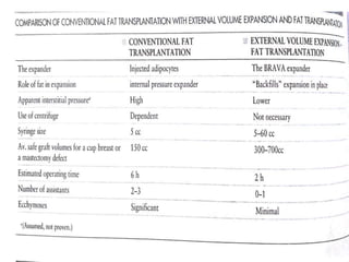
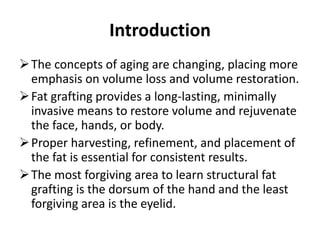
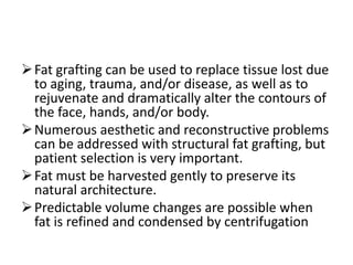
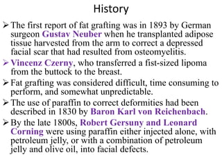
2. Introduction
The concepts of aging are changing, placing more
emphasis on volume loss and volume restoration.
Fat grafting provides a long-lasting, minimally
invasive means to restore volume and rejuvenate
the face, hands, or body.
Proper harvesting, refinement, and placement of
the fat is essential for consistent results.
The most forgiving area to learn structural fat
grafting is the dorsum of the hand and the least
forgiving area is the eyelid.
3. Fat grafting can be used to replace tissue lost due
to aging, trauma, and/or disease, as well as to
rejuvenate and dramatically alter the contours of
the face, hands, and/or body.
Numerous aesthetic and reconstructive problems
can be addressed with structural fat grafting, but
patient selection is very important.
Fat must be harvested gently to preserve its
natural architecture.
Predictable volume changes are possible when
fat is refined and condensed by centrifugation
4. History
The first report of fat grafting was in 1893 by German
surgeon Gustav Neuber when he transplanted adipose
tissue harvested from the arm to correct a depressed
facial scar that had resulted from osteomyelitis.
Vincenz Czerny, who transferred a fist-sized lipoma
from the buttock to the breast.
Fat grafting was considered difficult, time consuming to
perform, and somewhat unpredictable.
The use of paraffin to correct deformities had been
described in 1830 by Baron Karl von Reichenbach.
By the late 1800s, Robert Gersuny and Leonard
Corning were using paraffin either injected alone, with
petroleum jelly, or with a combination of petroleum
jelly and olive oil, into facial defects.
5. Cont……
Eugene Holländer proposed the use of fat injected through a
cannula to correct deformities.He noted considerable
reabsorption of the fat and therefore began mixing human fat
with fat from a ram in an attempt to stabilize it. This resulted
in a painful rash which lasted several days, however a good
cosmetic result was obtained.
Later, in 1919, Erich Lexer published a two-volume book
dedicated to the technique of fat grafting. In this book, he
presented a wide variety of conditions such as depressed scars,
breast asymmetry, knee ankylosis, tendon adhesions, and
micrognathia and the successful results after treatment with fat
grafting.
6. Cont…
Charles Miller also described the injection of transplanted fat
for the correction of facial folds and wrinkles in his 1926
publication, CannulaImplantsandReviewof ImplantationTechnicsin Esthetic
Surgery.
1950s Lyndon Peer studied the gross and microscopic
appearance of transplanted fat. He discovered that adipose
grafts lose approximately 45% of their weight and volume at 1
year due to cell rupture and subsequent death.
Graft size also appeared to play a role in survival. A graft the
size of a walnut was found to lose volume more rapidly than
multiple smaller grafts of similar weight, likely due to the
increased surface area of the smaller grafts.
7. Cont….
Coleman standardized the technique. This technique, called
Lipostructure emphasizes gentle extraction of fat,
centrifugation, and micro particle injections in multiple tissue
planes.
Dr Coleman has used this technique for more than 20 years
and has documented the longevity and stability of fat grafting
performed in this manner.
9. Aging and atrophy
In the face, our traditional response to the descent has been to resuspend the
tissues and to remove the excess skin.
one of the main reasons that the tissue starts to sag is lack of support, or
lack of volume beneath it. One component of this volume is fat.The goal
then should be to replace the missing fullness, which will support and
reposition the skin.
In most cases, the body supplies us with a significant source of filling
material to restore the volume lost with aging in the form of fat.
If there is a tremendous amount of sagging or excess skin, then manual
resuspension and trimming may be necessary.
10. The easiest way to
analyze a face to
determine how it has
changed is to study
photographs of patients
when they were younger
compared to their current
state. Depending on the
degree of aging, one can
often easily see that as
the face ages, typical
patterns emerge.
The model shown in this
picture exemplifies the
changes that occur, from
left to right, going from
age 20 to 50 to 70 years.
11. The 20-year-old model has a full,
smooth face. Her temples are flat and her
brow and glabella are unfurrowed.
Her upper eyelids are full beneath the
brow and there is a short distance
between the ciliary margin and the lid
crease. The lower eyelids are smooth and
there is minimal hollowing medially.
Her cheeks are round with the zygomatic
arches well covered with soft tissue.
There is a slight hollowing in the buccal
cheek.
She has mild nasolabial folds compared
with her full cheeks, but she does not
have deep folds or creases within the
folds.
The lips are full, pouty and everted and
the lower lip is slightly larger than the
upper lip.
Her jaw line and chin are well-defined
and smooth.
12. As we go across, we see hollowing of the
temples with skeletonization of the area.
Wrinkles become more and more
obvious in the forehead and glabella
secondary to loss of youthful fullness
over areas of muscular activity,
especially in the glabella.
The fullness that was previously present
beneath the brow is now diminished and
the upper eyelids appear to either
collapse and fold anteriorly or collapse
posteriorly to reveal the hollow orbit.
The lower eyelids begin to deflate,
making the orbital rim more apparent and
elongating the lid-cheek junction. The
tear trough extends diagonally into the
anterior cheek, breaking up the
continuous line of cheek fullness and
accentuating the nasolabial folds.
13. The zygomatic arches lose
their soft tissue covering to
reveal the bony outlines
beneath and hollow buccal
cheeks.
The vermillion of the lips
becomes thinner and thinner
with time and the lips begin
to invert
The anterior chin becomes
less well-defined and more
rounded like one button
instead of two defined
mounds with a central cleft.
14. patient presentation
Aging- These patients are
generally over 40 years of
age and present with loss of
facial fullness, resulting in
skin laxity, wrinkles, or a
gaunt, skeletal appearance.
The younger patient who
presents for facial fat
grafting is generally one
who is unhappy with a
facial feature such as the
cheeks, chin, or jaw line.
15. Patients may also present for facial fat
grafting for corrective purposes
Congenital deformities such as
hemifacial microsomia and Treacher
Collins syndrome.
Previous trauma resulting in
significant scarring or tissue loss.
Iatrogenic deformities such as hollow
upper and/or lower eyelids and
flattening of the posterior jaw line.
Facial atrophy from etiologies other
than aging , especially drug related
lipodystrophy seen in patients taking
antiretroviral and protease inhibitor
therapies.
16.
17.
18. Hands
There is a loss of volume, or soft tissue
coverage over the veins and tendons of
the dorsal hand.
The hands of a 20-year-old are
generally smooth and full, with the
extensor tendons and dorsal veins
barely visible. There is naturally little
fat over the dorsum of the hands, but
with thinning of the skin and loss of
interosseous muscle fullness, the hands
can take on a wasted appearance that
can be reversed with fat grafting.
19. As liposuction has become a more
common procedure, and there are
more deformities being created as a
result of liposuction and variations
of liposuction.
These liposuction deformities range
from very slight irregularities that
are barely perceptible, to large
indentations and even full thickness
skin loss.
Large defects, there can also be
significant disturbances of body
proportion, such as making the
feminine shape more boxy and
masculine or creating an unusually
deep buttock crease that makes the
buttock appear droopy
20. Patient selection
There are few contraindications to fat grafting .
The first contraindication is true for all procedures, which is poor patient
health prohibiting anesthesia.
Patients with unrealistic expectations are not good candidates .
Extremely thin patient who does not have sufficient fat for transfer.
For fat grafting to the hands, breasts or body, significantly more fat is
usually needed. Asking the patient to gain weight prior to the procedure
only makes sense if the patient is willing and able to maintain that weight
afterwards
In aging patients with a tremendous amount of loose,excess skin, fat
grafting can be disappointing.
21. Surgical technique
The Coleman method of fat grafting is essentially unchanged
since the original inception more than two decades ago. The
process involves-
harvesting the fat gently to preserve the delicate architecture,
refining the fat with centrifugation to remove nonviable
components and provide a predictable volume, and
placement of the fat in small aliquots to increase the surface
area and ensure a blood supply to the grafted tissue.
22.
23. Harvesting
The choice of donor site for fat grafting is dependent on the desires of the
patient and accessibility of the fat.
Posterior hip, back, and lateral thighs are more forgiving and do not have as
much potential to wrinkle as the abdomen and medial thighs.
Incisions are hidden in creases, scars, stretch marks, or hair-bearing areas, if
possible.
Through these incisions, local anesthetic solution is infiltrated using a blunt
Lamis infiltration cannula. For straight local cases, the local anesthetic solution
consists of 0.5% lidocaine with 1 : 200 000 epinephrine.
However for general anesthesia cases, where larger volumes of fat are
harvested, a solution containing 0.2% lidocaine with 1 : 400 000 epinephrine is
used.
The amount of solution infiltrated is essentially equal to the amount of fat
removed.
24. Fat is then harvested using a two-hole Coleman harvesting cannula
attached to a 10 cc syringe. This harvesting cannula is designed to
harvest intact fatty tissue parcels that are large enough to survive, but
small enough to pass through the standard infiltration cannula (17
gauge).
The plunger of the 10 cc syringe is pulled back only a few milliliters
during suctioning, so as not to create too much negative pressure and
rupture the fat cells.
25. Refinement
As fat is harvested, the first few
syringes occasionally have more
local anesthetic present than later.
After the 10 cc syringe is full, the
cannula is disconnected from the
syringe and a Luer-Lok plug is
used to cap the syringe. The
plunger is then removed, and the
syringe is placed into the
centrifuge.
Centrifugation at 1286 g for 2
min concentrates the fat so that
the aqueous components (the
local anesthetic and blood) can be
removed and discarded by
releasing the Luer-Lok plug.
26. Any ruptured fat cells that release their oil can be decanted off the top
and/or wicked away with Telfa pads.
The fat is then transferred to a 1 cc syringe for placement into the face and
hands or a 3 cc syringe for placement into the breasts or body.
The uncentrifuged sample will initially
appear to provide the proper
correction, but in a short period of
time, much of it will be reabsorbed
and the procedure will have been
considered a failure
27. Placement
Incision sites are anesthetized with 0.5%
lidocaine with 1 : 200 000 epinephrine and small
stab incisions are made for the placement of fat.
Small amounts of 0.5% lidocaine with 1 : 200
000 epinephrine are usually infiltrated into the
face for vasoconstriction of the vessels prior to
placement of the fat. This not only helps to
reduce bruising, but also decreases the chance of
accidental intravascular embolization of the fat.
The success of the fat grafting procedure
depends not only on the harvesting and
refinement, but also on the placement of the fat
in a manner that increases its chance for survival
and graft “take”. This means maximizing the
contact surface area of the fatty parcel with the
surrounding tissue, such that a blood supply can
be conferred to the newly grafted fat.
28. The fat is placed gently during
the withdrawal of a blunt
Coleman infiltration cannula.
Fat can be placed at different
levels to accomplish different
effects. For instance, grafting
fat immediately beneath the
dermis can improve the
quality of the skin, decreasing
wrinkles, decreasing pore size
and even reducing scarring.
Special care must be taken,
however, when placing fat
superficially, as irregularities
are more apt to be apparent in
this plane. This is especially
true in areas that have thin
skin, such as the lower eyelid.
29. Transferring large
globules of fat can result
in central necrosis of the
mass with subsequent
resorption and loss of
volume, or possibly even
cyst formation .
30.
31.
32.
33. To make changes in the shape of
the face or body that relate to the
underlying bony skeleton, fat can
be placed above the periosteum.
The structure should be
purposefully built up with tiny
aliquots of fat rather than
attempting to insert larger aliquots
and then mold the tissue after it is
placed.
Molding may displace the fat or
cause necrosis of some or all of the
fat in an area, resulting in uneven
contours.
The only time molding should be
considered is if an irregularity is
noted at the time of placement.
34.
35. Postoperative care
Initially, all areas of the face, except the upper
eyelids, were covered with Microfoam® tape,
forming a sort of mask.
Tegaderm®, which is more flexible, tolerable, and
socially acceptable can be use.
In areas of the face that were suctioned, 1/2”
Reston foam is usually applied, followed by
Tegaderm or Microfoam tape to compress the
foam.
In addition,cold therapy is usually recommended
for up to 72 h postoperatively.
36. The hands are generally still dressed with
Microfoam tape, and the donor sites are dressed
with a compression garment or abdominal binder.
The dressings are usually left in place for 3–4
days,. Body sutures at the donor sites and on the
hands are usually removed at 5–7 days.
Lymphatic drainage techniques using a very light
touch can be performed on the face and/or body,
but deep massage is to be avoided during the first
month.
37. Outcomes
Rigotti has grafted fat beneath irradiated, ulcerated breast skin
and has noted healing and normalization of the affected skin.
Mojallal et al. have also demonstrate improvement in adherent
scars, skin texture, skin suppleness, skin color and scar quality
in their patients.
38. complications
The most common complications,of fat grafting to the face,
hands, or body are aesthetic.
Swelling and bruising that occurs with the multiple passes of
the cannula used to place the fat.
Bruising generally resolves in 2–3 weeks, but there have been
a few cases of prolonged subcutaneous pigmentation that is
easily visible through the thin skin of the lower eyelids.This
has the appearance of “tea staining” and can take many months
to resolve.
subcutaneous irregularities, which can occur in both the
recipient and donor sites.
39. In the recipient sites, excess grafted fat will appear as a lump beneath
the skin. This can be the result of placement of a volume that was too
large just beneath thin skin, particularly in the periorbital region.
Potential remedies for irregularities caused by excess fat include
suctioning of the fat using the same cannula used for infiltration,direct
excision of the fat under visualization, and Lipodissolve,which is not
approved by the FDA in the United States.
Irregularities in the donor sites can also be problematic, particularly if
too much fat is removed.
The most catastrophic potential complication is an intravascular
embolization, which fortunately, is extremely rare
40. Infections in the recipient or donor site are also extremely rare,
but if they do occur, they can result in resorption of the grafted
fat and loss of the desired correction.
Significant changes in weight can result in concomitant
changes in the size of the area grafted, therefore patients are
encouraged to have the procedure performed when they are at
their ideal body weight and to maintain that weight, if
possible.
41. Secondary procedures
With fat grafting, secondary procedures, or touch up
procedures, are possible if the correct volume of fat was not
placed initially.
Due to the difficulty in the lower eyelid of removing fat, it
is far better to under correct the area and return to the
operating room for a second stage, if necessary, at a later
date.
In complicated liposuction deformity cases, a second stage
is often part of the original plan, as it can be very difficult
to make an area smooth enough in one procedure.
Generally, the goal of the first procedure is to fill in large
deficits with significant amounts of fat and later to come
back and refine the area.