
Recommended
More Related Content
What's hot
What's hot (20)
Similar to Airway Anatomy & Evaluation PPT.pptx
Similar to Airway Anatomy & Evaluation PPT.pptx (20)
Recently uploaded
Recently uploaded (20)
Airway Anatomy & Evaluation PPT.pptx
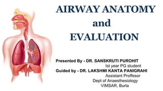
- 1. AIRWAY ANATOMY and EVALUATION Presented By - DR. SANSKRUTI PUROHIT Ist year PG student Guided by - DR. LAKSHMI KANTA PANIGRAHI Assistant Proffesor Dept of Anaesthesiology VIMSAR, Burla
- 2. WHY IS IT IMPORTANT FOR ANAESTHESIOLOGISTS? It is the fundamental responsibility of an anaesthesiologist to establish airway patency & to ensure adequate ventilation and oxygenation.
- 3. WHAT IS AIRWAY? Airway is the passage through which air passes into our body during respiration. or In the practice of airway management, airway is an artificial device with a lumen to aid ventilation or to serve as a conduit to endotracheal intubation.
- 5. CLASSIFICATION OF AIRWAY Upper Airway Oral Cavity Nasal Cavity Pharynx Larynx Lower Airway Trachea Bronchi Bronchioles Alveoli Upper Airway Lower Airway Oral Cavity
- 6. 1. ORAL CAVITY • Boundaries : Anteriorly - Lips Laterally - Cheeks Posteriorly - Faucial Pillars Roof - Hard & Soft Palate Floor - Tongue Posteriorly it communicates with oropharynx UPPER AIRWAYS
- 7. Anaesthetic importance of oral cavity: Difficult laryngoscopy & intubation - Contracture of mouth & lips Loose or buck teeth Loss of buccal pad of fat Large tongue During sleep, unconciousness or general anaesthesia, muscle tone decreases —> tongue can fall backwards —> obstruct the airway. Tongue is connected to mandible by Genioglossus muscle. Jaw thrust maneuver —> mandible & attached tongue anteriorly —> relieving the airway obstruction Ludwig’s Angina(cellulitis of sublingual tissues) —> elevation & posterior displacement of tongue —> airway obstruction
- 8. 2. NASAL CAVITY • Extends from naris (external openings) to choana (posterior openings) • Narrowest portion of upper airway • 2 nasal passages (right & left) by a nasal septum • External nose - formed by bones & cartilages Alae nasi(margins of nostrils) flare during airway obstruction & may be infolded while introduction of nasal tracheal tube. Distance from alae nasi to tragus used to estimate length of nasopharyngeal airway.
- 9. • Boundaries : Roof - Cribriform plate of ethmoid Floor - Palatine process of maxilla Horizontal plate of palatine bone Floor of nasal cavity is perpendicular to plane of face, so nasal tracheal tube/NG tubes should be inserted at right angles to face. Fracture of cribriform plate of ethmoid is a contraindication for nasal tracheal intubation & Ryle’s tube insertion.
- 10. Medial wall - Nasal septum (formed by septal cartilage anteriorly & ethmoid and vomer posteriorly) Lateral wall - 3 bony turbinates/conchae dividing the nasal passage into 3 meatuses - inferior, middle and superior Septal deviation is a common finding in adults, so more patent side should be used for instrumentation. Little’s area in anterior 1/3rd of nasal septum is highly vascular, so topical vasoconstrictor should be applied before insertion to minimise bleeding. Inferior meatus is the preffered pathway for passage of nasal airway devices. Conchae may obstruct passage of nasal airways leading to trauma. During nasal intubation, bevel of ETT should face medially to avoid injury to turbinates
- 11. LATERAL WALL MEDIAL WALL, ROOF & FLOOR
- 12. 3. PHARYNX • It is a muscular tube extending from base of skull to the level of cricoid cartilage. • Boundaries: Sup - base of skull Inf - continuous with esophagus Post - prevertebral fascia separating it from vertebral bodies Ant - communicates with nasal cavity, oral cavity & larynx • Divided into 3 parts: Nasopharynx Oropharynx
- 13. NASOPHARYNX • Extends from base of skull to soft palate • Ending of nasopharynx at soft palate is called Velopharynx Velopharynx is a common site of airway obstruction in both awake & anaesthetized patients. Adenoids in roof of nasopharynx can cause chronic nasal obstruction & difficulty in passage of airway devices if enlarged.
- 14. OROPHARYNX • Extends from soft palate to epiglottis • Includes tonsil, uvula & epiglottis • Lateral walls contain palatoglossal & palatopharyngeal folds aka anterior & posterior tonsillar pillars. Palatine tonsils present between these folds can cause airway obstruction if hypertrophied Space between base of tongue & epiglottis is called Vallecula - tip of laryngoscopic blade lies in vallecula during direct laryngoscopy
- 16. LARYNGOPHARYNX • Extends from epiglottis to the level of cricoid cartilage where it is continuous with esophagus • Deep depression in lateral walls of laryngopharynx is called Piriform fossa Piriform fossa is a catch point for foreign body.
- 17. 4. LARYNX • Situated in anterior midline of neck • Extends from upper border of epiglottis to lower border of cricoid cartilage (C3-C6 in adults, C1-C4 in children) • Made up of cartilages, muscles & ligaments.
- 18. • Cartilages of larynx : 9 in no.- 3 paired & 3 unpaired UnPaired cartilages - Epiglottis (leaf-like) Thyroid (shield-like) Cricoid (ring-like) Paired cartilages - Arytenoid (cup-shaped) Corniculate (horn-shaped) Cuneiform (wedge-shaped)
- 20. EPIGLOTTIS • Thin, leaf-like cartilage in anterior wall of upper part of larynx • Upper end- free, broad & projects upwards • Lower end- long, narrow & attached to thyroid cartilage by Thyroepiglottic ligament • Lateral margins -Aryepiglottic folds Thyroepiglottic muscle (to thyroid cartilage) • Ant surface - Median Glossoepiglottic fold (to tongue) Hyoepiglottic ligament (to hyoid bone)
- 21. THYROID CARTILAGE • Largest of all cartilages, V shaped in c/s. • Consists of 2 quadrilateral lamina • Ant borders of lamina - Make 90o in males & 120o in females - Lower part fuse & form median projection called Laryngeal Prominence - Upper part separated by Thyroid notch • Post borders - Free & prolong upwards & downwards as Superior & Inferior cornu. Thyroid notch & Laryngeal prominence serve as important landmarks for percutaneous airway techniques & larngeal nerve blocks
- 22. BURP - Used during laryngoscopy to improve view of glottis. - Backward Upward Rightward Pressure on Thyroid cartilage .
- 23. CRICOID CARTILAGE • Inferior limit of larynx. • Only complete cartilagenous ring in airway • Narrow anterior Arch & broad posterior Lamina. • Lamina projects upwards behind thyroid cartilage to articulate with Arytenoid cartilages superiorly. • Inferior cornu of thyroid cartilage articulates with cricoid cartilage at the junction of arch & lamina. Sellick’s maneuver - pressure over cricoid cartilage prevents passive regurgitation without airway obstruction in patients at risk of gastric aspiration
- 24. ARYTENOID CARTILAGE • Small pyramidal shaped cartilages on upper border of lamina of cricoid. • Apex - articulates with Corniculate • Base - prolonged to form Vocal process to which Vocal fold is attached & Vestibular fold is attached above Vocal process CORNICULATE CARTILAGE • Small, conical attached to apex of Arytenoids. • Present in post part of Aryepiglottic folds. CUNEIFORM CARTILAGE • Small rod-shaped cartilages in Aryepiglottic folds just anteior to corniculate cartilages
- 25. LIGAMENTS & MEMBRANES OF LARYNX • Extrinsic Thyrohyoid membrane Hyoepiglottic ligament Cricotracheal Ligament • Intrinsic Quadrate membrane-lower free border forms Vestibular fold and its upper border forms Aryepiglottic fold Cricovocal membrane-upper free border forms Vocal Fold and
- 26. CAVITY OF LARYNX • Extends from inlet of larynx to lower border of cricoid cartilage. • Inlet - open backwards & upwards - Boundaries : Ant - Epiglottis Post - Interarytenoid fold Sides - Aryepiglottic fold • Within the cavity, 2 folds : Upper fold - Vestibular fold Lower fold - Vocal fold
- 27. • Vestibular & Vocal folds divide laryngeal cavity into 3 parts : 1. Supraglottis / Vestibule - part above Vestibular fold 2. Ventricle / Sinus - part between Vestibular & Vocal fold 3. Infraglottis / Subglottis - part below Vocal cords
- 28. • Rima Vestibuli - space between right & left Vestibular folds • Rima Glottidis / Glottis - space between right & left Vocal folds - narrowest part of larynx in adults - Length = 24mm(males) 16mm(females) - Ant 3/5 = intermembranous part (between vocal folds) - Post 2/5 = intercartilagenous part (between arytenoid cartilages) *Sensory supply - upto Vocal cords by Internal Laryngeal N - below Vocal cords by Recurrent Laryngeal N
- 29. • Intrinsic muscles - attaches laryngeal cartilages to each other Acting on Vocal cord - Abductor - Posterior Cricoarytenoid Adductor - Lateral Cricoarytenoid Transverse & Oblique Arytenoid Tensor - Cricothyroid Relaxor - Thyroarytenoid Vocalis Acting on Laryngeal Inlet - Opener - Thyroepiglottic MUSCLES OF LARYNX
- 30. • Extrinsic Muscles - attach laryngeal cartilages to nearby structures Elevators - Thyrohyoid. - Mylohyoid Depressors - Sternothyroid - Sternohyoid *All muscles supplied by Recurrent laryngeal N except Cricothyroid which is supplied by Superior laryngeal N
- 32. DIFFERENCE BETWEEN ADULT & PEDIATRIC AIRWAY Epiglottis Shape (High up larynx)
- 34. TRACHEA • From lower border of cricoid cartilage(C6) & ends at Carina (T4 in supine,T6 in standing posteriorly and Sternal angle anteriorly) • Length - 10-11mm • Internal Diameter - 12mm in adults - 3mm in newborns-3yr then by 1mm/yr till 12yr LOWER AIRWAYS
- 35. • Trachea is made up of 16-20 C-shaped hyaline cartilage connected by fibroelastic membrane. • Posterior deficit part contains involuntary Trachealis muscle. • Trachea divides into Right and Left Bronchus at Carina at angle of 20-25o and 45-55o respectively ET Tube should be just above carina for equal ventilation of both lungs More chances of foreign body entry , aspiration or one lung ventilation in Right lungs b
- 36. Trachea Right & Left Bronchus Segmental Bronchioles Terminal bronchioles Respiratory bronchioles Alveolar Ducts Alveoli BRONCHI, BRONCHIOLES & ALVEOLI Cartilagenous No Cartilage Smooth Muscle+
- 38. Why is it necessary? To diagnose the potential for difficult airway for : 1. Optimal patient preparation 2. Proper selection of equipments & techniques 3. Participation of personnel experienced in difficult airway management
- 39. 1)FACIAL ANOMALIES - Maxillary hypoplasia, - Mandibular hypoplasia &hyperplasia 2)TMJ PATHOLOGY - Ankylosis or reduced movement 3)MOUTH & TONGUE ANOMALY - Microstomia (due to burns, trauma) - Tongue swelling in Ludwig’s angina - Tumors of mouth & tongue - Macroglossia (due to Down’s Syndrome) 4)TEETH PROBLEMS - Missing or Protruding upper incisors(buck tooth) 5)NOSE PATHOLOGY - Choanal atresia - Hypertrophied turbinate - Tumors & foreign bodies 6)PALATE PATHOLOGY - Narrow arched palate - Large cleft palate - Soft palatal swelling CAUSES OF DIFFICULT AIRWAY
- 40. 7) PHARYNX PATHOLOGY - Hypertrophied tonsils & adenoids - Tumors & abscesses 8) LARYNX PATHOLOGY - Epiglottitis & Laryngomalacia - Foreign bodies & tumors - Congenital/traumatic stenosis & Edema 9) TRACHEAL PATHOLOGY- Tracheitis, tracheoesophageal fistula, tracheal stenosis & webbing, tracheomalacia - Foreign bodies - Tracheal deviation due to mass/lung pathology 10) BRONCHIAL TREE PATHOLOGY- Mediastinal mass - Foreign body aspiration & bronchial tumors 11) NECK - Large goitre, skin contracture, ankylosing spondylitis 12) SPINE - Limitation of movement - Cervical spine instability
- 41. COMPONENTS of AIRWAY ASSESSMENT 1. History taking - H/O difficult airway in previous surgeries - H/O previous surgery, tumor, trauma, burns in & around the oral cavity, neck or cervical spine 2. General Examination - to rule out anatomic/pathologic factors that can lead to difficult airway 3. Assessment Tests/Indices - Individual Indices - Group Indices Mask Ventilation —> Laryngoscopy —> Intubation
- 42. PREDICTORS OF DIFFICULT MASK VENTILATION O B E S E - Obese ( BMI > 26kg/m2 ) Bearded individual Elderly ( age > 55yrs ) Snorers Edentulous M O A N S - Mask seal difficulty in patients with receding mandible, facial abnormalities, burn strictures etc Obesity ( BMI > 26kg/m2 ) Advanced age No teeth
- 43. PREDICTORS OF DIFFICULT LARYNGOSCOPY & TRACHEAL INTUBATION I. Individual Indices - 1.Physical examination indices 2.Radiological indices 3.Advanced indices II. Group indices - individual indices grouped & scoring systems done
- 45. A. ASSESSMENT OF CERVICAL & ATLANTO-OCCIPITAL FUNCTION 1. Direct Assessment a) Assessment of Neck Flexion & A-O Extension Asking patient to touch his manubrium with his chin ( If done => Neck flexion of 25-30o ) Asking patient to look at the ceiling without raising eyebrows( If done => A-O joint extension 85o ) *Grade III & IV Difficult Laryngoscopy GRADES REDUCTION OF A-O EXTENSION Grade I No Reduction Grade II 1/3rd reduction Grade III 2/3rd reduction Grade IV Complete reduction PHYSICAL EXAMINATION
- 46. b) Delilkan’s Test To assess movement of occiput on atlas during extension. Patient asked to look straight Place the index finger of left hand under the chin & the index finger of right hand on occipital tuberosity Patient is asked to look at the ceiling If the Left index finger higher than the Right => Normal If level of both fingers remains same or the Left finger remains lower than the Right => Abnormal extension
- 47. 2. Indirect Assessment a) PRAYER SIGN - A positive sign can be elicited if patient is unable to approximate their palms & cannot bend their fingers backwards - Seen in Diabetics with stiff joint syndrome - +ve Prayer sign => cervical spine stiffness limited A-O movement difficult laryngoscopy & intubation
- 48. b) PALM PRINT TEST - Joint rigidity in Diabetics also involve Laryngeal & Cervical joints leading to difficult larngoscopy. - Patient is asked to give their palm print on a white sheet of paper on a firm surface. Scoring is done as: GRADES PALM PRINTS Grade 0 Phalangeal area completely visible Grade I Interplangeal areas of 4th-5th digits partly visible Grade 2 Interplangeal areas between 2nd- 5th digits hardly visible Grade 3 Only fingertips printed * Higher the Score, more difficult is the Laryngoscopy
- 49. B. ASSESSMENT OF TEMPORO-MANDIBULAR JOINT (TMJ) FUNCTION • TMJ exhibits 2 functions : 1. Rotation of the condyle in the synovial cavity - 2-3cm mouth opening 2. Forward displacement of the condyle - further 2-3cm mouth opening • Tests : a) Interincisor Gap/Mouth Opening Mouth wide open Index, middle & ring finger in mouth vertically If > 5cm / > 3 fingers => Adequate for laryngoscopy
- 50. b) Sliding of Mandible Test Index finger in front of tragus & Thumb in front of lower part of mastoid process Asked to open his mouth as wide as possible Index finger in front of the tragus can be indented in its space and the thumb can feel the sliding of condyle Good Sliding function of mandible c) Calder Test Patient is asked to protrude the mandible Look for lower incisors alignment with respect to upper incisors
- 51. d) Upper Lip Bite test - Tests the range & freedom of mandibular movement & architecture of the teeth Class III of this test expected to have C-L Grade III & IV CLASS I Lower incisors can bite upper lip above vermilion line CLASS II Lower incisors can bite upper lip below vermilion line CLASS III Lower incisors cannot bite upper lip
- 52. C. ASSESSMENT OF MANDIBULAR SPACE This space determines how easily the laryngeal and pharyngeal axis will fall in line when the A-O joint is extended. 1. Thyromental distance - Distance between thyroid notch & symphysis menti when neck fully extended DISTANCE DIFFICULTY >6.5 cm NO PROBLEM WITH LARYNGOSCOPY & INTUBATION 6-6.5cm DIFFICULT LARYNGOSCOPY & INTUBATION >6.5cm LARYNGOSCOPY MAY BE IMPOSSIBLE
- 53. 2. Hyomental Distance - Distance between mentum & hyoid bone 3. Thyroid to floor of mouth distance - Indicates position of larynx in neck - Larynx position is normal if patient can place 2 fingers between top GRADES Distances GRADE I > 6cm GRADE II 4-6 cm GRADE III <4 cm Impossible laryngoscopy & intubation
- 54. 4. Sternomental Distance - Measured with head in full extension & closed mouth - Distance from sternal notch to mentum < 12.5 cm Difficult laryngoscopic intubation
- 55. D. TEST FOR ASSESSING ADEQUACY OF OROPHARYNX FOR LARYNGOSCOPY AND INTUBATION 1. Mallampatti grading - MC used test for predicting airway difficulty - Indicates amount of space within oral cavity to accomodate laryngoscope & ETT - Prerequisites - patient in sitting position & head neutral postion - mouth wide open - tongue protruded out without phonation - observer's eye at the level of patient's open mouth - Observe for structures visible
- 56. GRADES STRUCTURES VISIBLE GRADE I FAUCIAL PILLARS, UVULA, SOFT PALATE & HARD PALATE GRADE II BASE OF UVULA, SOFT PALATE & HARD PALATE GRADE III SOFT & HARD PALATE - DIFFICULT LARYNGOSCOPY GRADE IV ONLY HARD PALATE - DIFFICULT LARYNGOSCOPY
- 57. E. ASSESSMENT FOR QUALITY OF GLOTTIC VIEW DURING LARYNGOSCOPY 1. Indirect mirror laryngoscopic view - complete vocal cords visible - posterior commissure visible - epiglottis visible - no glottic structures visible 2. Direct laryngoscopy “awake look” - possible with sedation - gives estimate of forthcoming laryngoscopy & intubation - Cormack Lehane graded the laryngoscopy view
- 58. GRADE I ▪ The entire laryngeal aperture GRADE II ▪ Only the posterior laryngeal aperture GRADE III ▪ Only the epiglottis GRADE IV ▪ Only the soft palate CORMACK LEHANE GRADING
- 59. 3. Cook’s Modification of Cormack-Lehane’s grading GRADES LARYNGOSCOPIC VIEW GRADE 1 GLOTTIC OPENING CLEARLY VISIBLE ( POST COMMISURE & ENTIRE VOCAL CORDS SEEN ) GRADE 2a GLOTTIC OPENING PARTLY VISIBLE ( POST COMMISURE & PART OF VOCAL CORDS SEEN ) GRADE 2b POST COMMISURE SEEN BUT VOCAL CORDS NOT SEEN GRADE 3a ONLY EPIGLOTTIS SEEN & LIFTABLE GRADE 3b ONLY EPIGLOTTIS SEEN BUT NOT LIFTABLE GRADE 4 ONLY ROOT OF TONGUE SEEN
- 60. 4. Grading Ease of Intubation 5. POGO Scoring ( Percentage Of Glottic Opening ) GRADE I NO EXTRINSIC MANIPULATION OF LARYNX NEEDED GRADE II EXTERNAL MANIPULATION NECESSARY TO INTUBATE GRADE III INTUBATION POSSIBLE ONLY WHEN AIDED BY STYLET GRADE IV FAILED INTUBATION 100% ENTIRE GLOTTIC STRUCTURES VISIBLE 33% ONLY LOWER 1/3rd OF VOCAL CORD & ARYTENOID VISIBLE 0% NO GLOTTIC STRUCTURES VISIBLE
- 61. 6. Symmetry of Upper & Lower Face - Upper face : from bridge of nose to just below nasal septa at upper lip - Lower face : from just below nasal septa to chin - If lower face longer than upper face difficulty in straightening of airway structures during laryngoscopy
- 62. 1. X-RAYS - Lateral X-Ray of head & neck done. - Measurements predicting difficult laryngoscopy : 1. Effective mandibular length/Posterior depth of mandible < 3.6 2. Reduced distance between occiput & spinous process of C1 < 5mm 3. Increase in posterior depth of mandible >2.5cm 2. CT SCAN 3. MRI RADIOLOGICAL EXAMINATION
- 63. 1. FLOW VOLUME LOOPS - Helps to diagnose small airways vs large airways obstruction & intrathoracic vs extrathoracic obstruction - Helps to locate site of large airway obstruction - Helps to distinguish between fixed or variable obstruction ADVANCED INDICES
- 65. 2. ULTRASONOGRAPHY - Useful in airway assessment by : a) Identification of cricothyroid membrane b) Identification of intraoperative pneumothorax c) Localisation of trachea d) Quantifying anterior neck soft tissues - distance from skin to ant aspect of trachea measured at 3 levels: - vocal cords (zone I) - thyroid isthmus (zone II) - suprasternal notch (zone III) *Soft tissue thickness > 25mm in Zone I or > 28mm in avg of 3 zones indicates difficult laryngoscopy 3. ESOPHAGOGRAM
- 66. GROUP INDICES
- 67. BELLHOUSE’S CRITERIA - 3 parameters - Restricted A-O joint extension < 35 degree Reduced Mandibular space Enlarged tongue size with respect to pharynx WILSON’S SCORING SYSTEM - 5 parameters analysed simultaneously - Score 0, 1, 2 given to each - Easy / difficult laryngoscopy & intubation predicted from their sum total - 5 or less => easy laryngoscopy 6-7 => moderate difficulty 8-10 => severe difficulty
- 68. WILSON’S SCORING SYSTEM PARAMETER 0 1 2 WEIGHT(KG) < 90 90-110 >110 HEAD & NECK MOVEMENT >90o 90o <90o JAW MOVEMENT (INTER-INCISOR GAP) >5cm 5cm <5cm SLIDING MANDIBULAR BEYOND MAXILLARY INCISORS >0 0 <0 RECEDING MANDIBLE None Moderate Severe BUCK TEETH None Moderate Severe
- 69. BENUMOF’S 11 PARAMETER ANALYSIS - 11 step airway examination - Follows line of sight from upper incisors to glottis - 4-2-2-3 rule : 4 steps focusing on teeth 2 steps for oral cavity 2 steps for mandibular space 3 steps in neck examination
- 70. BENUMOF’S 11 PARAMETER ANALYSIS PARAMETER MIN ACCEPTABLE VALUE INTER-INCISOR GAP > 3cm BUCKED TOOTH no overriding LENGTH OF UPPER INCISORS short <1.5cm MANDIBLE PROTRUSION TEST mandibular teeth beyond maxillary teeth MALLAMPATI CLASS Class II or less PALATE CONFIGURATION no arching or narrowing THYROMENTAL DISTANCE >5cm or >3 finger breadth MANDIBULAR SPACE COMPLIANCE soft to palpation NECK LENGTH not too short NECK THICKNESS not too thick HEAD /NECK MOVEMENT neck flexion 35o & head extension 80o
- 71. ROCKE ET AL COMBINED MALLAMPATI GRADING - Rocke et al combined Mallampati grading with various other factors and showed correlation between classification of airway & laryngoscopic blade - Combined Mallampati with obesity short neck teeth abnormality facial edema swollen tongue NC/TMD RATIO - If >/= 5 —> cutoff point for difficult intubation in obese patients
- 72. ARNE’S SIMPLIFIED SCORE MODEL RISK FACTOR SCORE H/O DIFFICULT INTUBATION NO - 0 YES - 10 DISEASES ASSOCIATED WITH DIFFICULT INTUBATION NO - 0 YES - 5 SYMPTOMS OF AIRWAY PATHOLOGY NO - 0 YES - 3 IG & MANDIBULAR SUBLUXATION IG >/= 5cm or SLux > 0 —> 0 IG < 5-3.5cm or SLux = 0 —> 3 IG < 3.5cm or SLux < 0 —> 13 TMD >/= 6.5cm - 0 < 6.5cm - 4 MAX RANGE OF HEAD & NECK MOVEMENT > 100% - 0 90 +/- 10% - 2 < 80% - 5 MALLAMPATI SCORE Class I - 0 Class II - 2 Class III - 6 Class IV - 8
- 73. RAPID AIRWAY ASSESSMENT TEST - In emergency situation, 1-2-3 Finger Assessment test used - To assess - TMJ function - Mouth opening - Mandibular space - Can be done in 15sec 1-Finger Test : Index finger in front of tragus & Thumb in front of lower part of mastoid process Asked to open his mouth as wide as possible Index finger in front of the tragus can be indented in its space and the thumb can feel the sliding of condyle
- 74. 2-Finger Test : Ask patient to open his mouth wide Place 2 fingers i.e Index & Middle in the opening If done => >3cm & adequate for 2cm flange of laryngoscope 3-Finger Test : Ask patient to extend head Place 3 fingers i.e Index, Middle & Ring in submandibular space If done => adequate space infront of larynx and
- 75. LEMON LAW - represents 5 simple Rapid Assessment methods Look for anatomic features suggesting difficulties Examine airway anatomy using 3-3-2 method : Interincisor distance (3 fingers) Hyomental distance (3 fingers) Thyroid to floor of mouth (2fingers) Mallampati Grade Obstruction of Airway : location, type-fixed/mobile & progression Neck mobility - check neck flexion (chin on the chest) head extension (look at ceiling)
- 76. CRITERIA SCORE L Facial trauma 1 Large incisors 1 Beard/moustache 1 Large tongue 1 E Mouth opening </= 3 fingers 1 H-M distance </= 3 fingers 1 Thyroid to FoM distance </= 2 fingers 1 M Mallampati Score 3/4 1 O Obstruction of airway present 1 N Neck mobility decreased 1 Total 10 *Higher the score, more likely intubation will be difficult
- 77. 4 M’S WITH STOP SIGN Mallampati Measurement Movement Malformation of the skull, teeth, obstruction & Pathology S = Skull (Hydro and Microcephalus) T = Teeth (Buck, protruded & loose teeth, Macro and Micro mandibles) O = Obstruction (due to obesity, short bull neck & swellings around head & neck) P = Pathology (Craniofacial abnormalities & Syndromes) * Score >/= 8 ——> Difficult Intubation
- 78. SCORE 1 2 3 4 Mallampati Grade 1 Grade 2 Grade 3 Grade 4 Measurement Mouth open 3 fingers TMD 3 fingers HMD 2 fingers TMJ 1 finger Movement Left Right Flexion Extension Malformation Skull (Hydro and Microcephalus) Teeth (Buck, protruded & loose teeth) Obstruction Pathology
- 79. FOUR D'S OF DIFFICULT AIRWAY Dentition - prominent upper incisors, receding chin Distortion - edema, blood, vomits, tumor, infection Disproportion - short chin-to-larynx distance, bull neck, large tongue, small mouth Dysmobility - TMJ and cervical spine LM-MAP RULE Look externally for deformities Mallampati grading Measurements 3-3-2-1 A-O extension 35o Pathological obstruction
- 80. SAGHEI & SAFAVI’S test • For prolonged laryngoscopy time • Indices : Weight >80kg Tongue protrusion <3.2cm Mouth opening <5cm Upper incisor length >1.5cm Mallampati class >1 Head extension <70o Any 3 indices if present => Prolonged laryngoscopy
- 81. PREDICTORS OF DIFFICULT Supraglottic Airway Device PLACEMENT R O D S - Restricted mouth opening Obstructed upper airways Disrupted upper airway due to trauma & burns Stiff lungs PREDICTORS OF DIFFICULT SURGICAL AIRWAY B A N G - Bleeding tendency Agitated patient Neck scarring/flexion deformity Growth/vascular abnormalities in region of surgical airway
- 82. REFERENCES 1. Miller’s Anaesthesia 9th edition 2. Morgan & Mikhails Clinical anaesthesiology 3. Airway Management Manual by Rashid M Khan 4. B.D.Chaurasia’s Human Anatomy 5. Google search
- 83. THANK YOU