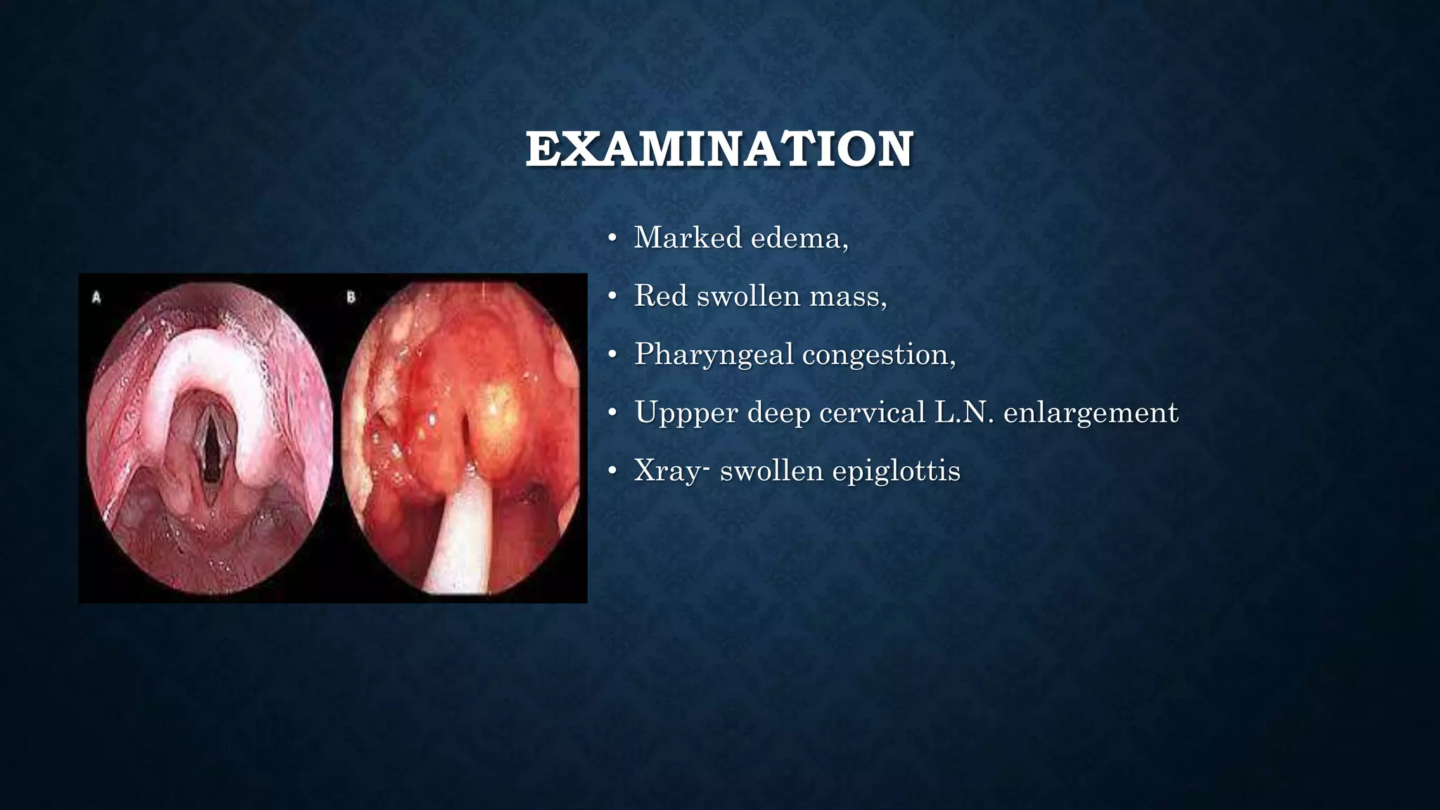
INFLAMMATIONS OF
LARYNX
Dr. Uzwal K. Jha
LARYNGITIS
Acute
A) Acute non specific laryngitis
-Acute laryngitis
-Acute Epiglottitis
-Acute laryngotracheobronchitis
B) Acute specific laryngitis
-Diphtheria
-Herpes Simplex
Chronic
• A) Chronic nonspecific laryngitis
-Simple diffuse chronic laryngitis
-Chronic diffuse hyperplastic laryngitis
(REINKE’s edema)
-chronic localized hyprtrophic laryngitis
-Keratosis/ leukoplakia
-Contact ulcer
B)Chronic specific laryngitis
-Laryngoscleroma,T.B.,syphilis,leprosy,mycosis
ACUTE LARYNGITIS
• Infectious/Non-infectious
• Etio-
-Non infectious -vocal abuse,allergy,burn,trauma
-Infectious -Usually follow URTI caused by – virus
(influenza,parainfluenza,rhino) later followed by bacteria such as-Strept. Pneum. ,H.
influenza e.t. al
CLINICAL FEATURE
• Hoarseness
• Discomfort/Pain
• Dry cough
• General symptoms
O/E—Erythema,edema,reddish,submucosal haemorrhages
TREATMENT
Voice rest,
Avoid smoking,Alcohol,
Steam inhalation
Cough sedatives
Antibiotics
Analgesics
steroids
ACUTE EPIGLOTTITIS
Causative org.- H. influenza type B
More common in children
CLINICAL PICTURE
• Starts by sore throat and followed by odynophagia and drooling
• High fever 40 C
• Associated features-malaise,headache,anorexia
• Muffled voica AKA Hot potato voice
• Rapidly progressive-stridor-life threatening
EXAMINATION
• Marked edema,
• Red swollen mass,
• Pharyngeal congestion,
• Uppper deep cervical L.N. enlargement
• Xray- swollen epiglottis
TREATMENT
• Hospitalization
• Intubation VS Tracheostomy
• Antibiotics
• Steroids
• O2
LARYNGOTRACHEOBRONCITIS
• AKA croup
• H. Streptococci (predominant)
CLINICAL PICTURE
symptoms
• Common cold,mild fever, rhinorrhea,
nasal obstruction
• May be followed by toxemic features
• Stridor (inspiratory) later biphasic
• cough
signs
• Mucopurulent exudates
• Subglottic edema
DIFFERENCE
TREATMENT
• Hospitalization
• Antibiotics
• Steroids
• Mucolytics
• Croupette
• ET VS TRACHE.
DIPTHERIA
• Usually secondary to Faucial diptheria
• Specific features-
Hoarseness,cough,stridor,dyspnea
O/E- greyish white, dirty, diptheric membrane
TREATMENT
CHRONIC LARYNGITIS(NON SPECIFIC)
• M/C in middle aged men
• Predisposing factors-
• Exogenous-recurrent acute laryngitis,smoking,chronic RTI,voice abuse,occupational
• Endogenous-hypothyroidism, vit.A def.
CLINICAL PICTURE
• Hoarseness
• Worse in morning
• +/- aphonia
• Dry cough
• pain
SIMPLE DIFFUSE CHRONIC LARYNGITIS
• Resembles acute laryngitis but prolonged
• Treatment-
• Avoid predisposing factors,voice rest,stem inh.,antibiotics
CHRONIC DIFFUSE HYPERPLASTIC
LARYNGITIS/REINKE’S
• Etio-same as above
• Laryngoscopy-
• Diffuse congestion of mucosa of larynx
• Diffuse B/L symmetrical congestion
• Edge of VC appear round not flat
• Incomplete approximation during phonation
TREATMENT
• Conservative
• Surgical-stripping
CHRONIC LOCALIZED HPERTROPHIC
LARYNGITIS
• Vocal cord nodules(singer’s)
• Localized area of hyperplasia of epithelium on free edge of VC or localized
hematoma
• Etio-
• Abuse
• GERD
• Follow viral
CLINICAL PICTURE
• Hoarseness
• Weakness and change in timber of voice
• Laryngoscopy-
• B/L small tiny nodules, pinkish,free margin of VC
• Site??
TREATMENT
• Voice rest
• Speech therapy
• Topical steroid
• MLS
POLYP
ETIO
• Voice abuse-injury to subepithelial connective tissue
• Allergy
Clinical picture—
Hoarseness
Aphonia
aspiration
TREATMENT
• ????????
LEUKOPLAKIA
• Hyperplasia and hyperkeratinization of epithelial lining
CONTACT ULCER AKA PACHYDERMA
LARYNGIS
SULCUS VOCALIS
T.B.
• Secondary to pulmonary tb
• Chronic specific inflammatory granuloma caused by TB bacilli
• Etio-
• Incd-less than 2-5% of all cases of pulmonary TB
• Organism??
PATHOLOGY
• Site – posterior part
• In order of frequency-(a) interarytenoid region (b) arytenoids (c) posterior part of
vocal cord ,,may affect ventricular folds ,epiglottis (laryngeal Surface)
• C/P and treatment
LARYNGOSCLEROMA
• Age
• Sex
• Geography
• Organism
• Clinical picture-
• Biphasic stridor, hoarseness of voice,cough
LARYNGOSCOPY
• Subglottic region-
• Pale,pinkish,smooth swelling on both side of subglottis with greenish crust
• Subglottic web and stenosis
• Lab investigation???
• Treatment???
SYPHILLIS
ETIO
• Treponema pallidum- spirochate
• Incidence-very rare
• Mode of infection---acquired vs congenital
• Site – anterior half of larynx, mainly epiglottis
CLINICAL PICTURE / TREATMENT
General- skin rashes,veneral wart ,generalized lymphadenopathy
Laryngeal-
Hoarseness, stridor, cough
Larynx—hperaemia of laryngeal mucosa,mucous patches,
ULCER- punched out edges, yellowish necrotic floor,indurated base
LAB INVESTIGATIONS
• Dark ground microscopy- direct smear from syphilitic lesion which shows—
spirochete
• Serology—
A)Non specific tests- wasserman rxn, VDRL
B)Specific test –TPI(trapenoma pallidum immobilization test)
-TPHA(T.P. haemagglutination test)
DL AND BX
• Treatment—
• Benzathine benzylpenicillin-100000 UI IM for—10-14 days
• +/- tracheostomy
DIAGNOSIS
Inflammations of larynx.pptx

Inflammations of larynx.pptx