USMLE CVS 005 Blood vessels – Arteries and veins.pdfAHMED ASHOUR
The major blood vessels in the human body form an extensive network that facilitates the transportation of blood, oxygen, and nutrients to various tissues and organs.
Understanding the anatomy and function of major blood vessels is essential for comprehending the circulatory system and diagnosing and treating cardiovascular conditions.
USMLE CVS 001 Mediastinum anatomy medical chest .pdfAHMED ASHOUR
The mediastinum is the central compartment of the thoracic cavity, located between the lungs.
It is a three-dimensional space that houses various structures within the chest.
The mediastinum extends from the sternum (front of the chest) to the vertebral column (back of the chest) and from the superior thoracic aperture (top of the chest) to the diaphragm (bottom of the chest).
Understanding the anatomy of the mediastinum is crucial for healthcare professionals to interpret diagnostic findings and manage conditions affecting this central compartment of the thoracic cavity.
Right Atrium of human heart
This PPT help to understand the external and internal structures of right atrium.
sulcus terminalis on external surface of rt atrium,
crista terminalis on internal side of rt. atrium,
interior is divided into rough anterior part and smooth posterior part ( sinus venarum)
superior and inferior venae cavae drains deoxygenated blood into rt. atrim
there is Eustachian valve to guard the opening of IVC and Thebesian valve to guard the opening of coronary sinus
septal wall presents fossa ovalis with its border limbus fossa ovalis
Students, digital devices and success - Andreas Schleicher - 27 May 2024..pptxEduSkills OECD
Andreas Schleicher presents at the OECD webinar ‘Digital devices in schools: detrimental distraction or secret to success?’ on 27 May 2024. The presentation was based on findings from PISA 2022 results and the webinar helped launch the PISA in Focus ‘Managing screen time: How to protect and equip students against distraction’ https://www.oecd-ilibrary.org/education/managing-screen-time_7c225af4-en and the OECD Education Policy Perspective ‘Students, digital devices and success’ can be found here - https://oe.cd/il/5yV
Welcome to TechSoup New Member Orientation and Q&A (May 2024).pdfTechSoup
In this webinar you will learn how your organization can access TechSoup's wide variety of product discount and donation programs. From hardware to software, we'll give you a tour of the tools available to help your nonprofit with productivity, collaboration, financial management, donor tracking, security, and more.
Read| The latest issue of The Challenger is here! We are thrilled to announce that our school paper has qualified for the NATIONAL SCHOOLS PRESS CONFERENCE (NSPC) 2024. Thank you for your unwavering support and trust. Dive into the stories that made us stand out!
Unit 8 - Information and Communication Technology (Paper I).pdfThiyagu K
This slides describes the basic concepts of ICT, basics of Email, Emerging Technology and Digital Initiatives in Education. This presentations aligns with the UGC Paper I syllabus.
Operation “Blue Star” is the only event in the history of Independent India where the state went into war with its own people. Even after about 40 years it is not clear if it was culmination of states anger over people of the region, a political game of power or start of dictatorial chapter in the democratic setup.
The people of Punjab felt alienated from main stream due to denial of their just demands during a long democratic struggle since independence. As it happen all over the word, it led to militant struggle with great loss of lives of military, police and civilian personnel. Killing of Indira Gandhi and massacre of innocent Sikhs in Delhi and other India cities was also associated with this movement.
We all have good and bad thoughts from time to time and situation to situation. We are bombarded daily with spiraling thoughts(both negative and positive) creating all-consuming feel , making us difficult to manage with associated suffering. Good thoughts are like our Mob Signal (Positive thought) amidst noise(negative thought) in the atmosphere. Negative thoughts like noise outweigh positive thoughts. These thoughts often create unwanted confusion, trouble, stress and frustration in our mind as well as chaos in our physical world. Negative thoughts are also known as “distorted thinking”.
The French Revolution, which began in 1789, was a period of radical social and political upheaval in France. It marked the decline of absolute monarchies, the rise of secular and democratic republics, and the eventual rise of Napoleon Bonaparte. This revolutionary period is crucial in understanding the transition from feudalism to modernity in Europe.
For more information, visit-www.vavaclasses.com
The Indian economy is classified into different sectors to simplify the analysis and understanding of economic activities. For Class 10, it's essential to grasp the sectors of the Indian economy, understand their characteristics, and recognize their importance. This guide will provide detailed notes on the Sectors of the Indian Economy Class 10, using specific long-tail keywords to enhance comprehension.
For more information, visit-www.vavaclasses.com
Sectors of the Indian Economy - Class 10 Study Notes pdf
large veins of thorax & abdomen.pptx
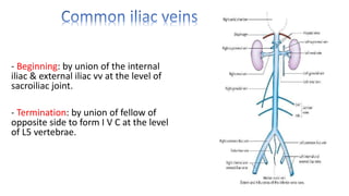
1. - Beginning: by union of the internal
iliac & external iliac vv at the level of
sacroiliac joint.
- Termination: by union of fellow of
opposite side to form I V C at the level
of L5 vertebrae.
2.
3. Beginning: by union of the right and left common
iliac veins, at the level L5 vertebrae.
- Course: The IVC lies along the right
anterolateral aspect of the vertebral column and
ascends upward passes through the central
tendon of the diaphragm around the level of T8
vertebrae.
- Termination: in the thorax by piercing the inferior
part of RT atrium 1/2 inch above the diaphragm
opposite 6th RT sternocostal junction.
4. Tributaries:
1. 2 common iliac veins.
2. 2 pairs of lumber veins (RT & LT
3RD & 4TH).
3. 2 renal veins (RT & LT).
4. 2 inferior phrenic vv (RT & LT).
5. 2 hepatic vv.
6. RT suprarenal v.
7. RT gonadal v
5.
6.
7. - Origin: By union of Rt. & Lt. brachio cephalic .vv. opposite Rt. 1st costal cartilage (at
it's lower border).
- Course & termination: It descends on Rt. Side of ascending aorta to open into post.
Part of Rt. Atrium; It's lower 1/2 lies within fibrous pericardium.
- Length: 2-3 inches long.
- Termination: Behind Rt. 3rd sternocostal. Junction by opening into the Rt.
Atrium.
- Tributaries: only arch of azygose. V into it's post. Aspect at level of 2nd costal
cartilage (Trachea).
- S.V.C drain upper 1/2 of body.
11. Course & termination
Ascends up, enters thorax- via aortic
opening of diaphragm
Passes upwards in thorax infront of
vertebral column ant to lower 8
thoracic vert
Opp T-4 arches above root of Rt lung
& ends in SVC
May pass via apex of rt lung; med
part of apex- lobe of azygos v
12. • Tributaries:
i Rt post IC veins except 1st vein (rt BC v)
ii. 2nd, 3rd, 4th IC vs unite to form the rt sup IC V which
enters arch of azygos v
iii. 5th to 11th end separately
iv. V formed by union of rt subcostal v & rt asc
lumbar v (may form azygos v itself)
v. Hemiazygos v at T-8 level
vi. Acc hemiazygos vein at T-7 level
vii. Rt bronchial v (last tribu)
viii. Oeso, pericardial & mediastinal vs
14. Hemiazygos vein
• Also k/a inferior hemiazygous vein Mirror
image of lower part of azygos v
• Begins-union of lt asc lumbar & lt subcostal vs
• may arise from post part of lt renal vein
• Pierces lt crus of diaph; ascends on lt side of vert column
(overlapped by aorta)
• At T-8 level turns to rt ; passes behind aorta, oeso &
thoracic duct ; joins azygos v
Tributaries:
- Lt asc lumbar v
- Lt subcostal v
- Lower 3-4 IC Vs of lt side
15. Accessory hemiazygos vein
• Also k/a Sup hemiazygos vein
• Cont’n of 4th post IC V of lt side
• Runs down on lt side of vert column
• At T-7 level turns rt – ends in azygos v (may join hemiazygos v)
• Tributaries:
– 5,6,7 & 8th post IC Vs of lt side
– Lt bronchial veins (at times)