Call Girls In Andheri East Call 9920874524 Book Hot And Sexy Girls
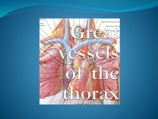
Great vessels artery
1.
2. GREAT VESSELS
IT INCLUDES:-
A. SVC
B. IVC
C. Aorta— (largest artery
of body)
Ascending Aorta
Arch Of Aorta
Descending Thoracic
Aorta
Abdominal Aorta
D. PULMONARY TRUNK
RT. <. pulmonary
artery
3. Aorta
The main arterial trunk of the
body
Carries oxygenated blood
Starts at the left ventricle
4. ASCENDING AORTA
It arises from upper end of Lt.
ventricle continues as arch of
aorta at sternal angle
Has--- 3 aortic sinuses
(dilatation)
1. Anterior aortic sinus--- RCA
2. Rt. Posterior aortic sinus---
non-coronary sinus
3. Lt. posterior aortic sinus----
LCA
BRANCHES
1)– Rt. Coronary artery
2)– Lt. coronary artery
Applied – aneurysm of ascending
aorta
6. ARCH OF AORTA
It is the continuation of
ascending aorta at the level of
sternal angle & continues as
descending thoracic aorta
Runs upwards, backwards and to
left then descends as DTA
Lies in superior mediastinum
BRANCHES—
1. Bracheocephalic Artery
2.Left Common Carotid Artery
3.Left Subclavian Artery
Applied--- coarctation of aorta
7. It is the continuation of the arch of aorta
Lies in the posterior mediastinum
Bigins at lower border of T4 vertebra &
ends at T12 vertebra ( continues as abd.
Aorta)
BRANCHES---
A. LATERAL BRACHES-
1. Posterior Intercostal Artery - 3rd-11th
2.Subcostal Artery
3.Superior Phrenic Artery
B. VISCERAL BRANCHES—
1. Pericardial Branches
2. Mediastinal Branches
3 . Left Bronchial Artery (Two)
4 . Esophageal Branches
Applied– dissecting aneurysm
Descending thoracic aorta
8. Located in the middle
mediastinum, enclosed in
the pericardium
Convey deoxygenated
blood to the lungs
Pulmonary trunk
9. Origin: Base of the right ventricle
Course: Runs upwards, backwards and
to the left
Length: 2 inches
Termination: Divides into right and left
pulmonary arteries.
Branches:
Right pulmonary artery
Left pulmonary artery
Pulmonary trunk
10. Relations:
Anterior: Thoracic wall, Internal
thoracic vessels, Right lung &
pleura
Posterior: Trachea, right vagus, root of
the right lung
Medial: Ascending aorta &
brachiocephalic artery
Lateral: Right phrenic, right lung &
pleura
Superior Vena cava
11. Lies in the superior mediastinum
Origin: continuation of the ascending aorta at
the level of the sternal angle (T4)
Begins posterior to the 2nd right sternocostal
joint
Termination:
Continues as the descending thoracic
aorta at the level of the sternal angle (T4)
Branches:
Brachiocephalic trunk
Left common carotid artery
Left subclavian artery
Arch of the Aorta
12. Relations:
Anterior and to the left:
• Left phrenic nerve
• Inferior cervical cardiac branch of left Vagus.
• Superior cervical cardiac branch of left sympathetic trunk.
• Left Vagus
• Left superior intercostal vein
• Mediastinal surface of left lung.
Posterior and to the right:
1. Deep cardiac plexus
2. Trachea
3. Left recurrent laryngeal nerve
4. Oesophagus
5. Thoracic duct
6. Vertebral column
Above:
Branches of the arch
Left brachiocephalic vein
Below:
1. Bifurcation of pulm. Trunk
2. Left principal bronchus
3. Ligamentum arteriosum
4. superficial cardiac plexus
5. Left recurrent laryngeal nerve
Arch of the Aorta