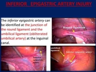
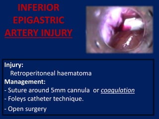
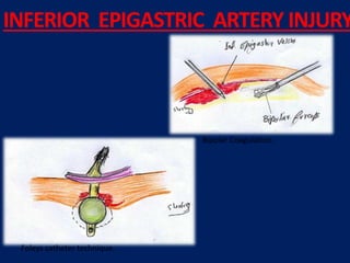
This document provides guidelines for laparoscopic entry techniques. It discusses positioning the patient and various methods for primary and secondary port entry. The preferred primary entry is through the umbilicus using a closed Veress needle technique. Guidelines are provided for Veress needle insertion including abdominal pressure, saline testing, and insufflation. Alternatives like Palmer's point or open Hasson technique should be considered if umbilical entry fails or is risky due to adhesions. Secondary ports should be inserted under direct vision at specific locations and angles to avoid injury.
Complications of hysteroscopy can be classified as those related to distention media, mechanical issues, electrocautery, anesthesia, or late effects. Distention media like glycine can be absorbed systemically during the procedure and cause electrolyte disturbances. Mechanical complications include cervical laceration and uterine perforation. Electrocautery risks include injury to intraperitoneal structures if the uterus is perforated. Positioning patients in lithotomy can potentially lead to compartment syndrome or neurologic injuries over time. Preventing complications involves using the lowest effective pressures, detecting fluid deficits, and avoiding overzealous electrocautery.
This document discusses complications that can occur during and after laparoscopy. It begins by stating that major complications are low at 1 in 1000 procedures, while complications related to initial abdominal access are less than 1%. It then describes various complications in more detail, including vascular injuries, gastrointestinal puncture, urinary injuries, nerve injuries, port-site hernias, and surgical site infections. Prevention strategies and treatment approaches are provided for each complication.
This document discusses the basic principles of laparoscopy. It describes the key differences between laparoscopic and open surgery for both patients and surgeons. For patients, laparoscopic surgery results in less pain, faster recovery times, and quicker return to normal activities due to smaller incisions. For surgeons, laparoscopy provides a magnified view but with altered tactile response and two-dimensional images. The document outlines the typical laparoscopic setup including the endoscope, light source, camera, monitor, insufflator, trocars and various instruments. It also lists some common laparoscopic procedures that can be used for diagnostic and operative purposes.
This document discusses various complications that can occur during and after laparoscopic surgery. It covers positioning-related injuries, access-related injuries such as bleeding and organ perforation, complications of pneumoperitoneum, and issues related to electrosurgery such as burns. Specific procedures like cholecystectomy and appendectomy have their own risks discussed as well, including bile duct injury and abscess formation. Prevention strategies are provided such as careful patient positioning, safe access techniques, monitoring vital signs, and inspecting equipment.
It has not changed the nature of disease
The basic principles of good surgery still apply,including appropriate case selection, excellent exposure,adequate retraction and a high level technical expertise
If a procedure makes no sense with conventional access, it will make no sense with a minimal access approach
The cleaner and gentler the act of operation, the less the patient suffers, the smoother and quicker his convalescence,the more exquisite his healed wound.
We actually do not know what is there stored for us, but we believe that laparoscopy is trending towards advancement and nano and robotic technology is going to replace in future.
3D cameras have come into existence and various newer technologies are being invented.
This document discusses the use of laparoscopy in gynecologic oncology. It notes that laparoscopy can be used for procedures like hysterectomy, node dissection, and bowel surgery. Studies show laparoscopy provides benefits like improved vision, less morbidity, shorter hospital stays, and better patient satisfaction compared to open surgery. However, laparoscopy requires a learning curve and is still being evaluated for oncologic outcomes in some cancers. The document reviews evidence for laparoscopy in endometrial, ovarian, and cervical cancers. It concludes laparoscopy is feasible and effective for gynecologic oncology when performed by trained specialists, though more research is still needed.
Hysterectomy is a common surgery but has risks and downsides. There are often alternatives to removing the uterus that can treat conditions like fibroids without surgery. Non-surgical options include medications, uterine artery embolization, MRI-guided focused ultrasound, and other procedures to destroy or remove fibroids. While hysterectomy may be necessary in some cases, it should not be the default option and removing just the disease rather than the whole organ is preferable when possible.
Baseball diamond concept for port position in laparoscopyJibran Mohsin
1) The document proposes the "Baseball Diamond Concept" for optimal port placement in laparoscopy, with three main principles: the primary port and working ports should be placed with half the instrument inside and half outside the abdomen; the primary port should be positioned between the working ports to allow for depth perception; and the manipulation angle between working instruments should be approximately 60 degrees.
2) It explains the rationales for each principle, including lever mechanics, elevation angles, avoidance of direct coupling between instruments and ports, and optimal depth perception with a contralateral primary port position.
3) Guidelines are provided for measuring and applying the concepts based on the target of dissection and average instrument lengths and hand sizes.
Entry technique with veress needle in LaparoscopyDrVarun Raju
The document summarizes the Veress needle technique for establishing pneumoperitoneum during laparoscopic surgery. It describes how Janos Veress first developed the spring-loaded needle in 1932 for tuberculosis treatment. Modern Veress needles are 12-15 cm long and have a blunt inner stylet and sharp outer cannula. Placement is typically at the umbilicus using various tests like aspiration and insufflation pressure to confirm intraperitoneal placement before trocar insertion. Complications can occur if not properly positioned.
The presentation covered key aspects of laparoscopic suturing including equipment, ergonomics, intracorporeal and extracorporeal suturing techniques, and the use of staplers. Proper port positioning, needle handling skills, knot tying, and attention to ergonomics were emphasized as important for achieving competent laparoscopic tissue approximation. Different suture materials, needle types, and knot styles were reviewed along with their applications in laparoscopic procedures.
Laparoscopic Suturing And Knotting Mob: 7289915430, www.drpradeepgargPradeep Garg
The document discusses laparoscopic suturing techniques. It recommends ipsilateral suturing as it is less fatiguing and more useful for procedures requiring many sutures, like myomectomy. Contralateral suturing is more fatiguing and can obstruct the camera and assistant. The document also provides recommendations for needle holders and suture materials used for laparoscopic suturing. It suggests box trainers are effective for teaching laparoscopic suturing skills as they provide realistic practice at a lower cost.
The document discusses complications that can occur during induction of pneumoperitoneum using the Veress needle for closed laparoscopic access. It describes injuries that can occur to the gastrointestinal tract, bladder, blood vessels, liver and spleen. It also mentions extra-peritoneal insufflation of gas, gas embolism, and strategies to prevent and manage these complications if they occur. Safety measures are outlined to minimize risks when using either closed Veress needle or open Hasson trocar techniques for establishing laparoscopic access.
Laparoscopy involves using small incisions and a camera to visualize the inside of the abdomen. It has several advantages over open surgery such as less pain, shorter hospital stays, and quicker recovery times. Some of the key equipment used in laparoscopy include rod lens systems and fiber optic cables for optics, trocars for abdominal access, and insufflators to inflate the abdomen with gas. Potential risks include injuries from trocars or pneumoperitoneum as well as effects of the pneumoperitoneum on respiratory and renal systems. Common procedures now performed laparoscopically include cholecystectomy, appendisectomy, hernia repair, and some cancer staging.
Laparoscopy involves using small incisions and a camera to perform abdominal surgeries. It was pioneered in the early 1900s and has since been used for procedures like cholecystectomy and appendectomy. Advantages include less pain, scarring and faster recovery compared to open surgeries. Proper patient positioning, insufflation, trocars and energy devices are required. Complications can include injuries from access and cautery. Recent advances include natural orifice translumenal endoscopic surgery and single-incision laparoscopic surgery.
NOTES (Natural Orifice Transluminal Endoscopic Surgery)- By Dr. Onkardronkarsingh
Natural Orifice Transluminal Endoscopic Surgery (NOTES) is an experimental surgical technique that performs abdominal operations through natural orifices like the mouth, urethra, anus or vagina without external incisions. NOTES aims to reduce surgical trauma and pain for patients by avoiding incisions. It also shortens recovery time and eliminates risks of complications from external incisions like infections and hernias. While still in development, NOTES shows promise as a less invasive future of surgery.
The document discusses various complications that can occur during laparoscopic gynecologic surgery and how to prevent, recognize, and manage them. It describes complications such as vascular injury, bowel injury, urinary tract injury, incisional hernia, gas embolism, shoulder pain, and more. Prevention techniques include careful insertion of trocars, use of safety checks, and positioning of the patient. Recognition involves direct visualization or signs of issues. Management may require suturing, conversion to laparotomy, or calling other specialists. Throughout, the emphasis is on safety and proper technique to minimize risks.
Ergonomics is the science of adapting work environments to maximize productivity by reducing physical and mental strain. In laparoscopic surgery, ergonomic challenges arise from its 2D view, fulcrum effect, and lack of depth perception compared to open surgery. Proper operating room setup aims to minimize strain, including monitor positioning, table height, and trocar placement. Instrument design can also impact ergonomics, such as handles that do not fit both hands. Addressing ergonomic issues is important to prevent physical constraints for surgeons like neck, back, shoulder, and hand pain from prolonged laparoscopic procedures.
Two years before surgery, the surgeon must prepare themselves through skills training, observing other surgeons, and ensuring the operating room (OR) and team are ready. One week before, the surgeon confirms the indication for surgery, reviews contraindications and counsels the patient. The day before, the patient receives instructions and the surgeon ensures the OR and team are prepared. On the day of surgery, the surgeon obtains consent, checks equipment and the anesthetized patient is brought to the OR. Special considerations are discussed for obese, pregnant and high risk patients.
The document provides information on safe entry techniques for gynecological laparoscopy. It discusses the risks of entry and various techniques such as the Veress needle technique, direct trocar insertion, and open laparoscopic entry. More than half of laparoscopy complications are related to the entry technique, with a risk of 0.3-1.0% for entry-related complications. The document outlines best practices for each entry method and factors to consider such as patient positioning, anatomy, and prior surgeries to safely establish pneumoperitoneum and insert trocars.
1) Natural orifice transluminal endoscopic surgery (NOTES) is a surgical technique that uses an endoscope passed through natural openings like the mouth, vagina, or anus to perform internal surgery without external incisions.
2) NOTES was first described in animal models in the early 2000s and the first human transgastric cholecystectomy was reported in 2007.
3) While offering advantages over laparoscopy by avoiding external incisions, NOTES faces challenges of developing improved flexible instruments, closing access sites without leaks, and standardizing safe techniques.
The document discusses proper trocar placement for various laparoscopic abdominal procedures. It presents a standardized approach of placing trocars in a semicircle around a target organ, while allowing for individual trocar positioning along an axis line. Various quadrants and procedures are reviewed, with exceptions for some extraperitoneal or combined cases where trocars may be placed in straight lines or other configurations. Proper trocar placement is emphasized as essential for optimizing visibility, ergonomics, safety and efficiency during laparoscopic surgery.
Laparoscopic hysterectomy is a minimally invasive surgical procedure to remove the uterus through small incisions in the abdomen using laparoscopic instruments and visualization. There are several types of laparoscopic hysterectomy depending on the extent of the procedure and whether it is assisted vaginally. Key advantages over traditional abdominal hysterectomy include less postoperative pain, shorter hospital stay, and faster recovery time. Important anatomical structures like the ureters must be carefully identified and protected during the procedure.
This document discusses techniques for performing a difficult vaginal hysterectomy. It identifies 5 keys to success: 1) ensuring adequate surgical experience, 2) obtaining adequate exposure through proper retraction and lighting, 3) entering the anterior cul-de-sac first to avoid bladder injury, 4) gaining uterine mobility through ligating supporting structures if needed, and 5) using proper morcellation techniques once the uterus is detached. The document emphasizes the importance of surgical experience and proper technique to overcome challenges and perform vaginal hysterectomy even in more difficult cases.
Optimising IVF results with good OPU and ET techniquesMangalaDevi9
This document provides guidance on optimizing oocyte pick-up (OPU) and embryo transfer (ET) procedures in IVF. It discusses key steps in OPU including prior assessments, equipment, the procedure itself, and complications. Factors affecting a successful ET are also reviewed such as embryo selection, catheter choice, ultrasound guidance, and embryo placement. The importance of minimizing trauma and having an experienced clinician is emphasized. Quality assurance measures like ongoing training and monitoring outcomes are recommended.
This document provides information about laparoscopy and hysteroscopy procedures. It begins with the basics of laparoscopy, including a definition, brief history, and descriptions of the instruments used. Advantages include reduced postoperative pain and recovery time compared to open surgery. Risks include potential injuries. Hysteroscopy allows direct visualization of the uterine cavity using a small telescope inserted through the cervix. Various devices and distension media options are described. Common indications for both procedures include diagnostic evaluation and treatment of conditions like endometriosis, cysts, and fibroids. Overall the document outlines the key elements of minimally invasive laparoscopic and hysteroscopic surgeries.
How to Deal with Access Injury: Digestive and VascularGeorge S. Ferzli
Access injuries during laparoscopic procedures can include damage to blood vessels, nerves, or internal organs like the bladder or bowel. Prevention is key by carefully choosing entry locations and using techniques like transillumination to identify deep blood vessels. If an injury does occur, prompt diagnosis and appropriate treatment depending on the structure involved, such as ligation of blood vessels or suturing of bowel/bladder injuries, can minimize morbidity and mortality. Conversion to open surgery may be needed for more severe injuries.
Laparoscopy is becoming more widely used in pediatric surgery due to its advantages over open surgery. It can be used diagnostically and for various procedures such as appendectomy, cholecystectomy, splenectomy, hernia repair, and treatment of undescended testes. Some challenges in pediatric laparoscopy include the thin abdominal wall and limited space. Care must be taken with trocar placement and insufflation pressures. Laparoscopy provides benefits like less pain, shorter hospital stays, and quicker recovery times for children. It is an important technique for pediatric surgeons to learn and incorporate into clinical practice.
This document discusses Natural Orifice Transluminal Endoscopic Surgery (NOTES), a new surgical technique. NOTES involves performing surgery using an endoscope inserted through natural openings like the mouth, vagina, or anus without external incisions. The document provides a brief history of NOTES, describes some procedures that have been performed, and discusses potential advantages as well as challenges to further development and acceptance of the technique.
MIS Complications: Managing the Emergency ConsultationGeorge S. Ferzli
This document discusses various situations in which an emergency consultation with an expert laparoscopic surgeon may be needed, including: before an operation begins if there are complications entering the abdomen; upon entry into the abdomen if there is a vascular injury; if there are difficulties visualizing structures; upon discovery of an injury to an intra-abdominal structure like the bowel, bladder, or ureter; and in the critical care setting for diagnostic laparoscopy. It provides details on managing different complications like vascular injuries, adhesions, and various organ injuries. The role of a laparoscopic consultant is to prevent injuries, aid in diagnosis and management of injuries, and improve the skills and learning of the consulting surgeon.
Laparoscopy can be performed safely during pregnancy to treat certain conditions like appendicitis and gallbladder disease. While it offers advantages over open surgery like faster recovery, there are also risks to the mother and fetus. Laparoscopy is considered safe in the first and early second trimester, but risks of complications increase in the late second and third trimester due to the enlarged uterus. As such, expertise is important and open surgery may be preferable if laparoscopic skills are lacking. With proper precautions and multidisciplinary care, laparoscopy can effectively treat several acute abdominal conditions during pregnancy while avoiding additional risks compared to open techniques.
1) Acute appendicitis is inflammation of the appendix that is most commonly seen in teenagers and young adults. It is usually caused by a blockage in the appendix that increases pressure and causes pain.
2) Symptoms include abdominal pain that starts around the belly button and shifts to the lower right side, nausea, loss of appetite, fever, and tenderness in the lower right abdomen.
3) Diagnosis is usually made clinically but imaging like ultrasound or CT scan can help if the diagnosis is uncertain. The Alvarado score is also used to evaluate the likelihood of appendicitis.
DR. NNC LAPAROSCOPY IN PREGNANCY IAGE VARANASI, 17TH MARCH 2024.pptxNiranjan Chavan
Our journey will navigate the evolution of laparoscopy in the context of pregnancy, detailing key milestones, breakthroughs, and advancements in technology and techniques. The presentation highlights how laparoscopy has revolutionized the diagnosis and treatment of conditions such as ectopic pregnancy, ovarian cysts and other gynecological disorders during pregnancy.
PREVENTIONandTreatment of Sleeve Gastrectomy Leaks
Dr Rutledge
Where does it occur?
ONE PLACE!
This is “Tiger Country” – remember that!
Managing ComplicationsFIRST Prevent Complications
Managing LeaksFirst Prevent Leaks!!
Examples of ComplacencySleeve Gastrectomy Leak
“Sleeve Gastrectomy & Risk of Leak: Systematic Analysis of 4,888 Patients”
“Risk of leak is low at 2.4%"
Surg Endosc. 2012 Jun;26(6):1509-15. Epub 2011 Dec 17. Aurora AR, Khaitan L, Saber AA. Department of Surgery, University Hospitals Case Medical Center, Cleveland, Ohio
This document discusses urinary tract injuries that can occur during laparoscopic gynecological surgery. It notes that bladder injury is the most common major complication. Prevention strategies include catheterization before trocar insertion, using the lowest effective power for electrosurgery, and identifying bladder boundaries. Injuries may be recognized intraoperatively through direct visualization, cystoscopy, or instilling dye. Postoperative recognition involves symptoms like pain and hematuria. Management often involves laparoscopic repair by a gynecologist or urologist to avoid additional morbidity of laparotomy.
The Obstetric Gynaecologis - 2014 - Minas - Urinary tract injuries in lapar...Amer Raza
This document discusses urinary tract injuries that can occur during laparoscopic gynecological surgery. It notes that bladder injury is the most common major complication. Prevention strategies include catheterization before trocar insertion, using caution during dissection near the bladder, and adhering to safe electrosurgery practices. Injuries may be recognized intraoperatively through direct visualization, cystoscopy, or detecting urine in trocar ports. Postoperative injuries present with symptoms like pain, hematuria, or oliguria and are diagnosed through imaging or cystoscopy. Most injuries can be repaired laparoscopically by suturing in one or two layers.
Diagnostic laparoscopy is a minimally invasive surgical
procedure that allows the visual examination of intraabdominal organs in order to detect any pathology. This
procedure allows the direct visual examination of intraabdominal organs including large surface areas of the
liver, gallbladder, spleen, peritoneum, pelvic organs, and
retroperitoneum. Biopsies, aspiration, and cultures can be
obtained, and laparoscopic ultrasound (US) may be used.
Diagnostic laparoscopy is safe and well tolerated and
can be performed in an outpatient or inpatient setting
under general anesthesia (Fig. 1A). There may also be
unique circumstances where office based diagnostic
laparoscopy may be considered under local anesthesia.
These circumstances should include only procedures where
complications and the need for therapeutic procedures
through the same access are extremely unlikely. Manipulation
and biopsy of the viscera is possible through additional ports.
Diagnostic laparoscopy is the most commonly performed
gynecological procedure today. Its greatest advantage is that
it has replaced exploratory laparotomy.
Diagnostic laparoscopy was first introduced in 1901,
when Kelling, performed a peritoneoscopy in a dog and was
called ‘‘celioscopy’’. A Swedish internist named Jacobaeus is
credited with performing the first diagnostic laparoscopy on
human in 1910. He described its application in patients with
ascites and for the early diagnosis of malignant lesions.
In last 10 years, laparoscopy has made a great difference
to the diagnosis of abdominal acute and chronic pain. It
has evolved as an informative and important method of
diagnosing a wide spectrum of both benign and malignant
diseases. Exploratory laparoscopy also allows tissue
biopsy, culture acquisition, and a variety of therapeutic
interventions. Elective diagnostic laparoscopy refers to the
use of the procedure in chronic intra-abdominal disorders.
Emergency diagnostic laparoscopy is performed in patients
presenting with acute abdomen
1) The document reviews various incision and closure techniques used in obstetrics and gynecology, including transverse (e.g. Pfannenstiel), vertical (e.g. midline), and laparoscopic incisions.
2) It discusses factors to consider when selecting an incision such as patient characteristics, pathology, and risk of adhesions or malignancy. It also reviews various suturing and closure methods like continuous versus interrupted sutures.
3) The ideal closure method provides good approximation with minimal risk of complications like infection, hemorrhage or wound dehiscence while allowing for the best cosmetic outcome. Layered versus mass closure techniques are evaluated.
This document discusses safe techniques for trocar insertion during laparoscopic surgery to minimize complications. It notes that blind entry may damage organs but elevated pneumoperitoneum allows for safer insertion. The first trocar should be placed in the paraumbilical region under direct vision. Placement of additional trocars depends on the procedure and must be done under direct vision to avoid injuring vessels. Common early complications include vascular injuries, bowel lesions, and bladder/ureter injuries which require prompt recognition and treatment to prevent long term issues. Proper training is needed to safely perform trocar placement and resolve any complications.
Complications of Laparoscopy Entry finaaaaal 2.pptxStevenWasef
This document discusses safe techniques for trocar insertion during laparoscopic surgery to minimize complications. It begins by emphasizing the importance of pneumoperitoneum for safe trocar insertion. Potential complications from trocar insertion include injury to blood vessels or organs. Alternative entry sites like Palmer's point can be used for patients with adhesions or obesity. Complications are classified as early injuries occurring during surgery or late injuries detected after. Early complications include major vessel injury, bowel injury, or bladder/ureter injury. Late complications involve secondary lesions from peritonitis, infection, or delayed detection of small vascular or ureteral injuries. Proper training in alternative techniques is important for resolving complications.
Complications of Laparoscopy Entry finaaaaal 2.pptxStevenWasef
This document discusses safe techniques for trocar insertion during laparoscopic surgery to minimize complications. It begins by emphasizing the importance of elevating the abdominal wall with pneumoperitoneum before trocar insertion. Primary trocar insertion should be visualized when possible to avoid injury. Additional trocars should only be placed under direct vision to avoid vascular or visceral damage. Common early complications include injury to major blood vessels or bowel. Late complications can include secondary bowel lesions or infections. Surgeons must be trained in alternative techniques to safely resolve any complications.
Similar to Lap Accsses technique and complications (20)
This document summarizes a journey through scales of size from microcosms to macrocosms. It begins at 1 meter and increases in size by factors of 10, observing perspectives from 10 meters to millions of light years away. It then returns rapidly, decreasing in size down to the scale of atoms and subatomic particles like quarks. The document notes that while traveling upward in scale had no apparent limits, traveling downward reaches the current limits of scientific understanding. It concludes by pondering humanity's place in the vast and largely unexplored universe.
Standard approach for focal disease
(<25% bowel involved)
Bowel resection with
primary anastomosis
For:
Case series showing feasibility and
safety in selected patients31–34
This document discusses various practical problems that can arise during laparoscopic surgeries and provides solutions to address them. It covers issues related to equipment, anesthesia, vision, suction/irrigation, expertise development, tissue retrieval, and more. The key challenges include ensuring proper OR setup and equipment functioning, developing hand-eye coordination, managing intraoperative bleeding, and selecting appropriate techniques for conditions like large uteri or adhesions. Attention to details and readiness to address technical issues are emphasized to help surgeons overcome challenges that come with new procedures.
1) This review examines the effectiveness of hepatitis B vaccines and immunoglobulin in preventing hepatitis B infection in newborn infants of mothers who test positive for hepatitis B.
2) The review finds that hepatitis B vaccines, immunoglobulin, and the combination of vaccines and immunoglobulin are all effective at reducing hepatitis B infection in newborns compared to placebo or no intervention.
3) Combining hepatitis B vaccines with immunoglobulin is more effective at preventing hepatitis B than vaccines alone. Adverse effects from the vaccines and immunoglobulin are rare and mostly mild.
Dr. Mohammad Abdul Quayyum is a chief consultant gynaecologist and endoscopic surgeon in Bangladesh. He was born in 1959 and received his MBBS in 1983 and FCPS in Gynaecology in 1994. He has over 25 years of experience and has held positions including assistant professor and chief consultant. He regularly participates in workshops and conferences on laparoscopic and endoscopic surgery techniques.
This document describes a technique for performing total laparoscopic hysterectomy (TLH). The key tools used are Kleppinger bipolar forceps for coagulation and desiccation of vessels, a Purandare uterine manipulator, and a mineral water bottle cap used as a cervico-vaginal delineator. Between 2005-2008, the author performed 505 laparoscopic hysterectomies, with 362 being TLHs. The simplified TLH technique presented allows for an intrafascial hysterectomy within 40-90 minutes using simple, safe tools and with minimal complications.
Muktapishti is a traditional Ayurvedic preparation made from Shoditha Mukta (Purified Pearl), is believed to help regulate thyroid function and reduce symptoms of hyperthyroidism due to its cooling and balancing properties. Clinical evidence on its efficacy remains limited, necessitating further research to validate its therapeutic benefits.
These lecture slides, by Dr Sidra Arshad, offer a quick overview of the physiological basis of a normal electrocardiogram.
Learning objectives:
1. Define an electrocardiogram (ECG) and electrocardiography
2. Describe how dipoles generated by the heart produce the waveforms of the ECG
3. Describe the components of a normal electrocardiogram of a typical bipolar lead (limb II)
4. Differentiate between intervals and segments
5. Enlist some common indications for obtaining an ECG
6. Describe the flow of current around the heart during the cardiac cycle
7. Discuss the placement and polarity of the leads of electrocardiograph
8. Describe the normal electrocardiograms recorded from the limb leads and explain the physiological basis of the different records that are obtained
9. Define mean electrical vector (axis) of the heart and give the normal range
10. Define the mean QRS vector
11. Describe the axes of leads (hexagonal reference system)
12. Comprehend the vectorial analysis of the normal ECG
13. Determine the mean electrical axis of the ventricular QRS and appreciate the mean axis deviation
14. Explain the concepts of current of injury, J point, and their significance
Study Resources:
1. Chapter 11, Guyton and Hall Textbook of Medical Physiology, 14th edition
2. Chapter 9, Human Physiology - From Cells to Systems, Lauralee Sherwood, 9th edition
3. Chapter 29, Ganong’s Review of Medical Physiology, 26th edition
4. Electrocardiogram, StatPearls - https://www.ncbi.nlm.nih.gov/books/NBK549803/
5. ECG in Medical Practice by ABM Abdullah, 4th edition
6. Chapter 3, Cardiology Explained, https://www.ncbi.nlm.nih.gov/books/NBK2214/
7. ECG Basics, http://www.nataliescasebook.com/tag/e-c-g-basics
TEST BANK For Community Health Nursing A Canadian Perspective, 5th Edition by...Donc Test
TEST BANK For Community Health Nursing A Canadian Perspective, 5th Edition by Stamler, Verified Chapters 1 - 33, Complete Newest Version Community Health Nursing A Canadian Perspective, 5th Edition by Stamler, Verified Chapters 1 - 33, Complete Newest Version Community Health Nursing A Canadian Perspective, 5th Edition by Stamler Community Health Nursing A Canadian Perspective, 5th Edition TEST BANK by Stamler Test Bank For Community Health Nursing A Canadian Perspective, 5th Edition Pdf Chapters Download Test Bank For Community Health Nursing A Canadian Perspective, 5th Edition Pdf Download Stuvia Test Bank For Community Health Nursing A Canadian Perspective, 5th Edition Study Guide Test Bank For Community Health Nursing A Canadian Perspective, 5th Edition Ebook Download Stuvia Test Bank For Community Health Nursing A Canadian Perspective, 5th Edition Questions and Answers Quizlet Test Bank For Community Health Nursing A Canadian Perspective, 5th Edition Studocu Test Bank For Community Health Nursing A Canadian Perspective, 5th Edition Quizlet Test Bank For Community Health Nursing A Canadian Perspective, 5th Edition Stuvia Community Health Nursing A Canadian Perspective, 5th Edition Pdf Chapters Download Community Health Nursing A Canadian Perspective, 5th Edition Pdf Download Course Hero Community Health Nursing A Canadian Perspective, 5th Edition Answers Quizlet Community Health Nursing A Canadian Perspective, 5th Edition Ebook Download Course hero Community Health Nursing A Canadian Perspective, 5th Edition Questions and Answers Community Health Nursing A Canadian Perspective, 5th Edition Studocu Community Health Nursing A Canadian Perspective, 5th Edition Quizlet Community Health Nursing A Canadian Perspective, 5th Edition Stuvia Community Health Nursing A Canadian Perspective, 5th Edition Test Bank Pdf Chapters Download Community Health Nursing A Canadian Perspective, 5th Edition Test Bank Pdf Download Stuvia Community Health Nursing A Canadian Perspective, 5th Edition Test Bank Study Guide Questions and Answers Community Health Nursing A Canadian Perspective, 5th Edition Test Bank Ebook Download Stuvia Community Health Nursing A Canadian Perspective, 5th Edition Test Bank Questions Quizlet Community Health Nursing A Canadian Perspective, 5th Edition Test Bank Studocu Community Health Nursing A Canadian Perspective, 5th Edition Test Bank Quizlet Community Health Nursing A Canadian Perspective, 5th Edition Test Bank Stuvia
Local Advanced Lung Cancer: Artificial Intelligence, Synergetics, Complex Sys...Oleg Kshivets
Overall life span (LS) was 1671.7±1721.6 days and cumulative 5YS reached 62.4%, 10 years – 50.4%, 20 years – 44.6%. 94 LCP lived more than 5 years without cancer (LS=2958.6±1723.6 days), 22 – more than 10 years (LS=5571±1841.8 days). 67 LCP died because of LC (LS=471.9±344 days). AT significantly improved 5YS (68% vs. 53.7%) (P=0.028 by log-rank test). Cox modeling displayed that 5YS of LCP significantly depended on: N0-N12, T3-4, blood cell circuit, cell ratio factors (ratio between cancer cells-CC and blood cells subpopulations), LC cell dynamics, recalcification time, heparin tolerance, prothrombin index, protein, AT, procedure type (P=0.000-0.031). Neural networks, genetic algorithm selection and bootstrap simulation revealed relationships between 5YS and N0-12 (rank=1), thrombocytes/CC (rank=2), segmented neutrophils/CC (3), eosinophils/CC (4), erythrocytes/CC (5), healthy cells/CC (6), lymphocytes/CC (7), stick neutrophils/CC (8), leucocytes/CC (9), monocytes/CC (10). Correct prediction of 5YS was 100% by neural networks computing (error=0.000; area under ROC curve=1.0).
Rasamanikya is a excellent preparation in the field of Rasashastra, it is used in various Kushtha Roga, Shwasa, Vicharchika, Bhagandara, Vatarakta, and Phiranga Roga. In this article Preparation& Comparative analytical profile for both Formulationon i.e Rasamanikya prepared by Kushmanda swarasa & Churnodhaka Shodita Haratala. The study aims to provide insights into the comparative efficacy and analytical aspects of these formulations for enhanced therapeutic outcomes.
share - Lions, tigers, AI and health misinformation, oh my!.pptxTina Purnat
• Pitfalls and pivots needed to use AI effectively in public health
• Evidence-based strategies to address health misinformation effectively
• Building trust with communities online and offline
• Equipping health professionals to address questions, concerns and health misinformation
• Assessing risk and mitigating harm from adverse health narratives in communities, health workforce and health system
8 Surprising Reasons To Meditate 40 Minutes A Day That Can Change Your Life.pptxHolistified Wellness
We’re talking about Vedic Meditation, a form of meditation that has been around for at least 5,000 years. Back then, the people who lived in the Indus Valley, now known as India and Pakistan, practised meditation as a fundamental part of daily life. This knowledge that has given us yoga and Ayurveda, was known as Veda, hence the name Vedic. And though there are some written records, the practice has been passed down verbally from generation to generation.
Osteoporosis - Definition , Evaluation and Management .pdfJim Jacob Roy
Osteoporosis is an increasing cause of morbidity among the elderly.
In this document , a brief outline of osteoporosis is given , including the risk factors of osteoporosis fractures , the indications for testing bone mineral density and the management of osteoporosis
Adhd Medication Shortage Uk - trinexpharmacy.comreignlana06
The UK is currently facing a Adhd Medication Shortage Uk, which has left many patients and their families grappling with uncertainty and frustration. ADHD, or Attention Deficit Hyperactivity Disorder, is a chronic condition that requires consistent medication to manage effectively. This shortage has highlighted the critical role these medications play in the daily lives of those affected by ADHD. Contact : +1 (747) 209 – 3649 E-mail : sales@trinexpharmacy.com
Histololgy of Female Reproductive System.pptxAyeshaZaid1
Dive into an in-depth exploration of the histological structure of female reproductive system with this comprehensive lecture. Presented by Dr. Ayesha Irfan, Assistant Professor of Anatomy, this presentation covers the Gross anatomy and functional histology of the female reproductive organs. Ideal for students, educators, and anyone interested in medical science, this lecture provides clear explanations, detailed diagrams, and valuable insights into female reproductive system. Enhance your knowledge and understanding of this essential aspect of human biology.
1. Prof. Dr. Mohammad Abdul Quayyum MBBS, FCPS
Gynecological Endoscopic surgeon,
Professor , Parkview Medical College , sylhet
Chief Consultant, Feni Pvt. Hospital and laparoscopy institute .
Bangladesh
Born in 1959 and passed MBBS in 1983 from SOMC
and FCPS(Gyn) from Bangladesh College of Physicians
and Surgeon in January 1994 .
He started his career as a Consultant (Gynae)
at Noakhali General Hospital in July 1995 .
•Appointed as Asstt. Professor at Cumilla
medical college, Bangladesh in March 1999.
•Promoted as Assoc.Professor at Faridpur
medical college, Bangladesh in Feb 2005.
Professor(Gyne).Park veiw medical college , BangladeshFounder member of IMAGES .
Member of AAGL and ISGE .
Observer in the AAGL Observer ship program at Winthrop University Hospital .
St.Luke’s‐Roosevelt Hospital Center U.S.A under the supervision of
Dr. NezhatMAY-2012.
Attended and presented in morethan32 national andinternational laparoscopic conferences.
2. ACCSSES TECHNIQUE AND ITS COMPLICATIONS
IN
LAPAROSCOPY
Dr. Mohammad Abdul Quayyum , FCPS
Chief Consultant (Gynae)
Gynecological Endoscopic surgeon
Feni Pvt. Hospital & Laparoscopy Institute
Park View Medical college , Sylhet ,Bangladesh .
FENI PVT HOSPITAL & LAPAROSCOPY INSTITUTE
3. FENIPVT.HOSPITALANDLAPAROSCOPICINSTITUTE
* Hospital is equipped with the latest technology and
infrastructure, supported by an experienced team .
* Performing all minor and major gynecological laparoscopic or
minimally invasive surgeries.
* Facilities for laparoscopic workshop and training
4. INTRODUCTION
1. Laparoscopic surgeries are currently being
increasingly used for wider application.
2. More surgeons are adopting this form of
management
3. New techniques are being developed .
4. Initially used as a diagnostic procedure in female infertility
and for tubal sterilization, it now allows one to perform
almost any surgery previously performed by laparotomy.
5. Entering the abdomen is the most dangerous part .
6. The complication rate can be expected to rise
5. 6.It is necessary tohaveaknowledge of itsequipments, basic
procedures, limitations and indications & complications
7.The learning curveforlaparoscopic procedures islengthy
8.Riskofcomplicationsisgreatestearlyinthesurgeonsexperience.
9 .The risk is higher when a new instrumentation.
or technique is utilized.
INTRODUCTION
6. LAPAROSCOPIC COMPLICATIONS
• Most complications during laparoscopy
occur during the surgeon’s first 100 cases.
Soderstrom RM et al.
Operative Laparoscopy: The Master’s Technique.
1993
7. • What is the Incidence of Laparoscopic
Complications?
1. Minor procedures :- 1.1% to 5.2%
2. Major procedures 2.5% to 6%
(Kane & Krejs, 1984).
INTRODUCTION
8. To reduce the prevalence of complications:-
1. Supervision Training programmes at all levels of
development.
2. There must be a high degree of awareness of the
potential risks of complications during
laparoscopic surgery.
INTRODUCTION
9. 1. Prior surgeries
2. Intra-abdominal disease: (endometriosis, & PID)
3. Extensive bowel distention
4. Very large pelvic or abdominal masses
5. Extensive pelvic/abdominal adhesions
6. Cardiopulmonary disease
7. Diaphragmatic hernia
Risk Factors for Complications
10. 1. ACCSSES OR PORT INSTRUMENTS .
2 . HANDS OR OPERATIVE INSTRUMENTS –
2A – RESUBLE INSTRUMENST
2B- DISPOSABLE INSTRUMENTS.
3 OPTICAL INSTRUMENTS
4 LIGHT INSTRUMENTS
5 INSUFFALATION INTRUMENTS
6 ENERGY SOURSE INSTRUMENTS –
LAPAROSCOPIC INSTRUMENTS
11. ABDOMINAL ACCESS INSTRUMENTS
A. Closed Technique
Blind
Co2 - Insufflated Veress Needle Entry
Non- Co2 -insufflated Direct Trocar Entry
Visual
- Optical Trocar insertion ( Layer by layer)
B- Open access
- Hasson Technique
12. 1. VERESS NEEDLE
• 3 SIZE - 8 cm = PEAD< 5 YRS
10 – 12 cm = ADULT
20 cm = BARIATRIC PATIENTS
• PARTS - NEEDLE AND STYLET, WITH EYE AT TIP AND SPRING VALVE
• Use of instruments.-
• 1 Pneumoperitonum
• 2 Aspiration of cyst overian or gall bladder
• 3 Port closure
• 4 Trans fascial mesh fixtion
2 .TROCAR CANULLA :- A – REUSEBLE B - DISPOSABLE
ACCSSES /PORT INSTRUMENTS
13. 1. Undue Long Needle
2. Premature Trendelnburg
3. Improper insertion
4. Distention:- stomach, colon or bladder
5. Adhesion
Risk Factors of vesses neddle
14. VERESS NEEDLE &PNEUMOPERITONEUM
Sharp, with a good and tested spring action.
INSERTION
Insertion sites
Insertion technique
Safety tests
Disposable Reusable
ACCESS RELATED INJURIES
15. Usual circumstances the Veress needle is inserted:-
In the umbilical area– Cosmatic area ,thinest part and
Central point location acceses of all quadantat
In the mid-sagittal plane
Unusual circumstances /Alternative Entry: When?
Suspected or known umbilical adhesions
History or presence of umbilical hernia
After 3 failed insufflations attempts at the umbilicus
What are Alternative Entries?
Left upper quadrant (Palmer’s point) 3 cm below
the left subcostal border in the midclavicular line.
Transuterine or Trans cul-de-sac
What are the Entry Sites of Choice?
ACCESS RELATED INJURIES
16. LEFT UPPER QUADRANT (PALMER’S ENTRY)
Elevation Of The Anterior Abdominal Wall- Veress Needle Insertion
Indication :-
Suspected or known periumbilical adhesions
History or presence of umbilical hernia
After three failed insufflation attempts at the umbilicus
obese as well as the very thin patient.
Prerequisites: -
Emptying of the stomach by nasogastric suction
No previous spleen or gastric surgery
No significant hepatosplenomegaly
ACCESS RELATED INJURIES
17. The veress needle can be inserted at 45°- in non-obese toward anus
90°- in very obese toward anus obese
Lift the abdominal wall maximum.and lax abdomen by muscle rextant .
Doule skin thikness plus 4cm
should be pushed in just sufficiently to penetrate the fascia and the peritoneum.
Two audible clicks are usually heard as these layers are penetrated.
INSERTION TECHNIQUE
ACCESS RELATED INJURIES
18. ACCORDING TO THE BMI OF THE PATIENT
THE UMBILICUS IS CAUDALLY TO THE AORTIC BIFURCATION .
(due to low shift of umbilicus)
ACCESS RELATED INJURIES
19. SAFETY TESTS:-
For determining the correct intra-peritoneal placement
1) Double click sound
2) Aspiration test
3) Hanging Drop of Saline test
4) “Hiss” sound test
5) Syringe test
What is the Most Reliable Safety Test?
The Veress intraperitoneal (VIP) Pressure Test:- ≤ 10 mm Hg is a
reliable indicator of correct intraperitoneal placement of the
Veress needle. ( single digite pressure ).
ACCESS RELATED INJURIES
20. ACCESS TECHNIQUE AND PORT PLACEMENT
1 All port are at and below umbilicus
2 Port Position depen upon pathology,H/o of surgeries,natureofoperation
Suturing techniques, hight of surgeon and surgeon comportness
3 Umbilicus port (primary) 10 or 5 mm - for telescope
4 Lateral Port (secondary) 5 or 3mm - for working instruments
-Bet. two instruments tips make 60 degree
5Difficult or extraperitoneal pneumoperitoneum-thenthrough Palmar’spoint
wrong port position cause of stressful surgery
21. .
ACCESS TECHNIQUE AND PORT PLACEMENT
Contraindications Of Umbilical Entry
Previous midline incision
Portal hypertension with recanalised umbilical artery
Umbilical abnormalities viz. Urachal cyst, sinus, hernia
22. Access technique and Port Placement
Pneumoperitoneum In Special Conditions
Obese Patients-Transumbilical perpendicular to abdominal wall
.
Assistant’shandinobesepatientscanhelpinintroductionoftrocar
Patient With Prior Abdominal Procedure - Choose site distant to
abdominal scar
24. COMPLICATIONS OF VERESSNEEDLE&PNEUMOPERITONEUM
1- Extra-peritoneal gas insufflation ( Common).
2- Pneumo-omentum
3- Pneumothorax
4- Mediastinal emphysema
5- Gas embolism
6- Blood vessel injury
7- Injury to gastro-intestinal tract
8- Bladder injury
9- Puncture of liver or spleen
10- Complications from the distension medium
ACCESS RELATED INJURIES
25. Management:-
Gas may be allowed to escape
Re-introduce through the same or another site.
Alternative :Open laparoscopy
Extra-peritoneal Gas Insufflation < 2%
Recognition:-
Typical telescopic appearance
Crepitus under the skin
ACCESS RELATED INJURIES
26. .
Usually occurs from laceration of the mesenteric vessels .
Small: Omental or mesenteric vessels. Major: Abdominal or pelvic large vessls
Recognition:
Blood returns up the open needle
Free blood in the peritoneal cavity or Hematoma
•Risk Groups: Adhesion Obese, thin or children
• Prevention
•Inserting only as much of the needle as necessary
Lifting the abdominal wall and Angling the needle towards the pelvis
Management
The needle should be left in place.
Minimal bleeding: - Controlled by bipolar coagulation or a laparoscopic suture
Severe bleeding: Laparotomy and compress the aorta - ( Call vascular surgery team)
ACCESS RELATED INJURIES
(Blood Vessel Injury )
27. ACCESS RELATED INJURIES
(Gastro-intestinal Tract Injury )
Predisposition:-
Upper abdominal site of insertion
Distension: (induction of anesthesia: Nasogastric T)
Adhesions of bowel to the abdominal
Recognition:-
Aspiration through the needle: GIT fluid
Belching, passing of flatus or a fecal odor
Management:-
If No tear:- Broad spectrum antibiotic and observation
Tear is seen:- Surgical repair
28. Usually it is simple puncture
Prevention:- Routine catheterization
Proper sitting of the needle
Recognition:- Pneumo-maturia
Management: - Conservative with postoperative
bladder catheter
ACCESS RELATED INJURIES
(Urinary Badder Injury )
30. They can cause the
most serious injuries
INTRODUCTION OF TROCARS & CANNULAE
31. Trocar- cannula
* Size – 3 mm , 5 mm 7 mm 10 mm & 12 mm
* Trocar with flap valve is better than trumpet valve
* Pyramidal tip is better than conical tip
* Introduction of primary trocar after
Pneumoperitoneum (20-22mm) Pressure is better .
*Introduction of Secondary trocar always under vision.
32. PRIMARY TROCAR INJURIES
Primary entry is blind
The injuries are similar to those of the Veress' needle.
But the magnitude of the injury is much greater.
• Risk Factors
Inadequate pneumoperitoneum
Peri-umbilical adhesions
Poor technique
What is the Adequate Pneumoperitoneum ?
Adequate pneumoperitoneum should be determined by a
pressure of 20 to 25 mm Hg .
33. IS THIS HIGH PRESSURE ENTRY SAFE?
Shift from 15 mm Hg ↓ pulmonary compliance by
20%
Transient high-pressure 20- 25 mm Hg causes minor
hemodynamic alterations of no clinical significance
34. THE HIGH INTRAPERITONEAL (15—20mm)
LAPAROSCOPIC ENTRY
The abdominal pressure may be increased
immediately prior to insertion of the first
secondary trocar with the patient flat.
The transient high intraperitoneal laparoscopic
entry technique does not adversely affect
cardiopulmonary function in healthy women.
35. ADEQUATEPNEUMOPERITONEUMPRESSURE ?
The distension pressure should be reduced to 12–15
mmHg once the insertion of the trocars is complete.
This gives adequate distension for operative
laparoscopy and allows the anesthetist to ventilate
the patient safely and effectively.
Once the laparoscope has been introduced through
the primary cannula.
It should be rotated through 360 degrees to check
visually for any adherent bowel and for any
evidence of hemorrhage, damage or
retroperitoneal hematoma
36. HOW SHOULD THE PRIMARY TROCAR BE INSERTED ?
(INSUSPECTED PERIUMBILICALADHESIONS)
)
Primary trocar site should be visualized from a
secondary 5 mm port
Bowel may be adherent under the umbilicus
37. A 5m Entry
Palmer site- Advocatedforpreviouslaparotomy .
Minilaparoscopy
Open laparoscopy (Hasson)
Optical trocar (Visual Entry Systems)
RISKFACTORSINSUSPECTED PERIUMBILICAL ADHESIONS
38. OPEN LAPAROSCOPIC ENTRY
A skin small incision at the umbilicus then the fascia,
then entering the peritoneal cavity under direct Vision.
The cannula is inserted with obturator with sutures on
either side of the cannula.
The laparoscope is then introduced and insufflation is
commenced.
At the end of the procedure the fascial defect and the
skin are closed.
(Hasson Technique71)
39. RISK FACTORS
(Poor Techniques)
Use of long trocar
Premature Trendelnburg
Uncontrolled sudden entry
Excessive force:
Improper Angle of Entry
Small umbilical incision
Scar tissue
Dull trocar
41. WHERE SHOULD THE PRIMARY
TROCAR BE INSERTED?
1.Theprimarytrocar shouldbeinsertedin acontrolledmannerat90degreestotheskin..
2.Insertionshouldbestoppedimmediatelythe trocarisinsidetheabdominalcavity.
3.Oneusefultechniqueistogentlytwistthetrocar whileexertingfirmdownward
pressure.
4.Excessive pressure to overcome skin or fascial resistance can lead to
uncontrolled trocar entry, increasing the risk of injury to bowel or othe
abdominal or retroperitoneal structures.
45. HOW SHOULD SECONDARY PORTS BE
INSERTED?
Secondary ports must be inserted under direct vision
perpendicular to the skin, with maintaining the
pneumoperitoneum at 20 mmHg
During insertion of secondary ports, the inferior
epigastric vessels should be visualized laparoscopically
to ensure the entry point is away from the vessels.
Any secondary punctures should be made medial or
lateral to the lateral edge of the rectus muscle
46. SUPERFICIAL EPIGASTRIC ARTERY INJURY
S.Circumflex Iliac
S Epigastric A
It arises from the femoral artery and
runs medially over the rectus muscle.
Prevention: Identified By
transillumination of the abdominal wall
Injury: subcutaneous haematoma
Management:
suture around the 5mm cannula
47. The inferior epigastric artery can
be identified at the junction of
the round ligament and the
umbilical ligament (obliterated
umbilical artery) at the inguinal
canal.
INFERIOR EPIGASTRIC ARTERY INJURY
50. HOW SHOULD SECONDARY PORTS
BE INSERTED?
Once the tip of the trocar has pierced the
peritoneum it should be angled towards
the anterior pelvis under careful visual
control until the sharp tip has been
removed.
RCOG Guideline No. 49 May 2008
51. SECONDARY PORTS REMOVAL
Secondary ports must be
removed under direct vision to ensure
that any hemorrhage can be observed
and treated, if present.
52. REMOVAL OF THE PRIMARY TROCAR
Primary port must be removed under direct vision to
ensure that bowel is not intraped
53. SMALL INTESTINE INJURY
Recognition
Early: (During operation)
Observation of lacerated area
Observation of the intestinal contents
Introduction of laparoscopy inside the
intestinal lumen
Late:
• 3rd, 4th post operative day fever, vomiting,
distension
54. Most common site is transverse colon
Diagnosis: -
• Direct observation
• Delayed: abdominal pain, distension,
fever, passage of fecal material from
abdominal wound
Treatment:-
• Exploration and repair, or colostomy
LARGE INTESTINE INJURIES
55. Omental and Richter's herniation
• May occur in 10 mm incisions and if cannula is withdrawn with its valve
closed, it is possible to draw a piece of omentum into the umbilical wound
by the negative pressure so produced.
• This is usually recognized immediately and the omentum is easily replaced.
Herniation may occur some hours after the operation.
• It is usually easy to replace it under local anesthesia and resuture the
wound.
• Herniation does not occur commonly with 5 mm skin incisions.
• Incisions greater than 7 mm should be sutured in layers to prevent
formation of a Richter's hernia.
56. Wound hematoma:-
Delayed bleeding from trocar sites with significant
drops in Hb and large ecchymoses conservative
Port site metastasis:-
If a patient with malignancy is explored after laparoscopy,
excision of port sites is a consideration if feasible.
Shoulder pain:-
Due to irritation of the diaphragm - positive pressure
pulmonary inflation 5 times, with port valves open at
Trendelenburg position OR intraperitoneal irrigation with 50
ml of 0.5% percent lidocaine
OTHERS COMPLICATIONS
57. LAPAROSCOPIC SKILLS
It requires 5 to 7 years to gain
adequate laparoscopic skills by
doing several procedures each
week, with gradually increasing
levels of complexity.
58. PATIENT SELECTION
Select appropriate patients for laparoscopy.
Cases that may pose greater risks than usual for laparoscopy
= Weight > 100 kg
= Previous bowel obstruction or peritonitis.
= > 2 previous subumbilical vertical incisions
59. Patient Counseling
Discuss with all patients, in simple
language and with documents, the risks
benefits and alternatives to
laparoscopy.
60. Operative Difficulties
• Consider conversion to laparotomy if difficulties are
encountered, or abandon the procedure if no harm
has been done and surgery is elective.
• Report technical difficulty in the operative record and
discuss complications postoperatively with the
patient.
61. Complications
Consult an appropriate colleague if a
complication occurs.
Could be another gynecologist,
General surgeon,
Vascular surgeon
Urologist.
62. CONCLUSIONS
- Appropriate patient selection,
- Early recognition of complications
- Full disclosure to patients
Minimize the physical, emotional, and
economic consequences of
laparoscopic complications.