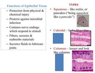
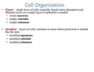
KILIMANJARO CHRISTIAN MMEDICAL UNIVERSITY
COLLEGE
Faculty of Nursing and Rehabilitation Medicine
Department of Anatomy and Neuroscience
Anatomy Lecture for BSc. Nursing and BSc. Physiotherapy
LECTURE 4, L5
TOPIC: EPITHELIAL TISSUES
Date: October, 2022, GYM – Physiotherapy School
Lecturer: J. S. Kauki, BSc, MSc, on PhD, Email: jskauki@gmail.com.
Office ext. 70 Block C, 3RD Floor, Anatomy dept.
TISSUES
• Histology:
– Is the study of tissues.
• Tissues:
– Simply groups of similar
cells that work together
performing the same task
– Greatest form of teamwork
in the body
There are four basic tissue
types:
– Epithelial Tissue
– Connective Tissue
– Muscular Tissue
– Nervous Tissue
Knowing the difference between normal and
abnormal tissue is the first step in diagnosis and
treatment of patients.
made of all four tissue types
Epithelial Tissue
• Makes up 3% of your body
weight
• Forms sheets lining body
surfaces and lumen
• Their cells are all touching one
another
• Of all tissues, they are the most
widely varied in structure and
function
Locations of Epithelial Tissues
• Covers the body (epidermis)
• Found on the inside of hollow
organs and the outside of all
organs
• Found above a connective tissue
layer (epi = above)
• Lines the cavities, tubes, ducts,
and blood vessels inside the body
Epithelial Anatomy
• Apical surface – upper surface that is free or exposed
to the “exterior”
• Basal surface – attached surface (below)
• May have surface modifications;
– Microvilli – small fingerlike extensions that increase the
surface area allowing for more work to be done
Functions of Epithelial Tissue
– Protection from physical &
chemical injury
– Protects against microbial
infection
– Contains nerve endings
which respond to stimuli
– Filters, secretes &
reabsorbs materials
– Secretes fluids to lubricate
joints
TYPES
• Squamous – like scales, or
pancakes (“being squashed
like a pancake”)
• Cuboidal – looks like cubes
• Columnar – longer and look
like columns
Cell Organization
• Simple – single layer of cells; typically found where absorption and
filtration occur or a single layer of epithelial is needed
• simple squamous
• simple cuboidal
• simple columnar
• Stratified – layers of cells; common in areas where protection is needed
like the skin
• stratified squamous
• stratified cuboidal
• stratified columnar
Two Types of Stratified Columnar
• Ciliated
• Non-ciliated
cilia
No cilia
Squamous Epithelium
• Simple – one cell thick
Forms solid layer of cells which line
blood vessels, body cavities and
covers organs in body cavities
Stratified – multiple layers
Forms epidermis
Cuboidal Epithelium
• Simple – one cell thick
• Roughly cube shaped
Line ducts in kidneys where re-
absorption and secretion activities
take place.
Duct
Cuboid Cells
Duct
Cuboid Cells
Columnar Epithelium
• Simple – one cell thick
• Column shaped (long and
narrow)
• Lines digestive tract where re-
absorption & secretion occurs.
Transitional Epithelium
• Transitional Epithelium – stratified tissue that can’t make up
its mind as to whether it is squamous or cuboidal
• Shape of cells varies, depends upon the amount of stretching
(ex: bladder)
Pseudostratified Epithelium
• “Pseudostratified Columnar Epithelium”
• Looks like it has more than one layer because of the position of the
nucleus
• Nuclei are positioned at differing levels
• Cells are narrow in the area without the nucleus
Epithelial Tissue in summary
Types of Epithelial Membranes
• Mucous membranes or mucosa – lining of tubes; moistens and
protects from enzymes (stomach, trachea, and vagina)
• Serous membrane or serosa – outside of organs; lubricates (eg.
Pleural and peritoneual membranes lining thoracic, abdominal and
pelvic organs)
• Cutaneous membrane or skin – body surface; protection
• Synovial membrane – synovial joints; lines and protects synovial
cavities (elbow, knee, hip, etc.)
Glandular Epithelium
A. Exocrine glands – Glands with secretory ducts
B. Endocrine glands – Glands with no secretory duct
Cell Junctions in Epithelial Tissue
A. Desmosome
B. Tight junction
C. Gap junction
Group Activity
Create 4 groups named as follows;
• Holocrine
• Apocrine
• Merocrine
• Endocrine
With examples, describe the mechanism of epithelial cell secretion,
the product secreted and distribution of those secretory cell in the
human body.
Take home Assignment
1. Describe different ways in which cells age?
2. What is cancer. Describe the causes of cancer
THANK YOU FOR YOUR ATTENTION

EPITHELIA TISSUES.pdf

  • 1.
    KILIMANJARO CHRISTIAN MMEDICALUNIVERSITY COLLEGE Faculty of Nursing and Rehabilitation Medicine Department of Anatomy and Neuroscience Anatomy Lecture for BSc. Nursing and BSc. Physiotherapy LECTURE 4, L5 TOPIC: EPITHELIAL TISSUES Date: October, 2022, GYM – Physiotherapy School Lecturer: J. S. Kauki, BSc, MSc, on PhD, Email: jskauki@gmail.com. Office ext. 70 Block C, 3RD Floor, Anatomy dept.
  • 2.
    TISSUES • Histology: – Isthe study of tissues. • Tissues: – Simply groups of similar cells that work together performing the same task – Greatest form of teamwork in the body There are four basic tissue types: – Epithelial Tissue – Connective Tissue – Muscular Tissue – Nervous Tissue Knowing the difference between normal and abnormal tissue is the first step in diagnosis and treatment of patients. made of all four tissue types
  • 3.
    Epithelial Tissue • Makesup 3% of your body weight • Forms sheets lining body surfaces and lumen • Their cells are all touching one another • Of all tissues, they are the most widely varied in structure and function Locations of Epithelial Tissues • Covers the body (epidermis) • Found on the inside of hollow organs and the outside of all organs • Found above a connective tissue layer (epi = above) • Lines the cavities, tubes, ducts, and blood vessels inside the body
  • 4.
    Epithelial Anatomy • Apicalsurface – upper surface that is free or exposed to the “exterior” • Basal surface – attached surface (below) • May have surface modifications; – Microvilli – small fingerlike extensions that increase the surface area allowing for more work to be done
  • 5.
    Functions of EpithelialTissue – Protection from physical & chemical injury – Protects against microbial infection – Contains nerve endings which respond to stimuli – Filters, secretes & reabsorbs materials – Secretes fluids to lubricate joints TYPES • Squamous – like scales, or pancakes (“being squashed like a pancake”) • Cuboidal – looks like cubes • Columnar – longer and look like columns
  • 6.
    Cell Organization • Simple– single layer of cells; typically found where absorption and filtration occur or a single layer of epithelial is needed • simple squamous • simple cuboidal • simple columnar • Stratified – layers of cells; common in areas where protection is needed like the skin • stratified squamous • stratified cuboidal • stratified columnar
  • 7.
    Two Types ofStratified Columnar • Ciliated • Non-ciliated cilia No cilia
  • 8.
    Squamous Epithelium • Simple– one cell thick Forms solid layer of cells which line blood vessels, body cavities and covers organs in body cavities Stratified – multiple layers Forms epidermis
  • 9.
    Cuboidal Epithelium • Simple– one cell thick • Roughly cube shaped Line ducts in kidneys where re- absorption and secretion activities take place. Duct Cuboid Cells Duct Cuboid Cells
  • 10.
    Columnar Epithelium • Simple– one cell thick • Column shaped (long and narrow) • Lines digestive tract where re- absorption & secretion occurs.
  • 11.
    Transitional Epithelium • TransitionalEpithelium – stratified tissue that can’t make up its mind as to whether it is squamous or cuboidal • Shape of cells varies, depends upon the amount of stretching (ex: bladder)
  • 12.
    Pseudostratified Epithelium • “PseudostratifiedColumnar Epithelium” • Looks like it has more than one layer because of the position of the nucleus • Nuclei are positioned at differing levels • Cells are narrow in the area without the nucleus
  • 13.
  • 14.
    Types of EpithelialMembranes • Mucous membranes or mucosa – lining of tubes; moistens and protects from enzymes (stomach, trachea, and vagina) • Serous membrane or serosa – outside of organs; lubricates (eg. Pleural and peritoneual membranes lining thoracic, abdominal and pelvic organs) • Cutaneous membrane or skin – body surface; protection • Synovial membrane – synovial joints; lines and protects synovial cavities (elbow, knee, hip, etc.)
  • 15.
    Glandular Epithelium A. Exocrineglands – Glands with secretory ducts B. Endocrine glands – Glands with no secretory duct
  • 16.
    Cell Junctions inEpithelial Tissue A. Desmosome B. Tight junction C. Gap junction
  • 17.
    Group Activity Create 4groups named as follows; • Holocrine • Apocrine • Merocrine • Endocrine With examples, describe the mechanism of epithelial cell secretion, the product secreted and distribution of those secretory cell in the human body.
  • 18.
    Take home Assignment 1.Describe different ways in which cells age? 2. What is cancer. Describe the causes of cancer
  • 19.
    THANK YOU FORYOUR ATTENTION