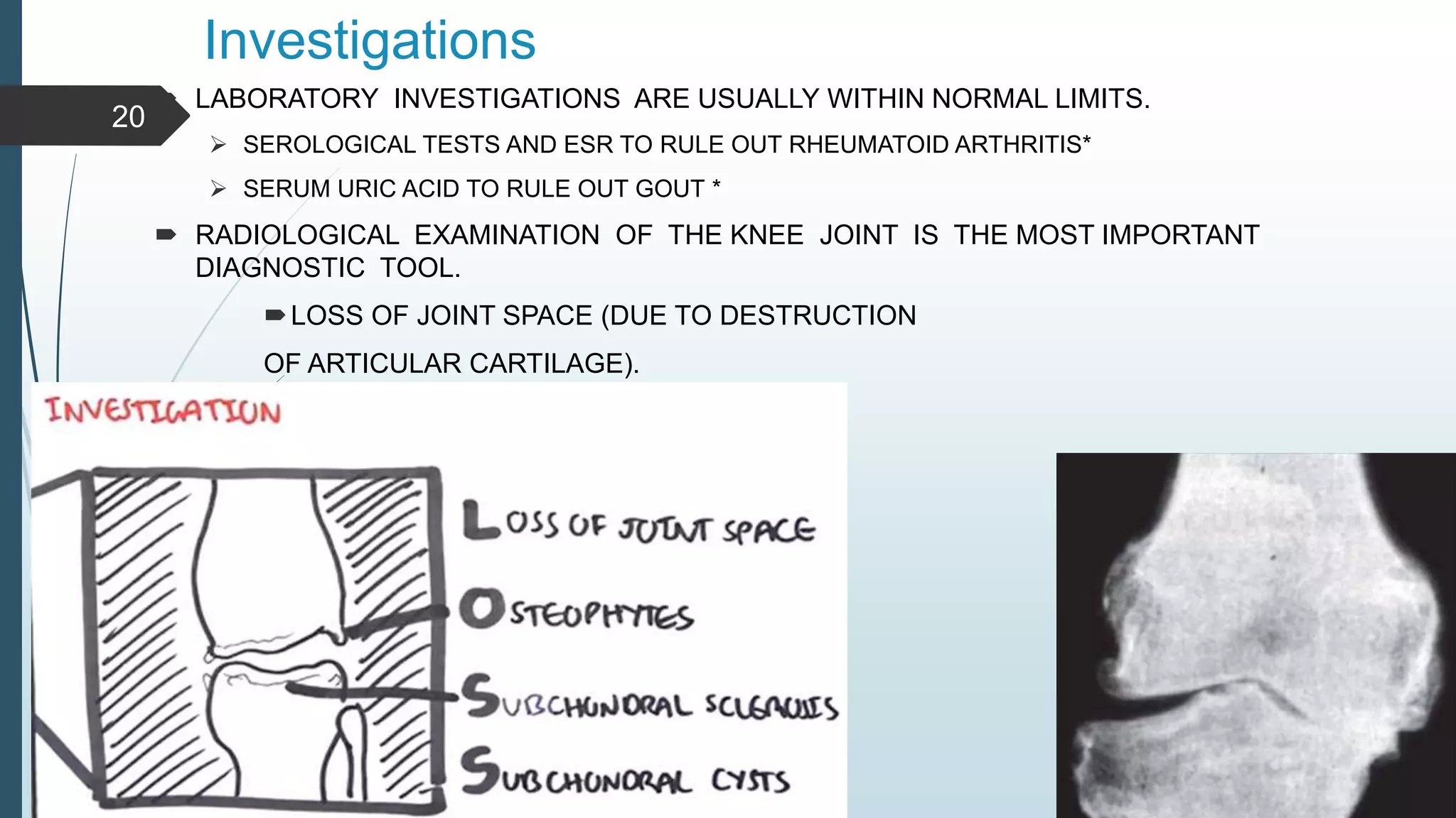
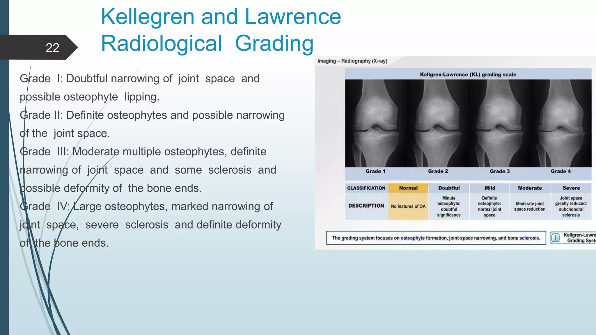
The document defines osteoarthritis as a degenerative joint disease characterized by destruction of articular cartilage and new bone formation at joint surfaces. It most commonly affects weight-bearing joints like the hip and knee. Treatment involves a combination of approaches to relieve pain, restore function, and reduce disability, including weight loss, exercises, braces, and medications like acetaminophen, NSAIDs, or opioids. Conservative treatment succeeds for about 50% of patients before considering surgical options.
Introduction:
Patellofemoral pain (PFP) is one of the most common disorders of the knee. The knee is involved in around 10% of all sporting injuries.
Tria and Alica, described Wiberg classification of patella facet shapes, and there is another classification based on Morphology ratio.
The purpose of this case control study is comparison between the different morphologic types of the patella (Wiberg classification and morphology Ratio) in patients with chondromalacia and normal persons.
Patients & Methods:
In this study we evaluated 30 limbs in 30 patients with chondromalacia (20 females, 10 males ). Medial and lateral facets were calculated on patellar knee view. Also patellar articular length and overall patellar length were calculated in knee joint in 30 flexion. The results were compared to values obtained from 30 limbs in 30 healthy volunteers.
Results:
In Wiberg classification, 57% of normal persons had patella type I while 17% of patients with chondromolacia had this type (p=0.01). Also 43% of normal persons had patella type II while 83% of patients had this type (p= 0.01).
In Morphology ratio classification, 40% of normal persons had patella type II while 13% of patients had this type (p=0.02). Also none of normal persons had patella type III while 13% of patients had this type (p=0.03).
Discussion:
A variety of sports commonly lead to chondromoalacia patella due to unusual compressive forces. Therefore young population specially athletes should pay attention to their patella shapes for selecting the sports types.
Knee osteoarthritis (OA), also known as degenerative joint disease, is typically the result of wear and tear and progressive loss of articular cartilage. It is most common in the elderly. Knee osteoarthritis can be divided into two types, primary and secondary. Primary osteoarthritis is articular degeneration without any apparent underlying reason. Secondary osteoarthritis is the consequence of either an abnormal concentration of force across the joint as with post-traumatic causes or abnormal articular cartilage, such as rheumatoid arthritis (RA).
Osteoarthritis is typically a progressive disease that may eventually lead to disability. The intensity of the clinical symptoms may vary for each individual. However, they typically become more severe, more frequent, and more debilitating over time. The rate of progression also varies for each individual. Common clinical symptoms include knee pain that is gradual in onset and worse with activity, knee stiffness and swelling, pain after prolonged sitting or resting, and pain that worsens over time. Treatment for knee osteoarthritis begins with conservative methods and progresses to surgical treatment options when conservative treatment fails. While medications can help slow the progression of RA and other inflammatory conditions, no proven disease-modifying agents for the treatment of knee osteoarthritis currently exist.
MCL. LCL.ALL injuries
To understand the relevant anatomy of the side ligaments of the knee
To study the mechanism of injury of each ligament and how to diagnose such injury
To highlight the different treatment options in acute or chronic situations
Avascular necrosis (AVN) or Aseptic Necrosis of the hip is caused by a disruption to the hip’s blood supply which results in the deterioration and often collapse of the ball of the thigh bone (femoral head). Early identification and treatment of the condition increases the likelihood that a patient’s hip will recover. Surgery may be required in severe cases to repair or revascularize (restore circulation) the hip or to replace the hip in neglected/end stage cases.
http://www.davidsfeldmanmd.com/specialties/avascular-necrosis-hip
Bicipital tendonitis is inflammation of long head of the biceps tendon under the bicipital groove.
In early stage, tendon becomes red and swollen, as tendonitis develops the tendon sheath can thicken.
In late stage, often become dark red in color due to inflammation.
28,000 ankle sprains occur daily in the US (Kaminski 2013)
Ankle is the 2nd most commonly injured body site. (Ferran 2006)
Ankle sprains are the most common type of ankle injury. (Ferran 2006)
A sprained ankle can happen to athletes and non-athletes,
children and adults.
Inversion injury most common mechanism (Ferran 2006)
Only risk factor is previous ankle sprain (Ferran 2006)
Sex , generalized joint laxity or anatomical foot types are
not risk factors. (Beynnon et al. 2002 )
Hallux rigidus:
A condition characterized by loss of motion of first MTP joint in adults due to degenerative arthritis
second most common condition affecting the big toe after hallux valgus
most common arthritic condition in the foot.
This is a short patient education and awarness presentation on tail bone pain (coccydynia). This presentation delivers a brief information on causes, diagnosis, investigations and treatment of tail bone pain,
Disclaimer:
This presentation is solely for educational purpose.
The Battle Sport Traumatology 2023 Castrocaro Terme FC.pdfNicola Taddio
In this presentation the author analyzes the various problems relating to the functional and mechanical instability of the ankle which has suffered a lesion of the lateral ligaments, the complications, failures and short and long term outcomes in order to have a 360 degree vision of the problem , the possible solutions and the correct management to avoid them.
Lung Cancer: Artificial Intelligence, Synergetics, Complex System Analysis, S...Oleg Kshivets
RESULTS: Overall life span (LS) was 2252.1±1742.5 days and cumulative 5-year survival (5YS) reached 73.2%, 10 years – 64.8%, 20 years – 42.5%. 513 LCP lived more than 5 years (LS=3124.6±1525.6 days), 148 LCP – more than 10 years (LS=5054.4±1504.1 days).199 LCP died because of LC (LS=562.7±374.5 days). 5YS of LCP after bi/lobectomies was significantly superior in comparison with LCP after pneumonectomies (78.1% vs.63.7%, P=0.00001 by log-rank test). AT significantly improved 5YS (66.3% vs. 34.8%) (P=0.00000 by log-rank test) only for LCP with N1-2. Cox modeling displayed that 5YS of LCP significantly depended on: phase transition (PT) early-invasive LC in terms of synergetics, PT N0—N12, cell ratio factors (ratio between cancer cells- CC and blood cells subpopulations), G1-3, histology, glucose, AT, blood cell circuit, prothrombin index, heparin tolerance, recalcification time (P=0.000-0.038). Neural networks, genetic algorithm selection and bootstrap simulation revealed relationships between 5YS and PT early-invasive LC (rank=1), PT N0—N12 (rank=2), thrombocytes/CC (3), erythrocytes/CC (4), eosinophils/CC (5), healthy cells/CC (6), lymphocytes/CC (7), segmented neutrophils/CC (8), stick neutrophils/CC (9), monocytes/CC (10); leucocytes/CC (11). Correct prediction of 5YS was 100% by neural networks computing (area under ROC curve=1.0; error=0.0).
CONCLUSIONS: 5YS of LCP after radical procedures significantly depended on: 1) PT early-invasive cancer; 2) PT N0--N12; 3) cell ratio factors; 4) blood cell circuit; 5) biochemical factors; 6) hemostasis system; 7) AT; 8) LC characteristics; 9) LC cell dynamics; 10) surgery type: lobectomy/pneumonectomy; 11) anthropometric data. Optimal diagnosis and treatment strategies for LC are: 1) screening and early detection of LC; 2) availability of experienced thoracic surgeons because of complexity of radical procedures; 3) aggressive en block surgery and adequate lymph node dissection for completeness; 4) precise prediction; 5) adjuvant chemoimmunoradiotherapy for LCP with unfavorable prognosis.
These simplified slides by Dr. Sidra Arshad present an overview of the non-respiratory functions of the respiratory tract.
Learning objectives:
1. Enlist the non-respiratory functions of the respiratory tract
2. Briefly explain how these functions are carried out
3. Discuss the significance of dead space
4. Differentiate between minute ventilation and alveolar ventilation
5. Describe the cough and sneeze reflexes
Study Resources:
1. Chapter 39, Guyton and Hall Textbook of Medical Physiology, 14th edition
2. Chapter 34, Ganong’s Review of Medical Physiology, 26th edition
3. Chapter 17, Human Physiology by Lauralee Sherwood, 9th edition
4. Non-respiratory functions of the lungs https://academic.oup.com/bjaed/article/13/3/98/278874
ARTIFICIAL INTELLIGENCE IN HEALTHCARE.pdfAnujkumaranit
Artificial intelligence (AI) refers to the simulation of human intelligence processes by machines, especially computer systems. It encompasses tasks such as learning, reasoning, problem-solving, perception, and language understanding. AI technologies are revolutionizing various fields, from healthcare to finance, by enabling machines to perform tasks that typically require human intelligence.
TEST BANK for Operations Management, 14th Edition by William J. Stevenson, Ve...kevinkariuki227
TEST BANK for Operations Management, 14th Edition by William J. Stevenson, Verified Chapters 1 - 19, Complete Newest Version.pdf
Ethanol (CH3CH2OH), or beverage alcohol, is a two-carbon alcohol
that is rapidly distributed in the body and brain. Ethanol alters many
neurochemical systems and has rewarding and addictive properties. It
is the oldest recreational drug and likely contributes to more morbidity,
mortality, and public health costs than all illicit drugs combined. The
5th edition of the Diagnostic and Statistical Manual of Mental Disorders
(DSM-5) integrates alcohol abuse and alcohol dependence into a single
disorder called alcohol use disorder (AUD), with mild, moderate,
and severe subclassifications (American Psychiatric Association, 2013).
In the DSM-5, all types of substance abuse and dependence have been
combined into a single substance use disorder (SUD) on a continuum
from mild to severe. A diagnosis of AUD requires that at least two of
the 11 DSM-5 behaviors be present within a 12-month period (mild
AUD: 2–3 criteria; moderate AUD: 4–5 criteria; severe AUD: 6–11 criteria).
The four main behavioral effects of AUD are impaired control over
drinking, negative social consequences, risky use, and altered physiological
effects (tolerance, withdrawal). This chapter presents an overview
of the prevalence and harmful consequences of AUD in the U.S.,
the systemic nature of the disease, neurocircuitry and stages of AUD,
comorbidities, fetal alcohol spectrum disorders, genetic risk factors, and
pharmacotherapies for AUD.
Explore natural remedies for syphilis treatment in Singapore. Discover alternative therapies, herbal remedies, and lifestyle changes that may complement conventional treatments. Learn about holistic approaches to managing syphilis symptoms and supporting overall health.
Tom Selleck Health: A Comprehensive Look at the Iconic Actor’s Wellness Journeygreendigital
Tom Selleck, an enduring figure in Hollywood. has captivated audiences for decades with his rugged charm, iconic moustache. and memorable roles in television and film. From his breakout role as Thomas Magnum in Magnum P.I. to his current portrayal of Frank Reagan in Blue Bloods. Selleck's career has spanned over 50 years. But beyond his professional achievements. fans have often been curious about Tom Selleck Health. especially as he has aged in the public eye.
Follow us on: Pinterest
Introduction
Many have been interested in Tom Selleck health. not only because of his enduring presence on screen but also because of the challenges. and lifestyle choices he has faced and made over the years. This article delves into the various aspects of Tom Selleck health. exploring his fitness regimen, diet, mental health. and the challenges he has encountered as he ages. We'll look at how he maintains his well-being. the health issues he has faced, and his approach to ageing .
Early Life and Career
Childhood and Athletic Beginnings
Tom Selleck was born on January 29, 1945, in Detroit, Michigan, and grew up in Sherman Oaks, California. From an early age, he was involved in sports, particularly basketball. which played a significant role in his physical development. His athletic pursuits continued into college. where he attended the University of Southern California (USC) on a basketball scholarship. This early involvement in sports laid a strong foundation for his physical health and disciplined lifestyle.
Transition to Acting
Selleck's transition from an athlete to an actor came with its physical demands. His first significant role in "Magnum P.I." required him to perform various stunts and maintain a fit appearance. This role, which he played from 1980 to 1988. necessitated a rigorous fitness routine to meet the show's demands. setting the stage for his long-term commitment to health and wellness.
Fitness Regimen
Workout Routine
Tom Selleck health and fitness regimen has evolved. adapting to his changing roles and age. During his "Magnum, P.I." days. Selleck's workouts were intense and focused on building and maintaining muscle mass. His routine included weightlifting, cardiovascular exercises. and specific training for the stunts he performed on the show.
Selleck adjusted his fitness routine as he aged to suit his body's needs. Today, his workouts focus on maintaining flexibility, strength, and cardiovascular health. He incorporates low-impact exercises such as swimming, walking, and light weightlifting. This balanced approach helps him stay fit without putting undue strain on his joints and muscles.
Importance of Flexibility and Mobility
In recent years, Selleck has emphasized the importance of flexibility and mobility in his fitness regimen. Understanding the natural decline in muscle mass and joint flexibility with age. he includes stretching and yoga in his routine. These practices help prevent injuries, improve posture, and maintain mobilit
263778731218 Abortion Clinic /Pills In Harare ,sisternakatoto
263778731218 Abortion Clinic /Pills In Harare ,ABORTION WOMEN’S CLINIC +27730423979 IN women clinic we believe that every woman should be able to make choices in her pregnancy. Our job is to provide compassionate care, safety,affordable and confidential services. That’s why we have won the trust from all generations of women all over the world. we use non surgical method(Abortion pills) to terminate…Dr.LISA +27730423979women Clinic is committed to providing the highest quality of obstetrical and gynecological care to women of all ages. Our dedicated staff aim to treat each patient and her health concerns with compassion and respect.Our dedicated group ABORTION WOMEN’S CLINIC +27730423979 IN women clinic we believe that every woman should be able to make choices in her pregnancy. Our job is to provide compassionate care, safety,affordable and confidential services. That’s why we have won the trust from all generations of women all over the world. we use non surgical method(Abortion pills) to terminate…Dr.LISA +27730423979women Clinic is committed to providing the highest quality of obstetrical and gynecological care to women of all ages. Our dedicated staff aim to treat each patient and her health concerns with compassion and respect.Our dedicated group of receptionists, nurses, and physicians have worked together as a teamof receptionists, nurses, and physicians have worked together as a team wwww.lisywomensclinic.co.za/
Pulmonary Thromboembolism - etilogy, types, medical- Surgical and nursing man...VarunMahajani
Disruption of blood supply to lung alveoli due to blockage of one or more pulmonary blood vessels is called as Pulmonary thromboembolism. In this presentation we will discuss its causes, types and its management in depth.
Ozempic: Preoperative Management of Patients on GLP-1 Receptor Agonists Saeid Safari
Preoperative Management of Patients on GLP-1 Receptor Agonists like Ozempic and Semiglutide
ASA GUIDELINE
NYSORA Guideline
2 Case Reports of Gastric Ultrasound
NVBDCP.pptx Nation vector borne disease control programSapna Thakur
NVBDCP was launched in 2003-2004 . Vector-Borne Disease: Disease that results from an infection transmitted to humans and other animals by blood-feeding arthropods, such as mosquitoes, ticks, and fleas. Examples of vector-borne diseases include Dengue fever, West Nile Virus, Lyme disease, and malaria.
These lecture slides, by Dr Sidra Arshad, offer a quick overview of physiological basis of a normal electrocardiogram.
Learning objectives:
1. Define an electrocardiogram (ECG) and electrocardiography
2. Describe how dipoles generated by the heart produce the waveforms of the ECG
3. Describe the components of a normal electrocardiogram of a typical bipolar leads (limb II)
4. Differentiate between intervals and segments
5. Enlist some common indications for obtaining an ECG
Study Resources:
1. Chapter 11, Guyton and Hall Textbook of Medical Physiology, 14th edition
2. Chapter 9, Human Physiology - From Cells to Systems, Lauralee Sherwood, 9th edition
3. Chapter 29, Ganong’s Review of Medical Physiology, 26th edition
4. Electrocardiogram, StatPearls - https://www.ncbi.nlm.nih.gov/books/NBK549803/
5. ECG in Medical Practice by ABM Abdullah, 4th edition
6. ECG Basics, http://www.nataliescasebook.com/tag/e-c-g-basics
Report Back from SGO 2024: What’s the Latest in Cervical Cancer?bkling
Are you curious about what’s new in cervical cancer research or unsure what the findings mean? Join Dr. Emily Ko, a gynecologic oncologist at Penn Medicine, to learn about the latest updates from the Society of Gynecologic Oncology (SGO) 2024 Annual Meeting on Women’s Cancer. Dr. Ko will discuss what the research presented at the conference means for you and answer your questions about the new developments.
Maxilla, Mandible & Hyoid Bone & Clinical Correlations by Dr. RIG.pptx
Knee joint Osteoarthritis
1.
2. Prepared by: Dr.Abdul Ahad Khoshal
PGR3Y, Orthopedic surgery
MRH, Kandahar, Afghanistan
Supervisor: Dr. Niamatullah shehzad
Trainer specialist, Orthopedic surgery
MRH, Kandahar, Afghanistan
Contact: daan4ig@gmail.com
3. D e f i n i t i o n
A DEGENERATIVE, NON-INFLAMMATORY
JOINT DISEASE CHARACTERIZED BY DESTRUCTION OF ARTICULAR
CARTILAGE AND FORMATION OF NEW BONE AT THE JOINT
SURFACES AND MARGINS.
THE TERM OSTEOARTHRITIS WAS COINED BY JOHN
SPENDON. HOWEVER, IT IS A MISNOMER AND THE RIGHT
TERM IS OSTEOARTHROSIS OR DEGENERATIVE JOINT DISEASE.
IT COULD BE PRIMARY OR SECONDARY AND THE FORMER IS
MORE COMMON
3
4. O s t e o a r t h r i t i s A ff e c t s T h e S y n o v i a l J o i n t s ,
T h o u g h
I t C a n A ff e c t A n y J o i n t , I t I s M o r e
C o m m o n I n T h e
We i g h t B e a r i n g J o i n t s L i k e T h e :
H i p
K n e e
S p i n e , E t c .
4
5. Primary Osteoarthritis Of The Knee
(Also Called Idiopathic)
Etiological causes for primary osteoarthritis:
Though exact cause is not known, the
following factors are
suspected to play an important role in the
causation
of primary osteoarthritis:
• Obesity
• genetics and
• Heredity
• occupation involving prolonged standing
• Sports
• multiple endocrinal disorders
• Multiple metabolic disorders.
5
6. SECONDARY OSTEOARTHRITIS OF THE KNEE
It Is Generally Observed That Secondary Osteoarthritis
Occurs In The Younger Age Groups And Is More Severe
Than The Primary.
6
7. The Causes For Secondary Osteoarthritis
The Knee Are As Follows:
•Obesity.
•Valgus And Varus Deformities Of The Knee.
•Intra-articular Fractures Of The Knee, Etc.
•Rheumatoid Arthritis, Infection, Trauma, Tb, Etc.
•Hyperparathyroidism.
•Hemophilia.
•Syringomyelia.
•Neurological Disease Like Diabetes.
•Overuse Of Intra-articular Steroid Therapy.
7
8. Features
•it commonly affects the knee joint.
•All races are susceptible.
•Common in older age group/middle-aged patients
• It causes varus deformity of the knee in the late stages
•Eighty percent of people are affected by 40 years,
but only 40 percent show symptoms.
8
9. Feature…..
. One in three people over 60 years are affected and
more than three in four persons over the age of
seventy show some radiographic evidence of the
condition
•Very rarely it can be seen in younger people.
• Women have a greater tendency than men do
• More than 50 percent have bilateral OA knee.
9
10. What are the typical symptoms of osteoarthritis?
• P A I N
• E A R L Y M O R N I N G S T I F F N E S S
• R E S T R I C T E D R A N G E O F J O I N T M O V E M E N T S
• S W E L L I N G O F T H E J O I N T S .
10
11. Remember the risk factors
o O – O B E S I T Y
o S – S E N I L I T Y O R O L D A G E
o T – T R AU M A
o E – E M O T I O N A L S T R E S S
o O – O S T E O P O R O S I S
o A – A L C O H O L
o R – R I G O R O U S L I F E S T Y L E S
o T – TA X I N G P R O F E S S I O N S
o H – H O R M O N A L I M B A L A N C E S
o R – R E P E T I T I V E I N J U R I E S
o I – I N D I A N C U LT U R A L H A B I T S
o T – A X I N G S P O R T S
o I – I M P R O P E R P O S T U R A L H A B I T S
o S – S M O K I N G
11
12. MECHANISMS FOR MAINTAINING
JOINT STABILITY
o Alignment of joint components
o Shape and fit of articular surfaces
o Adhesive property of synovial fluid
o Integrity of capsule and ligaments
o Muscle tone and power
o Neurological control of balance
12
13. Sequence of pathological events in
osteoarthritis
The disease process usually begins in the
anteromedial compartment of the knee joint.
Fibrillation due to loss of water of the weight bearing
articular cartilage is seen in early stages of the
disease followed by complete loss of
articularcartilage.
This puts enormous pressure on the underlying
bone, which causes sclerosis and later
eburnation.
Cysts may develop in the subchondral area due to
microfractures that degenerate.
New bone formation takes place and results in
osteophyte formatio
13
15. Clinical feature
PREDOMINANT SYMPTOM IS PAIN WHICH DECREASES ON WALKING.
THE PAIN IS POORLY LOCALIZED AND
IS DULL ACHING IN NATURE.
MILD SWELLING OF THE KNEE JOINT AND EARLY MORNING STIFFNESS.
EARLY MORNING STIFFNESS, WHICH SUBSIDES OVER THE DAY AFTER SOME
ACTIVITY.
MINIMAL TENDERNESS AND COARSE CREPITUS CANBE ELICITED.
IF THERE ARE LOOSE BODIES IN A JOINT,
THE PATIENT GIVES HISTORY OF LOCKING .
TERMINAL MOVEMENTS OF THE KNEE ARE RESTRICTED
THE PATIENT COMPLAINS OF GENU VARUM DEFORMITY MAY BE SEEN IN VERY
ADVANCED CASES
IN SOME CASES, OSTEOPHYTES MAY BE PALPABLE.
WASTING OF QUADRICEPS FEMORIS MUSCLE*
15
16. How to make a diagnosis?
• Physical examination
• Symptomatology
• Radiography
• Blood tests
• CT scan and MRI.
16
18. Criteria and Classification of OA Knee
(American College of Rheumatology—ACR)
Clinical:
1. Knee Pain For Most Days Of Prior Month.
2. Crepitus On Active Joint Motion.
3. Morning Stiffness Equal And Not More Than 30
Minutes In Duration.
4. Age Equal To More Than 38 Years.
5. Bony Enlargement Of The Knee On Examination.
Oa Is Present (Clinical)
1, 2, 3, 4 Or 1, 2, 5 Or 1, 4, 5
18
19. Clinical and Radiological
1. Knee pain for most days of the prior month.
2. Osteophytes at joint margins.
3. Synovial fluid typical of OA knee.
4. Age—40 years.
5. Morning stiffness equal and not more than 30
minutes.
6. Crepitus on active joint motion.
19
OA Present (Clinical And Radiological)
1, 2 Or 1, 3, 5, 6 Or 1, 4, 5, 6.
20. Investigations
LABORATORY INVESTIGATIONS ARE USUALLY WITHIN NORMAL LIMITS.
SEROLOGICAL TESTS AND ESR TO RULE OUT RHEUMATOID ARTHRITIS*
SERUM URIC ACID TO RULE OUT GOUT *
RADIOLOGICAL EXAMINATION OF THE KNEE JOINT IS THE MOST IMPORTANT
DIAGNOSTIC TOOL.
LOSS OF JOINT SPACE (DUE TO DESTRUCTION
OF ARTICULAR CARTILAGE).
20
21. 21
•S C L E R O S I S ( D U E T O I N C R E A S E C E L L U L A R I T Y
A N D B O N E
D E P O S I T I O N ) .
• S U B C H O N D R A L C Y S T S ( D U E T O S Y N O V I A L F L U I D
I N T R U S I O N
I N T O T H E B O N E ) .
• O S T E O P H Y T E S ( D U E T O R E VA S C U L A R I Z AT I O N O F
R E M A I N I N G C A R T I L A G E A N D C A P S U L A R
T R A C T I O N ) .
• B O N Y C O L L A P S E ( D U E T O C O M P R E S S I O N O F
W E A K E N E D
B O N E ) .
• L O O S E B O D I E S ( D U E T O F R A G M E N TAT I O N O F
O S T E O C H O N D R A L S U R FA C E ) .
• D E F O R M I T Y A N D M A L A L I G N M E N T ( D U E T O
D E S T R U C T I O N
O F C A P S U L E S A N D L I G A M E N T S ) .
22. Kellegren and Lawrence
Radiological Grading
Grade I: Doubtful narrowing of joint space and
possible osteophyte lipping.
Grade II: Definite osteophytes and possible narrowing
of the joint space.
Grade III: Moderate multiple osteophytes, definite
narrowing of joint space and some sclerosis and
possible deformity of the bone ends.
Grade IV: Large osteophytes, marked narrowing of
joint space, severe sclerosis and definite deformity
of the bone ends.
22
23. Radiological Classification of OA Knee
(Ahlbach)
AP weight bearing and Lateral Views
Type I : Joint space narrowing.
Type II : Total loss of joint space.
Type III : < 5 mm tibial erosion but posterior part of the
plateau intact.
Type IV : > 5 mm tibial erosion and erosion of posterior
plateau.
Type V : Subluxation.
Note: Grades IV and V: TKR is the line of treatment
23
24. Other Investigations
• Arthroscopic examination:
This allows direct inspection and visualization of the damaged joint
surfaces.
• But arthroscopy alone for diagnostic purposes is rarely used.
•Synovial fluid analysis shows non-inflammatory picture.
• Bone scan , MRI
and CT scan also helps to diagnose, subchondral cysts,
osteophytes, etc.
24
25. Treatment
Before beginning the treatment, the diagnosis of
OA is a must. ACR diagnostic criteria for OA
knee to be followed.
•Treatment to be individualized and tailored to
severity.
•Multiple strategies may be required in most of
the cases.
25
26. ACR Guidelines: Traditional Format
•KNEE PAIN.
•RADIOGRAPHIC OSTEOPHYTES.
•AT LEAST ONE OF THE FOLLOWING THREE:
–AGE GREATER THAN 50 YEARS.
–MORNING STIFFNESS LESS THAN OR EQUAL TO 30
MINUTES.
–CREPITUS ON MOTION.
26
27. Aims of Treatment of OA Knee
It can be best illustrated by 4 R’s:
•Relieve pain.
•Restore function.
•Reduce disability if any
•Rehabilitation.
27
28. Conservative Methods
This Forms The Mainstay Of Management In Osteoarthritis Of The Knee.
About 50 Percent Of Patients Respond To Conservative Treatment, Which
Consists Of The Following Measures.
28
29. Nonpharmacological Treatment
This is the initial and main stay of treatment in OA knees.
Self education—Educating the patient and his
relatives measures about the disease is the most
important aspect of the non-pharmacological
treatment and should be done first.
•Weight loss
•Physiotherapy
•Therapuetic exercises
•Assistive devices
•Occupational therapy
•Aerobic exercise program
•Strengthening of the quadriceps
•Supervised fitness walking program
•Swimming/hydrotherapy
•Modifications of activity of daily living
Avoidance of stress
29
30. Mechanical aids
They reduce the load on the knee joint and provides
support to the weak knees. The following are used
in OA knees:
•Cane
•Shoe inserts
•Shoe supplements: Good shock absorber, good
mediolateral support, adequate arch support>
•Lateral heel wedges: To reduce pain of medial
tibiofemoral joint OA.
•Knee brace and support in varus knees.
30
31. Components of Therapeutic Exercise
Range of motion and flexibility:
Soft tissue flexibility of both contractile (muscle, tendon) and noncontractile tissues (capsule,
ligaments) is affected by arthritis and inactivity. Joint stiffness and soft tissue shortening
can be reduced with:
appropriate range of motion (ROM) and
stretching exercises.
31
32. 32
• All aspects of muscle strength (strength, endurance, power) can be
impacted as a result of intra-articular and extra-articular inflammatory
processes, disuse, reflex
inhibition in response to pain and joint effusion,
decreased protective muscular reflexes, loss of
mechanical integrity around the joint, and even
medication side effects. Muscle strengthening
exercises helps to overcome these problems.
Quadriceps exercises strengthening of quadriceps
musculature with either isometric or isotonic,
resistive exercises was associated with significant
improvement in quadriceps strength, knee pain,
and function.
33. • Aerobic (Cardiovascular) exercise:
Persons with arthritis tend to be less fit than noninvolved peers. However, there is
strong evidence for the role of regular and vigorous exercise to improve
all components of physical fitness, including cardiovascular fitness and endurance even
in people with arthritis.
Most studies have limited their interventions to
walking,
Stationarybicycling
aerobic dancing
aquatic exercise
33
35. Avoidance of stress
Avoidance of stress and strain to the affected
joint in day-to-day activities. For example, a
patient with OA of the knee is advised to
avoid standing or running whenever possible.
Sitting cross legged and squatting is harmful
for OA of the knee.
35
37. Physiotherapy
Physical modalities that may contribute to pain relief include
the application of superficial heat (hot packs,heating pads, hot water bottles, or paraffin) and/or
cold (cold packs or ice packs).
37
38. Weight Loss
• Obesity is a risk factor for the development of OA, and is associated with radiological
progression of the disease, and disability.
• When people walk their body weight is transferred across the knee joint
• any excess weight should be multiplied by this factor to estimate the excess force across
the knee joint of overweight people.
• In managing OA, weight reduction should be a key goal. Exercise plays a role, but pain
and disability can make it difficult for patients to exercise
sufficiently to lose weight.
• Weight loss can be achieved with:
regular sessions with a dietitian who can provide instruction on reducing caloric intake
and the use of food diaries, and
cognitive-behavioral
modification to change dietary habits.
38
39. 39
Pharmacologic Drugs
• Nonopioid analgesics – E.g. Acetaminophen:
This is the drug of first choice. Up to 4 gm/day can be given.
• NSAIDs: If patients fail to respond to paracetamol or other
oral or topical analgesics, then the use of an NSAID is
indicated.
• Opioid analgesics: These can be tried if patients fail to
respond to paracetamol and NSAIDs
• Food supplementation: Glucosamine andChondroitin
sulfate: Can reduce 20-25 percent pain in mild to moderate
OA. Over the counter food supplements, 1500 mg/day for
at least 3months.
40. • Intra-articular steroids:
o This is indicated if there is effusion and there are signs of inflammation
o The basic intra-articular steroid injections are designed to provide 2 to 6 weeks of pain
relief for patients with knee osteoarthritis
o Such steroids are used to decrease the inflammatory reaction associated with
osteoarthritis
40
41. contraindicated in patients with:
• bacteremia,
• Sepsis
• periarticular or intra-articular infections (eg, septic arthritis, periarticular cellulitis,
osteomyelitis)
• significant skin breakdown at the target site
• known hypersensitivity to the steroid injection
• intraarticular or osteochondral fracture at the target site
• severe joint destruction
• joint prosthesis, or uncontrolled coagulopathy.
41
42. • Viscosupplementation: Injection of hyaluronic acid
into the joint.
• Once a week for 3 weeks. Adverse reactions in 2-3 percent.
• Topical analgesics: These are indicated in the following situations:
–If patients do not respond to oral analgesics.
–If patients do not wish to take systemic drugs.
–Can be used as a monotherapy or adjunct.
–Capsaicin cream – 4 times a day.
42
43. 43
Viscosuplementation in OA knee?
Viscosuplementation: (Intra-articular hyaluronan therapy)
This procedure consists of removal of pathologic
osteoarthritis synovial fluid
and replacement of hyaluronan-based products that restore
the molecular weight and concentration of hyaluronan to normal
values that is reduced in OA knee.
Hyaluronan helps in joint lubrication
buffers load transmission
imparts anti-inflammatory properties to synovial fluid.
Indications for Intra-articular Hyaluronic Acid Injection
•Failed conservative treatment
•If there are major risk factors for surgery
•Failed intra-articular steroid injections
•Advanced osteoarthritis.
45. Alternative Therapies
•Acupuncture
•Bio-feedback
• Aquatic physical therapy
•Massage
•Acupressure
•Tai Chi
•Balenotherapy
•Yoga
The proponents of alternative therapies claim
good results from their respective interventions. The
results are good in the hands of experts.
45
46. Surgery
Indications for surgery
•Pain refractory to conservative measures.
•History of frequent locking episodes.
•Hemarthrosis due to loose bodies or
osteochondral fractures.
•Deformity, usually genu varum.
•Joint instability.
•Progressive limitation of knee motion.
46
47. Surgical Methods
• Excision of osteophytes is rarely done alone.
• Excision of loose bodies, meniscectomy, synovectomy,
and reconstruction or joint debridement are best done
by arthroscopy.
47
48. • PROXIMAL TIBIAL OSTEOTOMY (SLOCUM’S):
Indicated for:
unicompartmental osteoarthritis of knee with pain
correct varus (less than 15°) or
valgus deformity (less than 12°).
Pain is decreased in 80 percent of the cases following surgery as
osteotomy changes the line of weightbearing and
brings the more normal surface
Mean
failure rate is 40 percent at 4 years.
48
49. • D i s t a l F e m o r a l O s t e o t o m y I s I n d i c a t e d W h e n Va r u s
O r Va l g u s D e f o r m i t y O f T h e K n e e I s M o r e T h a n 1 2 -
1 5 ° .
• C h o n d r a l R e s u r f a c i n g P r o c e d u r e
– A u t o l o g o u s C h o n d r o c y t e G r a f t i n g : A u t o l o g o u s
C h o n d r o c y t e s F r o m T h e P a t i e n t ’ s K n e e A r e
C u l t u r e d F o r Tw o W e e k s , R e i n s e r t e d U n d e r A
P a t c h O f P e r i o s t e u m .
M o s a i c P l a s t y : S p a r e A u t o l o g o u s H y a l i n e
C a r t i l a g e F r o m O t h e r A r e a s O f K n e e I s I n s e r t e d
I n t o T h e D e f e c t .
49
50. • Arthroscopic debridement: This is a successful
palliative, temporizing treatment of OA knee.
• Total knee arthroplasty: This is indicated when both
the compartments of the knee joint are destroyed
or if valgus or varus deformity is more than 15°.
It is also indicated in failed conservative treatment
50
51. • Arthrodesis is indicated less commonly than
arthroplasty. If the patient is young and involved
in heavy occupation, arthrodesis is indicated to
give him a stable and strong knee. However,
arthrodesis results in a stiff knee, which is a
severe disability.
• Patellectomy: It is rarely done.
• Unicompartmental knee arthroplasty (UKA): This is
again regaining its popularity over tibial
osteotomy in treating unicompartmental OA, as
it helps in early postoperative rehabilitation.
51
Jionts : Structure(materials): 1fibrous 2 cartiligenus 3 synovial
Synovial Joints:
Hange
Condyloid
Povit
Saddle
Plan
Ball and socket
Difference Between Genetic and Hereditary Diseases. The main difference between these two terms lies in the fact that hereditary diseases have the potential of being carried from one generation to another whereas a genetic disease can either be
hereditary or not, but there will always be a mutational change in the genome
Endocrine Dis: DM ,Cushing syndrome, Osteoporosis
Metabolic Dis: fish eye syndrome , Gaucher’s Disease
Arthritis in people with hemophilia is caused by frequent or inadequately treated bleeding into joints. It is sometimes called degenerative joint disease. It may be the most common complication of severe hemophilia. Arthritis can be avoided by properly taking care of joint bleeds. Once it occurs, it is difficult to treat
Syringomyelia is a rare neurogenic disease which can damage the spinal cord due to formation of a fluid-filled area in the form of a cyst (Syrinx), usually found in the high cervical spine. (Also found in lumbar area but this is very rare) The name derives from the word Syrinx, greek for a tube-formed object and the Myelum, referring to the spinal cord
Hemophilia is a rare disorder in which your blood doesn't clot normally because it lacks sufficient blood-clotting proteins (clotting factors). If you have hemophilia, you may bleed for a longer time after an injury than you would if your blood clotted normally.Small cuts usually aren't much of a problem. If you have a severe deficiency of the clotting factor protein, the greater health concern is deep bleeding inside your body, especially in your knees, ankles and elbows.
PATHOLOGY
Osteoarthritis is a degenerative condition primarily
affecting the articular cartilage. The first change
observed is an increase in water content and
depletion of the proteoglycans from the cartilage
matrix. Repeated weight bearing on such a cartilage
leads to its fbrillation. The cartilage gets abraded
by the grinding mechanism at work at the points
of contact between the apposing articular surfaces,
until eventually the underlying bone is exposed.
With further ‘rubbing’, the subchondral bone
becomes hard and glossy (eburnated). Meanwhile,
the bone at the margins of the joint hypertrophies to
form a rim of projecting spurs known as osteophytes.
A similar mechanism results in the formation of
subchondral cysts and sclerosis.
The loose flakes of cartilage incite synovial
infammation and thickening of the capsule, leading
to deformity and stiffness of the joint. Often one
compartment of a joint is affected more than the
other. For example, in the knee joint, the medial
compartment is affected more than the lateral,
leading to a varus deformity (genu varum).
Essential of Orthopeadics 5th Edition
Essential of orthopaedics 5th Edition
Pitfalls of X-rays in OA Knee
•Not reliable in about 15 percent of the cases.
•Weight bearing AP and lateral views are desired.
•Only 40 percent of the people with severe X-ray changes
experience pain.
( ځواکمنتیا، زغم، او قدرت)
By combining steroid injection with joint lavage,
OA patients get more effective pain relief than with
either therapy alone and pain could reduce for as
long as 24 weeks.
Tai Chi:Tai chi is an ancient Chinese tradition that, today, is practiced as a graceful form of exercise. It involves a series of movements performed in a slow, focused manner and accompanied by deep breathing. Tai chi, also called tai chi chuan, is a noncompetitive, self-paced system of gentle physical exercise and stretching
Balneotherapy is the presumed benefit from disease by bathing, a traditional medicine technique usually practiced at spas. While it is considered distinct from hydrotherapy, there are some overlaps in practice and in underlying principles. Balneotherapy may involve hot or cold water, massage through moving water, relaxation, or stimulation. Many mineral waters at spas are rich in particular minerals such as silica, sulfur, selenium, and radium. Medicinal clays are also widely used, a practice known as 'fangotherapy‘
a system of alternative medicine based on the theory that diseases can be successfully treated or prevented without the use of drugs, by techniques such as control of diet, exercise, and massage