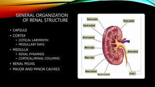
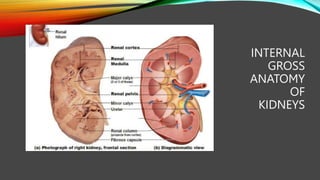
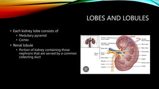
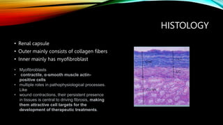
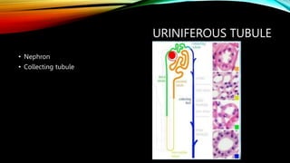
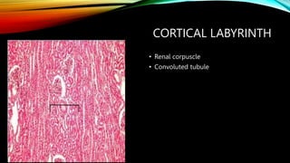
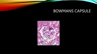
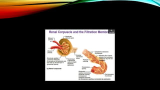
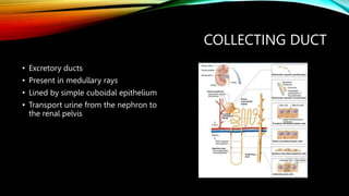
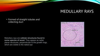
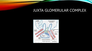
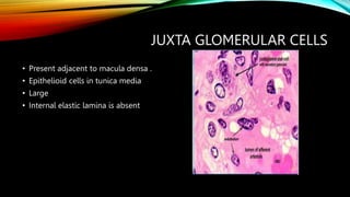
HISTOLOGY OF KIDNEY
Dr. Syeda Iqra Mushir
PGY-1
Histopathology
PIMS
GENERAL ORGANIZATION
OF RENAL STRUCTURE
• CAPSULE
• CORTEX
• COTICAL LABYRINTH
• MEDULLARY RAYS
• MEDULLA
• RENAL PYRAMIDS
• CORTICAL/RENAL COLUMNS
• RENAL PELVIS
• MAJOR AND MINOR CALYXES
INTERNAL
GROSS
ANATOMY
OF
KIDNEYS
LOBES AND LOBULES
• Each kidney lobe consists of
• Medullary pyramid
• Cortex
• Renal lobule
• Portion of kidney containing those
nephrons that are served by a common
collecting duct
Lobule
lobe
HISTOLOGY
• Renal capsule
• Outer mainly consists of collagen fibers
• Inner mainly has myofibroblast
• Myofibroblasts
• contractile, α-smooth muscle actin-
positive cells
• multiple roles in pathophysiological processes.
Like
• wound contractions, their persistent presence
in tissues is central to driving fibrosis, making
them attractive cell targets for the
development of therapeutic treatments.
URINIFEROUS TUBULE
• Nephron
• Collecting tubule
PARTS OF NEPHRON
CORTICAL LABYRINTH
• Renal corpuscle
• Convoluted tubule
BOWMANS CAPSULE
GLOMERULUS
RENAL CORPUSCLE
MESANGIAL CELLS
• Found within the glomerulus
• These cells secrete a matrix of
basement membrane–like material to
support the structure of the
glomerulus.
PROXIMAL CONVOLUTED TUBULE
• Longest and tortuous part of the
nephron
• Commences at urinary pole
• Occupies a major part of the cortex
• Lined by low columnar epithelium
• Show brush border
• Absorb sodium
LOOP OF HENLE
• The thin segment of Henle's loop leads into
the straight part of the distal tubule which is
formed by low cuboidal cells without a brush
border. A few short microvilli are present
• Epithelial cells in the ascending parts of the
intermediate and straight distal tubule cells
transport chloride ( active) and sodium ions (
passive) out of the tubular lumen into the
surrounding peritubular space.
• The epithelium can not be penetrated by
water. Consequently, the transport of ions
over the epithelium sets up a gradient in
osmotic pressure, which serves as a driving
force in the further concentration of the urine.
DISTAL CONVOLUTED TUBULE
• Slightly tortuous
• Lined by simple cuboidal epithelium
• Absorption of sodium and chloride
COLLECTING DUCT
• Excretory ducts
• Present in medullary rays
• Lined by simple cuboidal epithelium
• Transport urine from the nephron to
the renal pelvis
MEDULLARY RAYS
• Formed of straight tubules and
collecting duct
Medullary rays are cellular structures found in
some species of wood. They appear as radial
planar structures, perpendicular to the growth rings,
which are visible to the naked eye
JUXTA GLOMERULAR COMPLEX
MACULA DENSA
• Specialized region of DCT
• Cells are tall columnar
• Closely packed nuclei
• Thin and discontinuous basal lamina
• Release renin
JUXTA GLOMERULAR CELLS
• Present adjacent to macula densa .
• Epithelioid cells in tunica media
• Large
• Internal elastic lamina is absent
EXTRA GLOMERULAR MESANGIAL
CELLS
• Form a cushion of cells between
afferent and efferent arteriole
• Structurally resemble intraglomerular
cells
• Functional significance not clear
THANK YOU

Kidney Normal histology.pptx

  • 1.
    HISTOLOGY OF KIDNEY Dr.Syeda Iqra Mushir PGY-1 Histopathology PIMS
  • 2.
    GENERAL ORGANIZATION OF RENALSTRUCTURE • CAPSULE • CORTEX • COTICAL LABYRINTH • MEDULLARY RAYS • MEDULLA • RENAL PYRAMIDS • CORTICAL/RENAL COLUMNS • RENAL PELVIS • MAJOR AND MINOR CALYXES
  • 3.
  • 4.
    LOBES AND LOBULES •Each kidney lobe consists of • Medullary pyramid • Cortex • Renal lobule • Portion of kidney containing those nephrons that are served by a common collecting duct Lobule lobe
  • 5.
    HISTOLOGY • Renal capsule •Outer mainly consists of collagen fibers • Inner mainly has myofibroblast • Myofibroblasts • contractile, α-smooth muscle actin- positive cells • multiple roles in pathophysiological processes. Like • wound contractions, their persistent presence in tissues is central to driving fibrosis, making them attractive cell targets for the development of therapeutic treatments.
  • 7.
  • 8.
  • 9.
    CORTICAL LABYRINTH • Renalcorpuscle • Convoluted tubule
  • 10.
  • 11.
  • 12.
  • 16.
    MESANGIAL CELLS • Foundwithin the glomerulus • These cells secrete a matrix of basement membrane–like material to support the structure of the glomerulus.
  • 17.
    PROXIMAL CONVOLUTED TUBULE •Longest and tortuous part of the nephron • Commences at urinary pole • Occupies a major part of the cortex • Lined by low columnar epithelium • Show brush border • Absorb sodium
  • 18.
    LOOP OF HENLE •The thin segment of Henle's loop leads into the straight part of the distal tubule which is formed by low cuboidal cells without a brush border. A few short microvilli are present • Epithelial cells in the ascending parts of the intermediate and straight distal tubule cells transport chloride ( active) and sodium ions ( passive) out of the tubular lumen into the surrounding peritubular space. • The epithelium can not be penetrated by water. Consequently, the transport of ions over the epithelium sets up a gradient in osmotic pressure, which serves as a driving force in the further concentration of the urine.
  • 19.
    DISTAL CONVOLUTED TUBULE •Slightly tortuous • Lined by simple cuboidal epithelium • Absorption of sodium and chloride
  • 22.
    COLLECTING DUCT • Excretoryducts • Present in medullary rays • Lined by simple cuboidal epithelium • Transport urine from the nephron to the renal pelvis
  • 23.
    MEDULLARY RAYS • Formedof straight tubules and collecting duct Medullary rays are cellular structures found in some species of wood. They appear as radial planar structures, perpendicular to the growth rings, which are visible to the naked eye
  • 24.
  • 25.
    MACULA DENSA • Specializedregion of DCT • Cells are tall columnar • Closely packed nuclei • Thin and discontinuous basal lamina • Release renin
  • 26.
    JUXTA GLOMERULAR CELLS •Present adjacent to macula densa . • Epithelioid cells in tunica media • Large • Internal elastic lamina is absent
  • 27.
    EXTRA GLOMERULAR MESANGIAL CELLS •Form a cushion of cells between afferent and efferent arteriole • Structurally resemble intraglomerular cells • Functional significance not clear
  • 28.