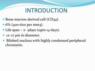
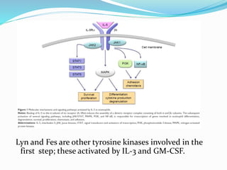
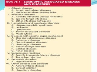
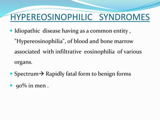
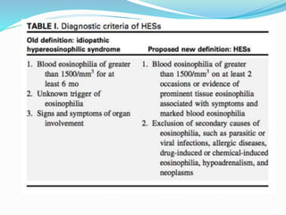
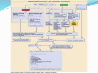
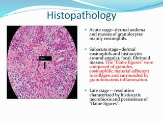
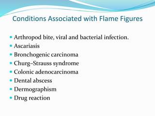
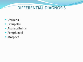
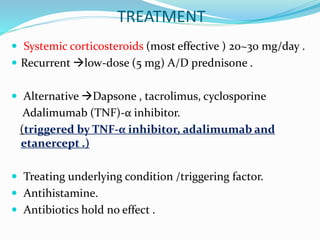
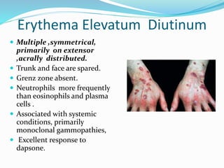
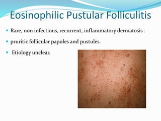
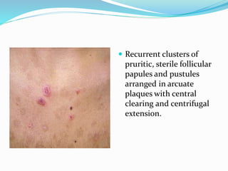
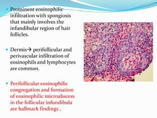
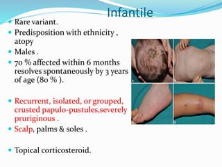
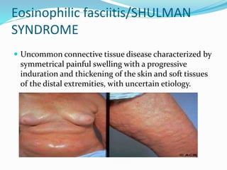
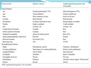
INTRODUCTION
 Bone marrow derived cell (CD34).
 6% (400-600 per mm3).
 Life span – 2- 5days (upto 14 days).
 12–17 μm in diameter.
 Bilobed nucleus with highly condensed peripheral
chromatin.
Lyn and Fes are other tyrosine kinases involved in the
first step; these activated by IL-3 and GM-CSF.
HYPEREOSINOPHILIC SYNDROMES
 Idiopathic disease having as a common entity ,
"Hypereosinophilia", of blood and bone marrow
associated with infiltrative eosinophilia of various
organs.
 Spectrum Rapidly fatal form to benign forms
 90% in men .
LYMPHOCYTIC HES
 Severe pruritus, eczematous , urticaria, and angioedema ,
lymphadenopathy rarely, endomyocardial fibrosis.
 Abnormal T-cell clones with unusual surface phenotypes.
 These T cells display activation markers, such as CD25, and secrete T
helper 2 cytokines, including high levels of interleukin 5 .
 Episodic angioedema and eosinophilia (Gleich Syndrome) and
nodules, eosinophilia, rheumatism, dermatitis, and swelling (NERDS)
syndrome have developed T-cell clones.
MYELOPROLIFERATIVE HES.
 Deletion on chromosome 4q12 that codes tyrosine kinase .
 FIP1L1-PDGFRA gene mutation form a distinct subset of HES, with
cardiomyopathy and endomyocardial fibrosis, that responds to imatinib.
 Elevated serum tryptase levels and atypical spindle shaped mast cells in bone
marrow. (clinical manifestations of systemic mastocytosis absent but patients
satisfy criteria for mastocytosis.)
The FIP1L1PDGFRA gene is detected
in mast cells, eosinophils,
neutrophils,
mononuclear cells
CLINICAL FINDINGS-
 Pruritic erythematous macules, papules, plaques, wheals, or
nodules (50% of patient).
 Head, trunk, and extremities.
 Fever, weight loss, malaise, hepatosplenomegaly.
 Mucosal ulcers (oropharynx or anogenital )an aggressive
clinical course.
 Untreated-- >death is likely within 2 years of presentation .
Cardiac manifestation--
 Subendocardial fibrosis restrictive cardiomyopathy .
 Tethering of chordae tendineae  valvular insufficiency.
 Cardiac abnormalities confined to intramural regions can
occur without peripheral blood eosinophilia.
 Splinter hemorrhages , nail fold infarcts may herald the
onset of thromboembolic disease.
 Nervous system, lungs, rarely, kidneys affected.
Evaluation--
 History-- Travel ,Ingestants ,Close contacts with itch.
 Physical examination
Cardiovascular .
Nails for splinter hemorrhage (medical emergency)
Hepatosplenomegaly , Lymphadenopathy
 Laboratory studies –
Complete blood counts , Liver, renal function tests .
Chromosomal study, FIP1L1-PDGFRA (CHIC2 gene)
Erythrocyte sedimentation rate ,C-reactive protein, Rheumatoid factor
Antiproteinase 3 and antimyeloperoxidase (c-ANCA and p-ANCA)
IgE level, Strongyloides IgG antibody
IL-5 serum level
 Muscle enzymes (creatine phosphokinase, aldolase)
 B12 serum level
 Mast cell/basophil tryptase (protryptase) level
 Coagulation factors Troponin (before imatinib )
 Serum protein electrophoresis
 Immuno fixation electrophoresis for monoclonal
proteins.
Imaging tests 2D Echo,CT chest, abdomen, and pelvis
GI endoscopy, Pulmonary function tests
 Bone marrow aspirate with staining for tryptase
 Tissue biopsy
 Direct immunofluorescence
 Immunostaining for eosinophil granule proteins
Differential diagnosis
 Parasitic infection
 Urticaria
 Hereditary angioedema
 Atopic dermatitis
 Contact dermatitis
 Drug reaction
 Fungal infection
 Mycosis fungoides
 Sézary syndrome
 Behçet syndrome
 Crohn disease
Ulcerative colitis
 Reiter syndrome
 Recurrent apthous
stomatitis
 Erythema multiforme
 Lichen planus
 Immunobullous
disease
 Herpes simplex
 Syphilis
TREATMENT
 Goal--- relieve symptoms & improve organ function while
keeping peripheral blood eosinophils at 1,000 to 2,000/mm3 .
 Myeloproliferative HES is responsive to imatinib.
(FIP1L1-PDGFRA +/-)
 Imatinib mesylate usually induces hematologic remission, but
endomyocardial disease may worsen during first several days .
 Monitor Troponin levels & Glucocorticoids should be given
before and with initiation of imatinib therapy
.
 In the absence of the gene mutation, first-line therapy
is prednisone.
 Failure to glucocorticoid monotherapy have worse
prognosis .
 Interferon (IFN)-α has been beneficial in both HES.
 Refractory disease may respond to infliximab
(antitumor necrosis factor-α)or alemtuzumab
(antiCD52)
Well syndrome/ Eosinophlic cellulitis
 Recurrent granulomatous dermatitis with eosinophilis.
 Unknown etiology.
 Triggering factor- arthropods bite, viral / bacterial
infection, heamatogenous/ non heamatogenous
malignancies.
 Pathogenesis-
Deregulated tissue eosinophil  increase IL-5 , IL- 2
• Mostly in adult , pediatrics also.
• Preceding pain, pruritis, malaise, fever.
• Extremities.
Clinical course
Cellulitic stage localised erythema and oedema
sometimes blistering
Granulomatous stage  gradual resolution from the
centre of lesions, which remains oedematous and slate
colored for several weeks.
Variant- plaque , annular granuloma , papulo vesicle ,
bullous , papulo nodular , fixed drug euption type
Histopathology
 Acute stage--dermal oedema
and masses of granulocytes
mainly eosinophils.
 Subacute stage--dermal
eosinophils and histiocytes
around angular, focal, fibrinoid
masses. The "flame figures" were
composed of granules,
eosinophilic material adherent
to collagen and surrounded by
granulomatous inflammation.
 Late stage -- resolution
characerised by histiocytic
necrobiosis and persistence of
"flame figures".
Conditions Associated with Flame Figures
 Arthropod bite, viral and bacterial infection.
 Ascariasis
 Bronchogenic carcinoma
 Churg–Strauss syndrome
 Colonic adenocarcinoma
 Dental abscess
 Dermographism
 Drug reaction
DIFFERENTIAL DIAGNOSIS
 Urticaria
 Erysipelas
 Acute cellulitis
 Pemphigoid
 Morphea
TREATMENT
 Systemic corticosteroids (most effective ) 20~30 mg/day .
 Recurrent low-dose (5 mg) A/D prednisone .
 Alternative Dapsone , tacrolimus, cyclosporine
Adalimumab (TNF)-α inhibitor.
(triggered by TNF-α inhibitor, adalimumab and
etanercept .)
 Treating underlying condition /triggering factor.
 Antihistamine.
 Antibiotics hold no effect .
Granuloma Faciale
 Unknown etiology.
 Localized chronic fibrosing vasculitis
 Extrafacial lesions isolated/ in conjunction with facial
lesions.
 Photoexacerbation of lesions.
 Immunoglobulins, fibrin and complement deposited
along dermal–epidermal junction in a granular pattern
and around blood vessels by direct immunofluorescence
 The lesions are typically
asymptomatic red,
brown, or violaceous
plaques that are soft
smooth , and well
circumscribed, often
showing follicular
accentuation and
telangiectasia
 Mixed infiltrate of
lymphocytes, histiocytes,
neutrophils, plasma cells,
and eosinophils. There is
sparing of a narrow grenz
zone between the
inflammatory
infiltrate and the overlying
epidermis.
Angiocentric eosinophil fibrosis
 It may accompany granuloma faciale in rare cases.
 Rare , bening , fibrosing condition.
 Involve sinonasal tract and upper respiratory tract.
 Variant of mucosal counterpart of GA.
DIFFERENTIAL DIAGNOSIS
 Face Sarcoidosis,Benign lymphocytic infiltrate of Jessner
,Rosacea, Lymphoma cutis ,Angiolymphoid hyperplasia with
eosinophilia , Tinea , Basal cell carcinoma,
Xanthogranuloma , Mastocytoma
 Extrafacial Erythema elevatum diutinum,Granuloma
annulare, Benign lymphocytic infiltrate of Jessner ,Fixed
drug eruption
Always Rule Out
 Face Discoid lupus erythematosus
 Trunk Erythema elevatum diutinum
Erythema Elevatum Diutinum
 Multiple ,symmetrical,
primarily on extensor
,acrally distributed.
 Trunk and face are spared.
 Grenz zone absent.
 Neutrophils more frequently
than eosinophils and plasma
cells .
 Associated with systemic
conditions, primarily
monoclonal gammopathies,
 Excellent response to
dapsone.
Treatment
 FIRST-LINE THERAPY-
Topical corticosteroids
Cryotherapy
Intralesional steroids
Pulsed dye laser
Dapsone, 50–100 mg/day
 SECOND-LINE THERAPY-
Topical tacrolimus ointment
Surgical excision
Eosinophilic Pustular Folliculitis
 Rare, non infectious, recurrent, inflammatory dermatosis .
 pruritic follicular papules and pustules.
 Etiology unclear.
 Associated with human immunodeficiency virus
(HIV) infection, malignancy.
 May occur as a consequence of immune
reconstruction syndrome ( IRIS ) rather than
immunodeficiency in HAART-treated cases / after
a bone marrow or stem cell transplantation
Pathogenesis  expression of limited to follicular
epithelium.
 intercellular adhesion molecule 1 (ICAM-1) ,
 vascular cell adhesion molecule 1 (VCAM-1),
 lymphocyte function-associated antigen 1 (LFA-1)
 Role of prostaglandin D2( good therapeutic
response to indomethacin )
VARIANTS
 Classic.
 Pediatric.
 Neonatal.
 HIV-associated.
Classic type / Ofuji disease
 Japanese population
 Adult(35–40 years)
 Recurrent.
 Typically last more than 1–2 weeks, involute and relapse
in average interval of every 3–4 weeks
 Initially on face, may also affect trunk, extremities, scalp,
palms or soles, where follicles are absent
 Recurrent clusters of
pruritic, sterile follicular
papules and pustules
arranged in arcuate
plaques with central
clearing and centrifugal
extension.
 Prominent eosinophilic
infiltration with spongiosis
that mainly involves the
infundibular region of hair
follicles.
 Dermis perifollicular and
perivascular infiltration of
eosinophils and lymphocytes
are common.
 Perifollicular eosinophilic
congregation and formation
of eosinophilic microabscess
in the follicular infundibula
are hallmark findings .
Infantile
 Rare variant.
 Predisposition with ethnicity ,
atopy
 Males .
 70 % affected within 6 months
resolves spontaneously by 3 years
of age (80 % ).
 Recurrent, isolated, or grouped,
crusted papulo-pustules,severely
pruriginous .
 Scalp, palms & soles .
 Topical corticosteroid.
Histology-
 Eosinophilic inflammatory infiltration is always present
but true follicular involvement is observed only in 62 %
of cases.
 Other cases present with perifollicular, interfollicular,
and/or periadnexal infiltrates instead 
NEONATAL EOSINOPHILIC PUSTULOSIS
 Occasionally Flame figures can be observed.
Immunosuppression-Associated
 HIV infection, hematological or other malignancies.
 A male predominance.
 Papular lesions are less likely to coalesce into plaques or be
arranged in an annular configuration (LIKE IN CLASSIC )
 Hematologic malignancy associated EPF shows relatively
better response.
 intensely itchy ,perifollicular
erythematous papules and
pustules.
 upper trunk, upper limbs,
head, and neck.
 In female patients
,predominantly affects face
and mimics acne excoriée .
 predominance of CD8+
lymphocytes in the lesional
infiltration
Differential diagnosis
Classic form –
 Acneiform eruptions
 Acne vulgaris
 Follicular eczema
 Folliculitis
 Pustural psoriasis.
 Subcorneal pustural
dermatosis.
 Alopecia mucinosa.
Pediatric form-
 Acropustulosis of
infancy
 Scabies
 Scalp pyoderma
Neonatal-
 Erythema toxicum
neonatorum
 Miliaria
 Neonatal acne
 Varicella
 Transient neonatal
pustular melanosis
HIV-associated-
 Pruritic papular
eruption of HIV
 Reactions to arthropod
bites and stings
 Follicular mycosis
fungoides.
 Photodermatitis
 Opportunistic
infections
TREATMENT
 Indomethacin, first-line and effective .
 Topical steroids /tacrolimus.
 Ultraviolet B ,PUVA
 Systemic corticosteroids
 Cyclosporine
 Anti histamine.
 Dapsone, minocycline ,colchicine, retinoids.
 IFNα-2b, and IFNγ,
 Transdermal nicotine patches (6.25– 12.5 mg) achieves
excellent therapeutic.
Recurrent Cutaneous Eosinophilic
Vasculitis
 Cutaneous necrotizing predominantly eosinophilic
vasculitis.
 Idiopathic ,may be a part of systemic disease CTD
 Recurrent, pruritic ,erythematous or purpuric papules
with peripheral blood eosinophilia and hypo
complementemia , generally responsive to
corticosteroids .
 Necrotizing vasculitis of small vessels in the dermis, with
exclusively eosinophilic infiltration.
 The small-vessel necrotizing vasculitis and fibrinoid
degeneration on small-vessel walls are featured for RCEV,
distinct from that of other eosinophilic cutaneous
disorders such as episodic angioedema with eosinophilia,
WS, and hypereosinophilic syndrome.
Differential Diagnosis--
 Churg– Strauss.
 Drug eruption.
 Urticaria/urticarial vasculitis
 Episodic angioedema with eosinophilia
 Wells syndrome
Treatment
 Systemic glucocorticoids --Prednisone at 15–30 mg daily,
or equivalent
 Adjuvant therapy Suplatast tosilate. ( TH2 INHIBITOR)
 Indomethacin
Eosinophilic fasciitis/SHULMAN
SYNDROME
 Uncommon connective tissue disease characterized by
symmetrical painful swelling with a progressive
induration and thickening of the skin and soft tissues
of the distal extremities, with uncertain etiology.
Etiology--
 Physical exertion or trauma (30– 46 %)
 Drugs( natalizumab ,influenza vaccination ,statins ,ACE
inhibitors ,phenytoin ,heparin, exposure to
trichloroethylene)
 Hematological disorders and malignancy
 Infections (borreliosis , Mycoplasma arginini )
 Physical factors(radiotherapy,burns)
 Stem cell transplantation.
 Autoimmune diseases (e.g., Hashimoto’s thyroiditis, Graves
disease, Sjögren syndrome, systemic lupus erythematous,
primary biliary cirrhosis),
 No age and sex predilection.
 Groove sign (depression appearance along the
course of a vein or between muscle groups).
 Peau d’orange sign with hyperpigmentation.
 Prayer sign( the inability to close a fist or restricted
joint movement)
 Morphea-like lesions are considered risk factor for
residual fibrosis
 Hide bound chest progressive respiratory
limitation due to the restriction of chest movement
and reduced expansibility of chest wall
 Spontaneous or provoked myalgia, weight loss, and
asthenia, are common
 Inflammatory polyarthritis causing arthralgia and
morning stiffness, carpal tunnel syndrome
 Visceral involvement is absent with very few exceptions
Laboratory and Imaging Findings
 Peperipheral eosinophilia, elevated serum levels of CRP
and ESR, hypergammaglobulinemia.
 Low titers of antinuclear antibodies (ANA) without
detectable anti-extractable nuclear antibody.
 MRI findings shows abnormal fascial signal intensity
and enhancement, both of which are directly
proportional to disease activity .
 Deposition of IgG and C3 in affected fascia, and
hypergammaglobulinemia
 Elevated serum levels of soluble CD40 ligand (sCD40L
(disease activity)
 Decreased Matrix metalloproteinase-13 (MMP-13)
 Predominancy of macrophages and CD8+ T lymphocytes
Histopathology
 Requires a full thickness, or skin-to-muscle, wedge biopsy.
 Epidermis, dermis are usually unaltered or mildly affected
 Inflammatory and fibrous thickening of fascia, usually 2 to
15 times thicker than normal, with inflammatory infiltrates
mainly composed of lymphocytes and/or eosinophils .
Differential Diagnosis
 Systemic sclerosis
 Eosinophilia-myalgia syndrome (EMS) after l-tryptophan ingestion
 Churg–strauss syndrome
 Hypereosinophilic syndrome
 Silica exposure
 Cutaneous metastatic malignancy, should also be ruled out
Treatment
 Systemic corticosteroid (0.5–1 mg/kg daily )
RESISTANCE /DEPENDENCY TO
CORTICOSTEROIDS
Methotrexate
Azathioprine
Cyclophosphamide
Cyclosporine
Dapsone
Rituximab, Anti TNF α (infliximab)
PUVA
ANGIOLYMPHOID HYPERPLASIA WITH
EOSINOPHILIA (EPITHELIOID HEMANGIOMA)
 Vasoproliferative, idiopathic condition.
 Manifests in adults ( female).
 Isolated or grouped papules, plaques,
or nodules in the skin of the head
and neck
KIMURA DISEASE
 Chronic inflammatory disorder.
 Unknown etiology.
 Painless lymphadenopathy or subcutaneous masses in
the head and neck region.
 Unusual granulation combined
with hyperplastic changes in lymphoid
tissue.
THANK
YOU

Eosiniphil in dermatology

  • 2.
    INTRODUCTION  Bone marrowderived cell (CD34).  6% (400-600 per mm3).  Life span – 2- 5days (upto 14 days).  12–17 μm in diameter.  Bilobed nucleus with highly condensed peripheral chromatin.
  • 5.
    Lyn and Fesare other tyrosine kinases involved in the first step; these activated by IL-3 and GM-CSF.
  • 8.
    HYPEREOSINOPHILIC SYNDROMES  Idiopathicdisease having as a common entity , "Hypereosinophilia", of blood and bone marrow associated with infiltrative eosinophilia of various organs.  Spectrum Rapidly fatal form to benign forms  90% in men .
  • 11.
    LYMPHOCYTIC HES  Severepruritus, eczematous , urticaria, and angioedema , lymphadenopathy rarely, endomyocardial fibrosis.  Abnormal T-cell clones with unusual surface phenotypes.  These T cells display activation markers, such as CD25, and secrete T helper 2 cytokines, including high levels of interleukin 5 .  Episodic angioedema and eosinophilia (Gleich Syndrome) and nodules, eosinophilia, rheumatism, dermatitis, and swelling (NERDS) syndrome have developed T-cell clones.
  • 12.
    MYELOPROLIFERATIVE HES.  Deletionon chromosome 4q12 that codes tyrosine kinase .  FIP1L1-PDGFRA gene mutation form a distinct subset of HES, with cardiomyopathy and endomyocardial fibrosis, that responds to imatinib.  Elevated serum tryptase levels and atypical spindle shaped mast cells in bone marrow. (clinical manifestations of systemic mastocytosis absent but patients satisfy criteria for mastocytosis.) The FIP1L1PDGFRA gene is detected in mast cells, eosinophils, neutrophils, mononuclear cells
  • 13.
    CLINICAL FINDINGS-  Pruriticerythematous macules, papules, plaques, wheals, or nodules (50% of patient).  Head, trunk, and extremities.  Fever, weight loss, malaise, hepatosplenomegaly.  Mucosal ulcers (oropharynx or anogenital )an aggressive clinical course.  Untreated-- >death is likely within 2 years of presentation .
  • 15.
    Cardiac manifestation--  Subendocardialfibrosis restrictive cardiomyopathy .  Tethering of chordae tendineae  valvular insufficiency.  Cardiac abnormalities confined to intramural regions can occur without peripheral blood eosinophilia.  Splinter hemorrhages , nail fold infarcts may herald the onset of thromboembolic disease.  Nervous system, lungs, rarely, kidneys affected.
  • 17.
    Evaluation--  History-- Travel,Ingestants ,Close contacts with itch.  Physical examination Cardiovascular . Nails for splinter hemorrhage (medical emergency) Hepatosplenomegaly , Lymphadenopathy  Laboratory studies – Complete blood counts , Liver, renal function tests . Chromosomal study, FIP1L1-PDGFRA (CHIC2 gene) Erythrocyte sedimentation rate ,C-reactive protein, Rheumatoid factor Antiproteinase 3 and antimyeloperoxidase (c-ANCA and p-ANCA) IgE level, Strongyloides IgG antibody IL-5 serum level
  • 18.
     Muscle enzymes(creatine phosphokinase, aldolase)  B12 serum level  Mast cell/basophil tryptase (protryptase) level  Coagulation factors Troponin (before imatinib )  Serum protein electrophoresis  Immuno fixation electrophoresis for monoclonal proteins.
  • 19.
    Imaging tests 2DEcho,CT chest, abdomen, and pelvis GI endoscopy, Pulmonary function tests  Bone marrow aspirate with staining for tryptase  Tissue biopsy  Direct immunofluorescence  Immunostaining for eosinophil granule proteins
  • 20.
    Differential diagnosis  Parasiticinfection  Urticaria  Hereditary angioedema  Atopic dermatitis  Contact dermatitis  Drug reaction  Fungal infection  Mycosis fungoides  Sézary syndrome  Behçet syndrome  Crohn disease Ulcerative colitis  Reiter syndrome  Recurrent apthous stomatitis  Erythema multiforme  Lichen planus  Immunobullous disease  Herpes simplex  Syphilis
  • 21.
    TREATMENT  Goal--- relievesymptoms & improve organ function while keeping peripheral blood eosinophils at 1,000 to 2,000/mm3 .  Myeloproliferative HES is responsive to imatinib. (FIP1L1-PDGFRA +/-)  Imatinib mesylate usually induces hematologic remission, but endomyocardial disease may worsen during first several days .  Monitor Troponin levels & Glucocorticoids should be given before and with initiation of imatinib therapy .
  • 22.
     In theabsence of the gene mutation, first-line therapy is prednisone.  Failure to glucocorticoid monotherapy have worse prognosis .  Interferon (IFN)-α has been beneficial in both HES.  Refractory disease may respond to infliximab (antitumor necrosis factor-α)or alemtuzumab (antiCD52)
  • 24.
    Well syndrome/ Eosinophliccellulitis  Recurrent granulomatous dermatitis with eosinophilis.  Unknown etiology.  Triggering factor- arthropods bite, viral / bacterial infection, heamatogenous/ non heamatogenous malignancies.  Pathogenesis- Deregulated tissue eosinophil  increase IL-5 , IL- 2
  • 25.
    • Mostly inadult , pediatrics also. • Preceding pain, pruritis, malaise, fever. • Extremities. Clinical course Cellulitic stage localised erythema and oedema sometimes blistering Granulomatous stage  gradual resolution from the centre of lesions, which remains oedematous and slate colored for several weeks.
  • 26.
    Variant- plaque ,annular granuloma , papulo vesicle , bullous , papulo nodular , fixed drug euption type
  • 27.
    Histopathology  Acute stage--dermaloedema and masses of granulocytes mainly eosinophils.  Subacute stage--dermal eosinophils and histiocytes around angular, focal, fibrinoid masses. The "flame figures" were composed of granules, eosinophilic material adherent to collagen and surrounded by granulomatous inflammation.  Late stage -- resolution characerised by histiocytic necrobiosis and persistence of "flame figures".
  • 28.
    Conditions Associated withFlame Figures  Arthropod bite, viral and bacterial infection.  Ascariasis  Bronchogenic carcinoma  Churg–Strauss syndrome  Colonic adenocarcinoma  Dental abscess  Dermographism  Drug reaction
  • 29.
    DIFFERENTIAL DIAGNOSIS  Urticaria Erysipelas  Acute cellulitis  Pemphigoid  Morphea
  • 30.
    TREATMENT  Systemic corticosteroids(most effective ) 20~30 mg/day .  Recurrent low-dose (5 mg) A/D prednisone .  Alternative Dapsone , tacrolimus, cyclosporine Adalimumab (TNF)-α inhibitor. (triggered by TNF-α inhibitor, adalimumab and etanercept .)  Treating underlying condition /triggering factor.  Antihistamine.  Antibiotics hold no effect .
  • 31.
    Granuloma Faciale  Unknownetiology.  Localized chronic fibrosing vasculitis  Extrafacial lesions isolated/ in conjunction with facial lesions.  Photoexacerbation of lesions.  Immunoglobulins, fibrin and complement deposited along dermal–epidermal junction in a granular pattern and around blood vessels by direct immunofluorescence
  • 32.
     The lesionsare typically asymptomatic red, brown, or violaceous plaques that are soft smooth , and well circumscribed, often showing follicular accentuation and telangiectasia
  • 33.
     Mixed infiltrateof lymphocytes, histiocytes, neutrophils, plasma cells, and eosinophils. There is sparing of a narrow grenz zone between the inflammatory infiltrate and the overlying epidermis.
  • 34.
    Angiocentric eosinophil fibrosis It may accompany granuloma faciale in rare cases.  Rare , bening , fibrosing condition.  Involve sinonasal tract and upper respiratory tract.  Variant of mucosal counterpart of GA.
  • 35.
    DIFFERENTIAL DIAGNOSIS  FaceSarcoidosis,Benign lymphocytic infiltrate of Jessner ,Rosacea, Lymphoma cutis ,Angiolymphoid hyperplasia with eosinophilia , Tinea , Basal cell carcinoma, Xanthogranuloma , Mastocytoma  Extrafacial Erythema elevatum diutinum,Granuloma annulare, Benign lymphocytic infiltrate of Jessner ,Fixed drug eruption Always Rule Out  Face Discoid lupus erythematosus  Trunk Erythema elevatum diutinum
  • 36.
    Erythema Elevatum Diutinum Multiple ,symmetrical, primarily on extensor ,acrally distributed.  Trunk and face are spared.  Grenz zone absent.  Neutrophils more frequently than eosinophils and plasma cells .  Associated with systemic conditions, primarily monoclonal gammopathies,  Excellent response to dapsone.
  • 37.
    Treatment  FIRST-LINE THERAPY- Topicalcorticosteroids Cryotherapy Intralesional steroids Pulsed dye laser Dapsone, 50–100 mg/day  SECOND-LINE THERAPY- Topical tacrolimus ointment Surgical excision
  • 38.
    Eosinophilic Pustular Folliculitis Rare, non infectious, recurrent, inflammatory dermatosis .  pruritic follicular papules and pustules.  Etiology unclear.
  • 39.
     Associated withhuman immunodeficiency virus (HIV) infection, malignancy.  May occur as a consequence of immune reconstruction syndrome ( IRIS ) rather than immunodeficiency in HAART-treated cases / after a bone marrow or stem cell transplantation
  • 40.
    Pathogenesis  expressionof limited to follicular epithelium.  intercellular adhesion molecule 1 (ICAM-1) ,  vascular cell adhesion molecule 1 (VCAM-1),  lymphocyte function-associated antigen 1 (LFA-1)  Role of prostaglandin D2( good therapeutic response to indomethacin )
  • 41.
    VARIANTS  Classic.  Pediatric. Neonatal.  HIV-associated.
  • 42.
    Classic type /Ofuji disease  Japanese population  Adult(35–40 years)  Recurrent.  Typically last more than 1–2 weeks, involute and relapse in average interval of every 3–4 weeks  Initially on face, may also affect trunk, extremities, scalp, palms or soles, where follicles are absent
  • 43.
     Recurrent clustersof pruritic, sterile follicular papules and pustules arranged in arcuate plaques with central clearing and centrifugal extension.
  • 44.
     Prominent eosinophilic infiltrationwith spongiosis that mainly involves the infundibular region of hair follicles.  Dermis perifollicular and perivascular infiltration of eosinophils and lymphocytes are common.  Perifollicular eosinophilic congregation and formation of eosinophilic microabscess in the follicular infundibula are hallmark findings .
  • 45.
    Infantile  Rare variant. Predisposition with ethnicity , atopy  Males .  70 % affected within 6 months resolves spontaneously by 3 years of age (80 % ).  Recurrent, isolated, or grouped, crusted papulo-pustules,severely pruriginous .  Scalp, palms & soles .  Topical corticosteroid.
  • 46.
    Histology-  Eosinophilic inflammatoryinfiltration is always present but true follicular involvement is observed only in 62 % of cases.  Other cases present with perifollicular, interfollicular, and/or periadnexal infiltrates instead  NEONATAL EOSINOPHILIC PUSTULOSIS  Occasionally Flame figures can be observed.
  • 47.
    Immunosuppression-Associated  HIV infection,hematological or other malignancies.  A male predominance.  Papular lesions are less likely to coalesce into plaques or be arranged in an annular configuration (LIKE IN CLASSIC )  Hematologic malignancy associated EPF shows relatively better response.
  • 48.
     intensely itchy,perifollicular erythematous papules and pustules.  upper trunk, upper limbs, head, and neck.  In female patients ,predominantly affects face and mimics acne excoriée .  predominance of CD8+ lymphocytes in the lesional infiltration
  • 49.
    Differential diagnosis Classic form–  Acneiform eruptions  Acne vulgaris  Follicular eczema  Folliculitis  Pustural psoriasis.  Subcorneal pustural dermatosis.  Alopecia mucinosa. Pediatric form-  Acropustulosis of infancy  Scabies  Scalp pyoderma
  • 50.
    Neonatal-  Erythema toxicum neonatorum Miliaria  Neonatal acne  Varicella  Transient neonatal pustular melanosis HIV-associated-  Pruritic papular eruption of HIV  Reactions to arthropod bites and stings  Follicular mycosis fungoides.  Photodermatitis  Opportunistic infections
  • 51.
    TREATMENT  Indomethacin, first-lineand effective .  Topical steroids /tacrolimus.  Ultraviolet B ,PUVA  Systemic corticosteroids  Cyclosporine  Anti histamine.  Dapsone, minocycline ,colchicine, retinoids.  IFNα-2b, and IFNγ,  Transdermal nicotine patches (6.25– 12.5 mg) achieves excellent therapeutic.
  • 52.
    Recurrent Cutaneous Eosinophilic Vasculitis Cutaneous necrotizing predominantly eosinophilic vasculitis.  Idiopathic ,may be a part of systemic disease CTD  Recurrent, pruritic ,erythematous or purpuric papules with peripheral blood eosinophilia and hypo complementemia , generally responsive to corticosteroids .
  • 53.
     Necrotizing vasculitisof small vessels in the dermis, with exclusively eosinophilic infiltration.  The small-vessel necrotizing vasculitis and fibrinoid degeneration on small-vessel walls are featured for RCEV, distinct from that of other eosinophilic cutaneous disorders such as episodic angioedema with eosinophilia, WS, and hypereosinophilic syndrome.
  • 54.
    Differential Diagnosis--  Churg–Strauss.  Drug eruption.  Urticaria/urticarial vasculitis  Episodic angioedema with eosinophilia  Wells syndrome Treatment  Systemic glucocorticoids --Prednisone at 15–30 mg daily, or equivalent  Adjuvant therapy Suplatast tosilate. ( TH2 INHIBITOR)  Indomethacin
  • 55.
    Eosinophilic fasciitis/SHULMAN SYNDROME  Uncommonconnective tissue disease characterized by symmetrical painful swelling with a progressive induration and thickening of the skin and soft tissues of the distal extremities, with uncertain etiology.
  • 56.
    Etiology--  Physical exertionor trauma (30– 46 %)  Drugs( natalizumab ,influenza vaccination ,statins ,ACE inhibitors ,phenytoin ,heparin, exposure to trichloroethylene)  Hematological disorders and malignancy  Infections (borreliosis , Mycoplasma arginini )  Physical factors(radiotherapy,burns)  Stem cell transplantation.  Autoimmune diseases (e.g., Hashimoto’s thyroiditis, Graves disease, Sjögren syndrome, systemic lupus erythematous, primary biliary cirrhosis),
  • 57.
     No ageand sex predilection.  Groove sign (depression appearance along the course of a vein or between muscle groups).  Peau d’orange sign with hyperpigmentation.  Prayer sign( the inability to close a fist or restricted joint movement)  Morphea-like lesions are considered risk factor for residual fibrosis  Hide bound chest progressive respiratory limitation due to the restriction of chest movement and reduced expansibility of chest wall
  • 59.
     Spontaneous orprovoked myalgia, weight loss, and asthenia, are common  Inflammatory polyarthritis causing arthralgia and morning stiffness, carpal tunnel syndrome  Visceral involvement is absent with very few exceptions Laboratory and Imaging Findings  Peperipheral eosinophilia, elevated serum levels of CRP and ESR, hypergammaglobulinemia.  Low titers of antinuclear antibodies (ANA) without detectable anti-extractable nuclear antibody.
  • 60.
     MRI findingsshows abnormal fascial signal intensity and enhancement, both of which are directly proportional to disease activity .  Deposition of IgG and C3 in affected fascia, and hypergammaglobulinemia  Elevated serum levels of soluble CD40 ligand (sCD40L (disease activity)  Decreased Matrix metalloproteinase-13 (MMP-13)  Predominancy of macrophages and CD8+ T lymphocytes
  • 61.
    Histopathology  Requires afull thickness, or skin-to-muscle, wedge biopsy.  Epidermis, dermis are usually unaltered or mildly affected  Inflammatory and fibrous thickening of fascia, usually 2 to 15 times thicker than normal, with inflammatory infiltrates mainly composed of lymphocytes and/or eosinophils .
  • 62.
    Differential Diagnosis  Systemicsclerosis  Eosinophilia-myalgia syndrome (EMS) after l-tryptophan ingestion  Churg–strauss syndrome  Hypereosinophilic syndrome  Silica exposure  Cutaneous metastatic malignancy, should also be ruled out
  • 64.
    Treatment  Systemic corticosteroid(0.5–1 mg/kg daily ) RESISTANCE /DEPENDENCY TO CORTICOSTEROIDS Methotrexate Azathioprine Cyclophosphamide Cyclosporine Dapsone Rituximab, Anti TNF α (infliximab) PUVA
  • 65.
    ANGIOLYMPHOID HYPERPLASIA WITH EOSINOPHILIA(EPITHELIOID HEMANGIOMA)  Vasoproliferative, idiopathic condition.  Manifests in adults ( female).  Isolated or grouped papules, plaques, or nodules in the skin of the head and neck
  • 66.
    KIMURA DISEASE  Chronicinflammatory disorder.  Unknown etiology.  Painless lymphadenopathy or subcutaneous masses in the head and neck region.  Unusual granulation combined with hyperplastic changes in lymphoid tissue.
  • 69.