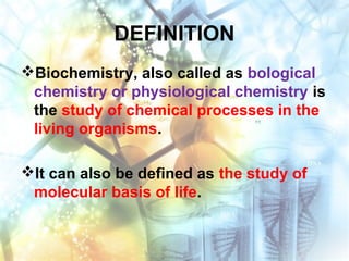
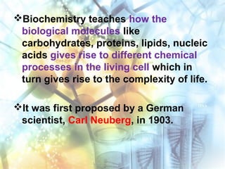
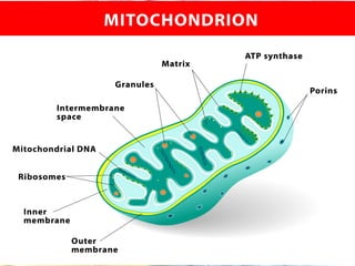
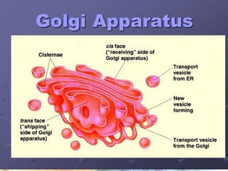
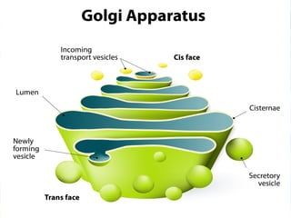
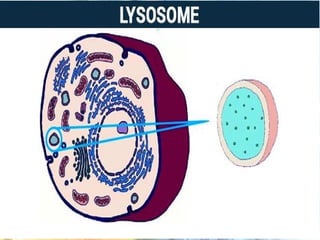
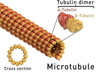
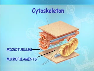
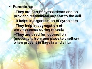
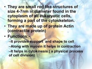
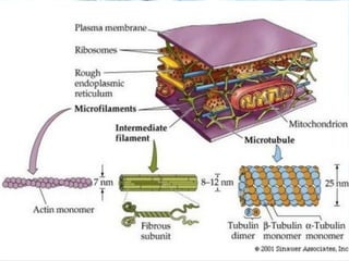
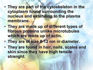
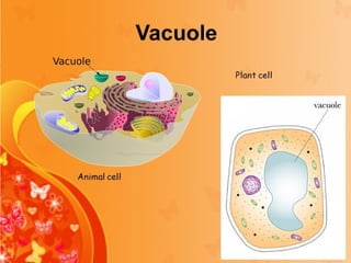
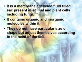
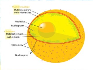
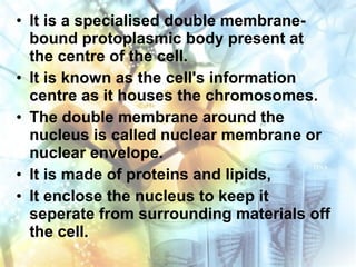
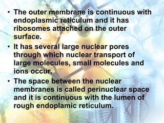
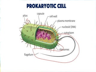
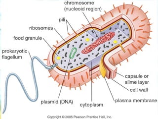
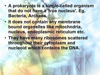
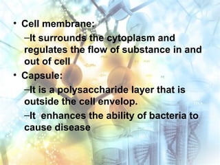
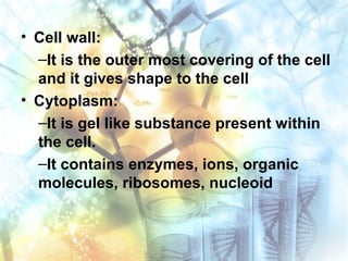
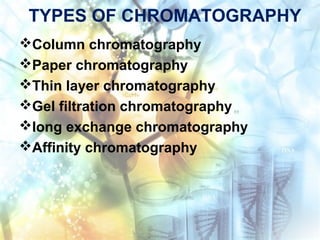
SlideShare a Scribd company logo
1 of 115
Download to read offline
BIOCHEMISTRY
BY, MS.PRIYANKA GOHIL
M.Sc. (N)
Nursing Tutor, MBNC
UNIT- 1
INTRODUCTION
DEFINITION
Biochemistry, also called as bological
chemistry or physiological chemistry is
the study of chemical processes in the
living organisms.
It can also be defined as the study of
molecular basis of life.
Biochemistry teaches how the
biological molecules like
carbohydrates, proteins, lipids, nucleic
acids gives rise to different chemical
processes in the living cell which in
turn gives rise to the complexity of life.
It was first proposed by a German
scientist, Carl Neuberg, in 1903.
SIGNIFICANCE IN NURSING
As the definition says that biochemistry
is the study of chemical processes in
the living cell, it is very essential that a
nurse being a medical professional,
study and understand biochemistry in
order to care for their patients.
SIGNIFICANCE IN NURSING cont..
For example, a patient was admitted to
the hospital and during the stay in the
hospital he suddenly comes up with
certain symptoms like confusion,
dizziness, shaky feeling, pounding
heart, racing pulse,sweating, trembling
and weakness.
SIGNIFICANCE IN NURSING cont..
The medical history show that he has
diabetes mellitus and was taking
insulin.
A nurse will be able to understand
immediately that the cause of his
symptoms were due to hypoglycaemia
and she will be understand the changes
that can occur in the body and will be
able to give the timely treatment.
SIGNIFICANCE IN NURSING cont..
(Hypogycaemia is a serious condition in
day to day life if not taken care, as is
may lead to life threatening
complications affecting cardiac and
central nervous system. There is also a
chance that the patient may go into
seizure and coma.)
SIGNIFICANCE IN NURSING cont..
So, a thorough understanding of the
biochemical processes taking place in
the body will help the nurse to have a
patient-centred approach in the
treatment.
SIGNIFICANCE IN NURSING cont..
 Some of the importance of biochemistry
in nursing:-
Knowing the normal processes that occur
within the body will helps us to identify the
alterations that can occur during disease
conditions so that an effective medical
treatment can be facilitated.
 Biochemistry teaches about homeostasis and
electrolyte balance and a nurse should
understand the importance of it in the body.
All the organs and tissues in the body function to
maintain this homeostasis.
Certain disease conditions may create an imbalance
of one or more electrolytes which is a serious
condition.
The biochemistry teaches about the normal and
abnormal metabolite levels and this knowledge helps
the nurse to take special care of the patient by
maintaining the fluid and electrolytes.
• Homeostasis :-
–The ability or tendency of a living
organism, cell, or group to keep the
conditions inside it the same despite any
changes in the conditions around it, or this
state of internal balance.
 The various biochemical tests like blood test,
kidney function test, liver function test, lipid
profile, will help the nurse to diagnose
diseases and the test results will help in
assessing the needs of the patient to provide
an effective care.
 Every drug has a biochemical action and this
knowledge will help in selecting the
appropriate doses and the duration of
treatment a patient requires.
 Nutritional biochemistry teaches the relation
between health, diet and disease.
It focuses on how each nutrient functions
metabolically and biochemically.
Using biochemical tools nutritional deficiency
deseases can be diagnosed.
In a nutshell, Biochemistry is important in
nursing because it gives knowledge about
normal chemical process within the living cells
and any change in it helps in the diagnosis of
disease, thereby helping the nurse to provide
an effective care to the patient.
THE CELL
All organisms are made up of cells.
“ It is the basic structural, functional and
biological unit of life.”
It was discovered by Robert Hooke in
1665.
The word cell comes from a latin word
'cella' meaning small room.
Cells are the building block of life,
where all the chemical processes
occur.
They take up the biological molecules,
convert them in to energy or use them
to carry out specailized functions.
It also contains the hereditary material
which determine the genetic
characteristics.
The cell theory developed by Mathias
Jacob and Theodar Schwann states
that..
“All organisms are composed of one
or more cells, that all cells come
from pre-existing cells, the vital
functions of an organism occur
within the cells and that all cells
contain hereditary information
necessary for cell funtions and for
transmitting from one generation to
next.”
Every living things (animals, plants,
bacteria, fungi, protozoans) are made
up of cell.
Some organisms are made up of just
one single cell. ( Unicellular organism
e.g. Bacteria)
While some organisms are made up of
many cells. (multi cellular organisms
e.g. Animal cell)
The human body is built of about one
trillion cells specialising in different
functions.
Many identical cells joins together and
forms a tissue.
Various tissues, that perform a
particular function organize together to
form an organ.
Various organs joins together to form
an organ system.
TYPES OF CELL
1. Eukaryotic cell
2. Prokaryotic cell
1. EUKARYOTIC CELL
• Eukaryotic cells are those cells which
have a true nucleus.
• It has a nuclear membrane within which
there is well defined chromosomes.
• It has other membrane bound
organelles like mitochondria,
endoplasmic reticulum, golgi bodies
etc.
• Organisms with eukaryotic cells are
called as Eukaryotes.
• They may be single-celled or
multicellular organisms. e.g.cells of
plant, animals, fungi.
2. PROKARYOTIC CELL
• Prokaryotic cells are those cells whose
nucleus is not distinct and their DNA is
not organized in to chromosomes.
• They lack most of the membrane bound
organelles.
• The organisms with prokaryotic cells
are called as Prokatryotes.
• They are usually unicellular organisms.
e.g. bacteria.
EUKARYOTIC CELL
• A typical Eukaryotic cell, as seen under
light microscope has two major parts:
• The different substances that make up
the cell are collectively called as
Protoplasm.
The plasma membrane
The cytoplasm and its
organelles
1. PLASMA MEMBRANE
• It is a thin, elastic, semi permeable
membrane of 7.5 to 10 nanometres
thickness.
• It is a living membrane made up of
phospholipid bilayer embedded with
proteins.
• It is a flexible membrane and so it can
fold in or out.
• It is made up of 55 % proteins, 25 %
phospholipids, 13 % cholesterol, 4%
lipids 3% carbohydrates.
Functions of plasma membrane
• It protects the cell contents from the
surrounding environmement.
• It provides a shape to the cell.
• It is semi permeable and allows
transport of certain substances in to
and out of the cell.
• It helps in forming cell junctions.
2. CYTOPLASM AND ITS ORGANELLES
• In eukaryotes, the protoplasm
surrounding the nucleus is called
cytoplasm.
• It is a clear gelatinous fluid that fills the
cell and surrounds the organelle.
• It contains 90 % water, dissolved
substances, minerals, sugar, iorns,
vitamins, amino acids, proteins and
enzymes.
• Cytoplasm is the seat for many
pathways like glycolysis and HMP
(Hexose mono phosphate) pathway.
• The cytoplasm is also called as
cytosol.
• It contains well organized structures
called organelles which vary in size
from a few nanometres to many
micrometers and they are specialized
to carry out one or more vital functions
of the cell.
• The organelles include...
–Mitochondria
–Endoplasmic reticulum
–Golgi apparatus
–Ribosomes
–Lysosomes
–Peroxisomes
–Centriole
–Vacuole
–Nucleus
• These are tiny, sausage shaped
structures of diameter 0.5 to 1
micrometre.
• It is called as “ Power house of the
cell” as it generates energy in the form
of AdenosineTriphosphate (ATP) which
is required by all the cells.
• It is found both in plant and animal
cells.
• It has a double membrane envolope: an
inner membrane and an outer
membrane.
• The inner membrane is folded and
pleated (double fold) and it is called
cristae.
• It provides large surface area for
different biochemical processes as it
contains many oxidative enzymes.
• Within this membrane are the proteins
involved in electron transport chain,
ATP synthase and transport proteins.
• It is impermeable to molecules and
ions but allows the free passage of
carbon dioxide, oxygen and water.
• The metabolites are transported across
the membrane with the help of
transport proteins.
• The outer membrane is a smooth
phospholipid bilayer enveloping the
mitochondria.
• It had enzymes like monoamine
oxidase and NADH reductase. (NADH-
Necotinamide adenine dinucleotide)
• An intrinsic protein called porin form
the channels that makes the membrane
permeable to solutes and metabolites.
• It allows the free passage of
substances with molecular weight less
than 10,000.
• The membranes create two
compartments...
–The space between the outer and inner
membrane is called the intermembrane
space.
–It is here that oxidative phosphorylation
occus. (Releasing energy by oxidise
nurients for ATPsyntehsis)
–The inner cavity of mitochondria is called
as matrix.
• It is packed with many enzymes like
pyruvate dehydrogenease, pyruvate
arboxylase, enzymes for oxidation of
fatty acids, aminoacids and enzymes of
citric acid cycle.
• It also contains mitochondrial genome,
mitochondrial ribosomes, tRNAs,
dissolved oxygen, carbon dioxide and
water.
• FUNCTIONS:-
–It is the seat for Kreb's cycle (cytric
acid cycle)
–It contains enzymes for Oxidative
phosphorylation which helps in
producing energy rich ATP molecules
–It provides intermediates for
synthesis of cytochrome, chlorophyl,
hemoglobin and steroids
–Aminoacids like glutamate are
synthesised in it from alpha
ketoglutarate and oxalo acetate
–Many fatty acids are synthesised in
the matrix
–Calcium can be stored in the
mitochondria and released whenever
required.
• It is a network of tubular and vascular
structures extending from outer
membranes of nucleus to the plasma
membrane.
• It is seen to be spread throughout the
cytoplasm and it provides a large
surface area for various physiological
activities.
• The inside of the vesicles and tubules
is filled with an endoplasmic matrix.
• When the ribosomes are attached on
the outer surface of mebrane of the
endoplasmic reticulum, it is called as
Rough endoplasmic reticulum (RER).
• It lies adjacent to the cell nucleus and
its membrane is continuous with the
membrane of the nucleus.
• When there are no ribosomes attached
to the endoplasmic reticulum, it is
called as Smooth endoplasmic
reticulum (SER).
• Functions:-
–RER helps in transporting proteins from
ribosomes to golgi bodies.
–Proteins that enters RER undergoes
processing, folding and sorting
–SER is involved in the synthesis of lipids,
including cholesterol and phospholipids
–In some cells, SER helps in the synthesis
of steroid hoemones from cholesterol
–In the cells of liver, SER helps in
detoxifying drugs and harmful chemicals.
• It is a stack of membranous sac, like a pile of
disc.
• It is present between endoplasmic reticulum
and plasma membrane.
• Like endoplasmic reticulum it is a single
mebrane bound structure.
• In animal cells it is present around the
nucleus while in plant cell it is scattered
throughout the cell.
• These cells organelles which pack and sort
the proteins before they are sent to their
destinations.
• Functions:-
–The proteins that enter it from RER is
modified, processed, sorted and transported
in the form of vesicles to the plasma
membrane and other destinations.
–Glycolipids, sphingomyelin are synthesised
within it.
–In plant cells it help in the synthesis of
polysaccharides needed by the cell wall.
–It has role in synthesis of carbohydrates
like galactose.
–Primary lysosomes develop from mature
Golgi bodies.
–It plays an important role in lipid trafficking.
• They are spherical shaped organelles
seen either free in the cytoplasm or
attached to RER.
• They are found in eukaryotes and
prokaryotes.
• They are synthesised by nucleolus.
• The ribosomes link the amino acids
together in the order that is specified
by the messengers RNA.
• They are made up of two subunits - a
small sub unit and a large sub unit.
• The small sub unit reads the mRNA
while the large sub unit assembles the
amino acids to form large polypeptide.
• The ribosomal sub units are made up
of one or more eRNA and proteins.
• Function:-
–It is the site for protein synthesis.
• They are tiny sac like organelle of size
0.5 to 1.5 Âľm, which are membrane
bound and found in hudreds in a single
cell.
• They are formed from golgi bodies as
small vesicles which bud off from
them.
• Within the sac there are several
hydrolytic enzymes that breakdown
macromolecules like nucleic acid,
proteins and polysaccharides.
• They are also called as “suicidal begs”
as enzymes contained in term can digest
the cell's own material when damaged or
dead.
• The important enzymes in it are DNA-
ase, RNA-ase, protease,
lipase,glycosidase, phosphatase,
sulphatase which are synthesised in the
endoplasmic reticulum and then
transported to the golgi bodies.
• Functions:-
–It helps in digestion of food releasing
enzymes.
–They digest worn out organelles
–It helps in defence by digesting germs
–It helps sperm cells in entering the egg by
breaking through the egg membrane
–It provides energy during cell starvation by
digestion of cells own parts.
• It is a type of microbody which is small,
spherical shaped, with single membrane
and of size 0.5-1.5 Âľm.
• They are found in both animal and plant
cells.
• Just like lysosomes they contain many
enzymes which help in biological
reactions.
• They are formed from endoplasmic
reticulum unlike lysosomes which are
formed from golgi bodies.
• Functions:-
–The enzymes found in peroxisomes are
usually used for different metabolic reactions
and for digesting different materials in the
cell.
–They help in oxidation of many substances
resulting in the formation of hydrogen
peroxide as a by product.
–But, it contains enzymes peroxidase or
catalase which decomposes this harmful
hydrogen peroxide into water and oxygen or
uses it to oxidize other organic compounds
like phenol, alcohol, formaldehyde, etc.
–They are also involved in catabolism of fatty
acids (beta oxidation), D- aminoacids and
polyamines.
–They are needed in the synthesis of
plasmalogens (type of ether phospholipid
needed for functioning of brain and lungs.)
–They participate in synthesis of cholesterol,
bile acids and myelin.
–In plants it helps in photorespiration and
symbolic nitrogen fixation.
• They are long, hollow cylindrers of size
24 nm in diameter and can grow up to a
length of 50 Âľm.
• They are found in eukaryotic cell.
• They are made up of
two globular
proteins namely
Îą-tubulin and
β-tubulin.
• Along the
microtubule axis
tubulins are joined
end to end to form
protofilaments.
• They are important component of
cytoskeleton and so they found
throughout the cytoplasm.
• They are organised by microtubule
organising structures, primarily the
centrioles.
• Functions:-
–They are part of cytoskeleton and so
provides mechanical support to the cell
–It helps in organisation of cytoplasm
–They help in segregation of
chromosomes during mitosis
–They are used for locomotion
(movement from one place to another)
when present in flagella and cilia)
• They are small rod like structures of
size 4-7nm in diameter found in the
cytoplasm of all eukaryotic cells,
forming a part of the cytoskeleton.
• They are made up of protein Actin
(contractile protein)
• Functions:-
–It provides support and shape to cell
–Along with myosin it helps in contraction
–It helps in cytokinesis ( a physical process
of cell division)
• They are part of the cytoskeleton in the
cytoplasm found surrounding the
nucleus and extending to the plasma
membrane.
• They are made up of different types of
fibrous proteins unlike microtubules
which are made up of actin.
• They are of size 8-12 nm in diameter.
• They are found in hair, nails, scales and
skin since they have high tensile
strenght.
• Due to their rope like structure they
provide mechanical strength to the
cells and help cells with stand stress
like stretching and changing shape. Eg.
Keratin filaments in skin and epithelial
cells.
• It is cylindrical in shape and of length
0.5 micrometer.
• it is present in all animal cells just
outside the nucleus.
• It does not have a membrane.
• All centrioles are made up of protein
strands called tubulin.
• Each centriole has 9 sets of inter
connected peripheral tubules and each
set has 3 micro tubules arranged at
definite angles making the shape of a
cylinder.
• It has its own DNA nad RNA and
therefore it is self duplicating.
• Two centrioles when oriented at a right
angle, forms centrosome.
Functions:-
It is involved in cell division. They are seen
in the process of both meiosis and mitosis.
It helps in the formation of cilia and flagella.
It helps in organization and alignment of
microtubules within the cell.
• It is a membrane enclosed fluid filled
sac present in animal and plant cells
including fungi.
• It contains organic and inorganic
molecules within it.
• They do not have particular size or
shape but adjust themselves according
to the need of the cell.
Functions:-
It helps in removing waste products from
the cell
It isolates substances that are harmful to
the cell
It holds water and waste prodcuts within it
It helps in maintaining the internal pH of cell
It helps to maintain hydrostatic pressure
within the cell
It plays a major role in autophagy by
maintaining a balance between biogenesis
and degradation
• It is a specialised double membrane-
bound protoplasmic body present at
the centre of the cell.
• It is known as the cell's information
centre as it houses the chromosomes.
• The double membrane around the
nucleus is called nuclear membrane or
nuclear envelope.
• It is made of proteins and lipids,
• It enclose the nucleus to keep it
seperate from surrounding materials off
the cell.
• The outer membrane is continuous with
endoplasmic reticulum and it has
ribosomes attached on the outer
surface.
• It has several large nuclear pores
through which nuclear transport of
large molecules, small molecules and
ions occur.
• The space between the nuclear
membranes is called perinuclear space
and it is continuous with the lumen of
rough endoplamic reticulum.
• Within the nuclear membrane is a jelly
like substance called karyolymph or
nucleoplasm.
• Within it there is a network of chromatin
fibrils which condence to form
chromosomes during cell division
• The nucleolus is present within the
nucleus.
• It does not have a membrane around it.
• It synthesises rRNA and assembles.
• It regulates the synthetic activity of
nucleus.
Functions:-
It controls the hereditary charateristics of
an organism
Protein synthesis, cell division, growth and
dfferentiation occurs in it
Stores heredity materials in the form of DNA
It is a site for transcription in protein
synthesis
Nucleolus helps in the synthesis of
ribosomes
It regulates the integrity of genes and gene
expression
• A prokaryote is a single-celled organism
that do not have a 'true nucleus'. Eg.
Bacteria, Archaea.
• It does not contain any membrane
bound organelles like mitochndria,
nucleus, endoplasmic reticulum etc.
• They have many ribosomes scattered
throughout their cytoplasm and
nucleoid which contains the DNA.
Parts of prokaryotic cell:
• Flagellum:
–It is long whip like structure that helps
in locomotion
• Pili:
–Small hair like structure present on the
surface which helps in attaching to the
surface of other bacterias.
• Cell membrane:
–It surrounds the cytoplasm and
regulates the flow of substance in and
out of cell
• Capsule:
–It is a polysaccharide layer that is
outside the cell envelop.
–It enhances the ability of bacteria to
cause disease
• Cell wall:
–It is the outer most covering of the cell
and it gives shape to the cell
• Cytoplasm:
–It is gel like substance present within
the cell.
–It contains enzymes, ions, organic
molecules, ribosomes, nucleoid
• Ribosome:
–It is organelle which helps in protein
synthesis.
• Nucleoid:
–It contains the genetic material
•Plasmid:
–A small DNA molecule within the cell
that can replicate itself.
–They are small double stranded and
circular in shape.
–They are usually found in bacteria.
MICROSCOPY
• It is the technical field of using
microscopes to view samples and
objects that cannot be seen with the
unaided eye.
• It is the science of investing small
objects using microscopes.
MICROSCOPE
• Microscope is an instrument used to
see objects that are too small for the
naked eye.
• Identification of minute organisms are
necessary for diagnosis and
treatment.
TYPES OF MICROSCOPE
Optical light microscope
Electron microscope
Dark field or ultra-microscope
Phase contrast and differential
interference contrast microscope
Fluorescent microscope
Ultraviolet microscope
1.OPTICAL/LIGHT MICROSCOPE
It a type of microscope which uses
visible light and a system of lenses to
magnify images of small samples.
It was invented by Hans Janssess
and Zacharias in 1550.
The eyepiece, objective lenses,
reflector, condenser and stage is first
cleaned by a tissue.
The specimen slide is then placed on
the stage with help of clips at the
centre.
Observe through the eyepiece and
focus the object with the help of
coarse or fine adjustment.
The reflector can be adjusted to get
proper light.
The light shining through the
specimen is focused by the lense so
that a magnified image can be seen
through the eyepiece.
2. ELECTRON MICROSCOPE
It was designed by Ernst Ruska and
Max Knoll in 1932 in Germany.
It is a type of microscope that uses a
beam of electrons to illuminate a
specimen and produce a magnified
image.
The wave length of lens as when compared
to light is smaller and so they helps to
magnify even very small objects.
It has high magnifying power when
compared to light microscope as electrons
have shorter wavelength than visible light.
It uses electrostatic and electro magnetic
lenses to control electron beam and
focuses in to form an image.
It is used to see micro organism, cell
organelles, large molecules, biopsy
samples, etc.
TYPES OF ELECTRON MICROSCOPE
Transmission Electron microscope
Scanning Electron microscope
Reflection Electron microscope
Scanning Transmission electron
microsope
Low voltage electron microscope
Transmission Electron Microscope:-
It uses high voltage electron beam to
create an image.
The beam of electron is sent through
the specimen.
Some electrons are reflected while
others pass through it creating an image
of the specimen.
Scanning Electron Microscope:-
It does not produce a complete image
of the specimen.
It scans the surface of the specimen
and forms an image by detecting
electron that are reflected or absorbed.
Reflection Electron Microscope:-
Similar TEM, the reflected electrons are
detected to get the information about
the surface of the specimen.
Scanning Transmission Electron
Microscope:-
It combines high magnification of TEM
with surface details of SEM. It helps to
perform a complex analysis of the
specimen.
Low Voltage Electron Microscope:-
It operates at accelerating voltage of a
few kilo electrovolts or less.
3. DARK MICROSCOPE
It is a microscope which has special
consider with stop disc to scatter the
light as it reaches the specimen.
Here reflected light is used in place of
transmitted light.
The oblique beams of refracted and
defracted light coming from the sides
passes into and over the specimen to
illuminte it.
The object looks bright in the dark
background.
It is useful in observing small living objects
and small organelles like nucleus,
mitochodria, vacuole, etc.
It is usally used to see unstained objects.
4. PHASE CONTROL MICROSCOPE
It is a microscope which helps to see
unstained micro organisms.
It is the technique that converts
phase shifts in light passing through
a transparent specimen to brighness
changes in the image.
The light passing through two
different materials with different
refractive index will undergo a change
in the phase of light.
These phase differences are converted to
difference in intensity of light, making
image appear dark against a light
background.
Phase contrast microscopy improves the
contrast and make the structures visible.
5. FLUORESCENT MICROSCOPE
It is an optical microscope that uses
fluoresce and phosphorescence to
study the peoperties of organic and
inorganic substances.
Special dyes like fluorescein,
rhodamine and auramine are used.
6. ULTRAVIOLET MICROSCOPE
It is a microscope that has quartz lens
and slides uses ultra violet light as
illumination instead of common light.
The shorter wavelength (180-400 nm)
of ultraviolet rays compared to
common light, brings about higher
resolution of objects.
CELL FRACTIONATION
• It is the technique of rupturing the cell
to seperate various cell components
while preserving their individual
functions in order to study their
structure and chemistry.
• Cells can be broken down by many
ways like subjecting it to osmotic
shock or ultrassonic vibration or
ground in a small blender.
• These procedures break up the cell in
to fragments and nuclei,
mitochondria, golgi body, lysosomes,
peroxysomes, etc can be seperated.
• The cells are first suspended in 0.25
molal sucrose solution at 0-4℃.
• Cells are then ground well to form an
isotonic slurry called homogenate.
• The homogenate is then subjected to
different values of centrifugal force.
• This helps in seperating the cell
components by size and density.
• At reletively a low speed the large
components like nuclei sediment to
form a pellet at the bottom of the
centrifuge tube, at slightly higher
speed, a pellet of mitochondria is
formed, and at even higher speeds
and with longer period of
centrifugation, first the small closed
vesicles and then the ribosomes can
be collected.
GEL ELECTROFORESIS
• It is a method of separating
macromolecules like DNA,RNA and
proteins based on their size and
charge, by passing it through a gel
medium namely agarose and by
applying an electric field.
• The smaller molecules move faster
than the larger ones through the
pores of the gel and the molecules in
the gel can be stained to make them
visible.
• Ethidium bromide is the most
commonly used stain to make DNA or
RNA strands visible.
CHROMATOGRAPHY
• It is the technique used for separation
of a mixture by dissolving it in a
solution or suspension and allowing
it to pass through a medium in which
the components move at different
rates.
• In this technique there is a stationary
phase and mobile phase.
• The various constituents of the
mixture travel at different speeds,
causing them to separate.
TYPES OF CHROMATOGRAPHY
Column chromatography
Paper chromatography
Thin layer chromatography
Gel filtration chromatography
Iong exchange chromatography
Affinity chromatography

More Related Content

Similar to Introduction about biochemistry @nursing

Cell: it’s structure and functions
Cell: it’s structure and functionsCell: it’s structure and functions
Cell: it’s structure and functionsdigvijay3344
 
Fundamental Units of Life. ppt
Fundamental Units of Life. pptFundamental Units of Life. ppt
Fundamental Units of Life. pptJanzaib
 
Biochemistry unit 1.pptx
Biochemistry unit 1.pptxBiochemistry unit 1.pptx
Biochemistry unit 1.pptxGayatriPatra14
 
Cell-biology-1.pptx (1).pdf
Cell-biology-1.pptx (1).pdfCell-biology-1.pptx (1).pdf
Cell-biology-1.pptx (1).pdfSachinPokhrel2
 
Basic biology for nurses
Basic biology for nursesBasic biology for nurses
Basic biology for nursesDr Hakim Mehenni
 
INTRODUCTION TO BIOCHEMISTRY.
INTRODUCTION TO BIOCHEMISTRY.INTRODUCTION TO BIOCHEMISTRY.
INTRODUCTION TO BIOCHEMISTRY.SUJATA WANKHEDE
 
Introduction to biochemistry
Introduction to biochemistry Introduction to biochemistry
Introduction to biochemistry SSMV2016
 
Cell for Yoga Net jrf by Dr Shivam Mishra.pptx
Cell for Yoga Net jrf by Dr Shivam Mishra.pptxCell for Yoga Net jrf by Dr Shivam Mishra.pptx
Cell for Yoga Net jrf by Dr Shivam Mishra.pptxDr Shivam Mishra
 
1) Introduction physio.pptx
1) Introduction physio.pptx1) Introduction physio.pptx
1) Introduction physio.pptxHgjUy7
 
introduction to Biochemistry.pptx
introduction to Biochemistry.pptxintroduction to Biochemistry.pptx
introduction to Biochemistry.pptxKomalNanaware6
 
Unit 1 Humans and Animals Cells. 4TH OF ESO
Unit 1 Humans and Animals Cells. 4TH OF ESOUnit 1 Humans and Animals Cells. 4TH OF ESO
Unit 1 Humans and Animals Cells. 4TH OF ESOmirareche
 
Chapter 1 Introduction to Biochemistry-1.ppt
Chapter 1 Introduction to Biochemistry-1.pptChapter 1 Introduction to Biochemistry-1.ppt
Chapter 1 Introduction to Biochemistry-1.pptNebiyatSintayehu
 
Foundations of biochemistry
Foundations of biochemistryFoundations of biochemistry
Foundations of biochemistryPharmacy Universe
 
Cell the fundamental unit of life
Cell  the fundamental unit of lifeCell  the fundamental unit of life
Cell the fundamental unit of lifeArindam Sain
 

Similar to Introduction about biochemistry @nursing (20)

Cell: it’s structure and functions
Cell: it’s structure and functionsCell: it’s structure and functions
Cell: it’s structure and functions
 
Cell.pptx
Cell.pptxCell.pptx
Cell.pptx
 
Fundamental Units of Life. ppt
Fundamental Units of Life. pptFundamental Units of Life. ppt
Fundamental Units of Life. ppt
 
Biochemistry unit 1.pptx
Biochemistry unit 1.pptxBiochemistry unit 1.pptx
Biochemistry unit 1.pptx
 
Cell-biology-1.pptx (1).pdf
Cell-biology-1.pptx (1).pdfCell-biology-1.pptx (1).pdf
Cell-biology-1.pptx (1).pdf
 
Basic biology for nurses
Basic biology for nursesBasic biology for nurses
Basic biology for nurses
 
INTRODUCTION TO BIOCHEMISTRY.
INTRODUCTION TO BIOCHEMISTRY.INTRODUCTION TO BIOCHEMISTRY.
INTRODUCTION TO BIOCHEMISTRY.
 
HUMAN CELL
HUMAN CELLHUMAN CELL
HUMAN CELL
 
Introduction to biochemistry usp
Introduction to biochemistry uspIntroduction to biochemistry usp
Introduction to biochemistry usp
 
Introduction to biochemistry
Introduction to biochemistry Introduction to biochemistry
Introduction to biochemistry
 
Cell for Yoga Net jrf by Dr Shivam Mishra.pptx
Cell for Yoga Net jrf by Dr Shivam Mishra.pptxCell for Yoga Net jrf by Dr Shivam Mishra.pptx
Cell for Yoga Net jrf by Dr Shivam Mishra.pptx
 
1) Introduction physio.pptx
1) Introduction physio.pptx1) Introduction physio.pptx
1) Introduction physio.pptx
 
introduction to Biochemistry.pptx
introduction to Biochemistry.pptxintroduction to Biochemistry.pptx
introduction to Biochemistry.pptx
 
Unit 1 Humans and Animals Cells. 4TH OF ESO
Unit 1 Humans and Animals Cells. 4TH OF ESOUnit 1 Humans and Animals Cells. 4TH OF ESO
Unit 1 Humans and Animals Cells. 4TH OF ESO
 
Chapter 1 Introduction to Biochemistry-1.ppt
Chapter 1 Introduction to Biochemistry-1.pptChapter 1 Introduction to Biochemistry-1.ppt
Chapter 1 Introduction to Biochemistry-1.ppt
 
Class8 Science Chapter8 Cell
Class8 Science Chapter8 Cell Class8 Science Chapter8 Cell
Class8 Science Chapter8 Cell
 
Cm1 cu1-cell
Cm1 cu1-cellCm1 cu1-cell
Cm1 cu1-cell
 
Cell Organelles ppt
Cell Organelles pptCell Organelles ppt
Cell Organelles ppt
 
Foundations of biochemistry
Foundations of biochemistryFoundations of biochemistry
Foundations of biochemistry
 
Cell the fundamental unit of life
Cell  the fundamental unit of lifeCell  the fundamental unit of life
Cell the fundamental unit of life
 

Recently uploaded

History Class XII Ch. 3 Kinship, Caste and Class (1).pptx
History Class XII Ch. 3 Kinship, Caste and Class (1).pptxHistory Class XII Ch. 3 Kinship, Caste and Class (1).pptx
History Class XII Ch. 3 Kinship, Caste and Class (1).pptxsocialsciencegdgrohi
 
Final demo Grade 9 for demo Plan dessert.pptx
Final demo Grade 9 for demo Plan dessert.pptxFinal demo Grade 9 for demo Plan dessert.pptx
Final demo Grade 9 for demo Plan dessert.pptxAvyJaneVismanos
 
Full Stack Web Development Course for Beginners
Full Stack Web Development Course  for BeginnersFull Stack Web Development Course  for Beginners
Full Stack Web Development Course for BeginnersSabitha Banu
 
Capitol Tech U Doctoral Presentation - April 2024.pptx
Capitol Tech U Doctoral Presentation - April 2024.pptxCapitol Tech U Doctoral Presentation - April 2024.pptx
Capitol Tech U Doctoral Presentation - April 2024.pptxCapitolTechU
 
Presiding Officer Training module 2024 lok sabha elections
Presiding Officer Training module 2024 lok sabha electionsPresiding Officer Training module 2024 lok sabha elections
Presiding Officer Training module 2024 lok sabha electionsanshu789521
 
Alper Gobel In Media Res Media Component
Alper Gobel In Media Res Media ComponentAlper Gobel In Media Res Media Component
Alper Gobel In Media Res Media ComponentInMediaRes1
 
ECONOMIC CONTEXT - LONG FORM TV DRAMA - PPT
ECONOMIC CONTEXT - LONG FORM TV DRAMA - PPTECONOMIC CONTEXT - LONG FORM TV DRAMA - PPT
ECONOMIC CONTEXT - LONG FORM TV DRAMA - PPTiammrhaywood
 
Introduction to AI in Higher Education_draft.pptx
Introduction to AI in Higher Education_draft.pptxIntroduction to AI in Higher Education_draft.pptx
Introduction to AI in Higher Education_draft.pptxpboyjonauth
 
“Oh GOSH! Reflecting on Hackteria's Collaborative Practices in a Global Do-It...
“Oh GOSH! Reflecting on Hackteria's Collaborative Practices in a Global Do-It...“Oh GOSH! Reflecting on Hackteria's Collaborative Practices in a Global Do-It...
“Oh GOSH! Reflecting on Hackteria's Collaborative Practices in a Global Do-It...Marc Dusseiller Dusjagr
 
Hierarchy of management that covers different levels of management
Hierarchy of management that covers different levels of managementHierarchy of management that covers different levels of management
Hierarchy of management that covers different levels of managementmkooblal
 
DATA STRUCTURE AND ALGORITHM for beginners
DATA STRUCTURE AND ALGORITHM for beginnersDATA STRUCTURE AND ALGORITHM for beginners
DATA STRUCTURE AND ALGORITHM for beginnersSabitha Banu
 
Framing an Appropriate Research Question 6b9b26d93da94caf993c038d9efcdedb.pdf
Framing an Appropriate Research Question 6b9b26d93da94caf993c038d9efcdedb.pdfFraming an Appropriate Research Question 6b9b26d93da94caf993c038d9efcdedb.pdf
Framing an Appropriate Research Question 6b9b26d93da94caf993c038d9efcdedb.pdfUjwalaBharambe
 
Incoming and Outgoing Shipments in 1 STEP Using Odoo 17
Incoming and Outgoing Shipments in 1 STEP Using Odoo 17Incoming and Outgoing Shipments in 1 STEP Using Odoo 17
Incoming and Outgoing Shipments in 1 STEP Using Odoo 17Celine George
 
Enzyme, Pharmaceutical Aids, Miscellaneous Last Part of Chapter no 5th.pdf
Enzyme, Pharmaceutical Aids, Miscellaneous Last Part of Chapter no 5th.pdfEnzyme, Pharmaceutical Aids, Miscellaneous Last Part of Chapter no 5th.pdf
Enzyme, Pharmaceutical Aids, Miscellaneous Last Part of Chapter no 5th.pdfSumit Tiwari
 
How to Make a Pirate ship Primary Education.pptx
How to Make a Pirate ship Primary Education.pptxHow to Make a Pirate ship Primary Education.pptx
How to Make a Pirate ship Primary Education.pptxmanuelaromero2013
 
ECONOMIC CONTEXT - PAPER 1 Q3: NEWSPAPERS.pptx
ECONOMIC CONTEXT - PAPER 1 Q3: NEWSPAPERS.pptxECONOMIC CONTEXT - PAPER 1 Q3: NEWSPAPERS.pptx
ECONOMIC CONTEXT - PAPER 1 Q3: NEWSPAPERS.pptxiammrhaywood
 
EPANDING THE CONTENT OF AN OUTLINE using notes.pptx
EPANDING THE CONTENT OF AN OUTLINE using notes.pptxEPANDING THE CONTENT OF AN OUTLINE using notes.pptx
EPANDING THE CONTENT OF AN OUTLINE using notes.pptxRaymartEstabillo3
 

Recently uploaded (20)

History Class XII Ch. 3 Kinship, Caste and Class (1).pptx
History Class XII Ch. 3 Kinship, Caste and Class (1).pptxHistory Class XII Ch. 3 Kinship, Caste and Class (1).pptx
History Class XII Ch. 3 Kinship, Caste and Class (1).pptx
 
Final demo Grade 9 for demo Plan dessert.pptx
Final demo Grade 9 for demo Plan dessert.pptxFinal demo Grade 9 for demo Plan dessert.pptx
Final demo Grade 9 for demo Plan dessert.pptx
 
Full Stack Web Development Course for Beginners
Full Stack Web Development Course  for BeginnersFull Stack Web Development Course  for Beginners
Full Stack Web Development Course for Beginners
 
Capitol Tech U Doctoral Presentation - April 2024.pptx
Capitol Tech U Doctoral Presentation - April 2024.pptxCapitol Tech U Doctoral Presentation - April 2024.pptx
Capitol Tech U Doctoral Presentation - April 2024.pptx
 
Presiding Officer Training module 2024 lok sabha elections
Presiding Officer Training module 2024 lok sabha electionsPresiding Officer Training module 2024 lok sabha elections
Presiding Officer Training module 2024 lok sabha elections
 
OS-operating systems- ch04 (Threads) ...
OS-operating systems- ch04 (Threads) ...OS-operating systems- ch04 (Threads) ...
OS-operating systems- ch04 (Threads) ...
 
Alper Gobel In Media Res Media Component
Alper Gobel In Media Res Media ComponentAlper Gobel In Media Res Media Component
Alper Gobel In Media Res Media Component
 
ECONOMIC CONTEXT - LONG FORM TV DRAMA - PPT
ECONOMIC CONTEXT - LONG FORM TV DRAMA - PPTECONOMIC CONTEXT - LONG FORM TV DRAMA - PPT
ECONOMIC CONTEXT - LONG FORM TV DRAMA - PPT
 
Introduction to AI in Higher Education_draft.pptx
Introduction to AI in Higher Education_draft.pptxIntroduction to AI in Higher Education_draft.pptx
Introduction to AI in Higher Education_draft.pptx
 
Model Call Girl in Tilak Nagar Delhi reach out to us at 🔝9953056974🔝
Model Call Girl in Tilak Nagar Delhi reach out to us at 🔝9953056974🔝Model Call Girl in Tilak Nagar Delhi reach out to us at 🔝9953056974🔝
Model Call Girl in Tilak Nagar Delhi reach out to us at 🔝9953056974🔝
 
“Oh GOSH! Reflecting on Hackteria's Collaborative Practices in a Global Do-It...
“Oh GOSH! Reflecting on Hackteria's Collaborative Practices in a Global Do-It...“Oh GOSH! Reflecting on Hackteria's Collaborative Practices in a Global Do-It...
“Oh GOSH! Reflecting on Hackteria's Collaborative Practices in a Global Do-It...
 
Hierarchy of management that covers different levels of management
Hierarchy of management that covers different levels of managementHierarchy of management that covers different levels of management
Hierarchy of management that covers different levels of management
 
DATA STRUCTURE AND ALGORITHM for beginners
DATA STRUCTURE AND ALGORITHM for beginnersDATA STRUCTURE AND ALGORITHM for beginners
DATA STRUCTURE AND ALGORITHM for beginners
 
Framing an Appropriate Research Question 6b9b26d93da94caf993c038d9efcdedb.pdf
Framing an Appropriate Research Question 6b9b26d93da94caf993c038d9efcdedb.pdfFraming an Appropriate Research Question 6b9b26d93da94caf993c038d9efcdedb.pdf
Framing an Appropriate Research Question 6b9b26d93da94caf993c038d9efcdedb.pdf
 
Incoming and Outgoing Shipments in 1 STEP Using Odoo 17
Incoming and Outgoing Shipments in 1 STEP Using Odoo 17Incoming and Outgoing Shipments in 1 STEP Using Odoo 17
Incoming and Outgoing Shipments in 1 STEP Using Odoo 17
 
Enzyme, Pharmaceutical Aids, Miscellaneous Last Part of Chapter no 5th.pdf
Enzyme, Pharmaceutical Aids, Miscellaneous Last Part of Chapter no 5th.pdfEnzyme, Pharmaceutical Aids, Miscellaneous Last Part of Chapter no 5th.pdf
Enzyme, Pharmaceutical Aids, Miscellaneous Last Part of Chapter no 5th.pdf
 
How to Make a Pirate ship Primary Education.pptx
How to Make a Pirate ship Primary Education.pptxHow to Make a Pirate ship Primary Education.pptx
How to Make a Pirate ship Primary Education.pptx
 
ECONOMIC CONTEXT - PAPER 1 Q3: NEWSPAPERS.pptx
ECONOMIC CONTEXT - PAPER 1 Q3: NEWSPAPERS.pptxECONOMIC CONTEXT - PAPER 1 Q3: NEWSPAPERS.pptx
ECONOMIC CONTEXT - PAPER 1 Q3: NEWSPAPERS.pptx
 
EPANDING THE CONTENT OF AN OUTLINE using notes.pptx
EPANDING THE CONTENT OF AN OUTLINE using notes.pptxEPANDING THE CONTENT OF AN OUTLINE using notes.pptx
EPANDING THE CONTENT OF AN OUTLINE using notes.pptx
 
TataKelola dan KamSiber Kecerdasan Buatan v022.pdf
TataKelola dan KamSiber Kecerdasan Buatan v022.pdfTataKelola dan KamSiber Kecerdasan Buatan v022.pdf
TataKelola dan KamSiber Kecerdasan Buatan v022.pdf
 

Introduction about biochemistry @nursing

  • 3. DEFINITION Biochemistry, also called as bological chemistry or physiological chemistry is the study of chemical processes in the living organisms. It can also be defined as the study of molecular basis of life.
  • 4. Biochemistry teaches how the biological molecules like carbohydrates, proteins, lipids, nucleic acids gives rise to different chemical processes in the living cell which in turn gives rise to the complexity of life. It was first proposed by a German scientist, Carl Neuberg, in 1903.
  • 5. SIGNIFICANCE IN NURSING As the definition says that biochemistry is the study of chemical processes in the living cell, it is very essential that a nurse being a medical professional, study and understand biochemistry in order to care for their patients.
  • 6. SIGNIFICANCE IN NURSING cont.. For example, a patient was admitted to the hospital and during the stay in the hospital he suddenly comes up with certain symptoms like confusion, dizziness, shaky feeling, pounding heart, racing pulse,sweating, trembling and weakness.
  • 7. SIGNIFICANCE IN NURSING cont.. The medical history show that he has diabetes mellitus and was taking insulin. A nurse will be able to understand immediately that the cause of his symptoms were due to hypoglycaemia and she will be understand the changes that can occur in the body and will be able to give the timely treatment.
  • 8. SIGNIFICANCE IN NURSING cont.. (Hypogycaemia is a serious condition in day to day life if not taken care, as is may lead to life threatening complications affecting cardiac and central nervous system. There is also a chance that the patient may go into seizure and coma.)
  • 9. SIGNIFICANCE IN NURSING cont.. So, a thorough understanding of the biochemical processes taking place in the body will help the nurse to have a patient-centred approach in the treatment.
  • 10. SIGNIFICANCE IN NURSING cont..  Some of the importance of biochemistry in nursing:- Knowing the normal processes that occur within the body will helps us to identify the alterations that can occur during disease conditions so that an effective medical treatment can be facilitated.
  • 11.  Biochemistry teaches about homeostasis and electrolyte balance and a nurse should understand the importance of it in the body. All the organs and tissues in the body function to maintain this homeostasis. Certain disease conditions may create an imbalance of one or more electrolytes which is a serious condition. The biochemistry teaches about the normal and abnormal metabolite levels and this knowledge helps the nurse to take special care of the patient by maintaining the fluid and electrolytes.
  • 12. • Homeostasis :- –The ability or tendency of a living organism, cell, or group to keep the conditions inside it the same despite any changes in the conditions around it, or this state of internal balance.
  • 13.  The various biochemical tests like blood test, kidney function test, liver function test, lipid profile, will help the nurse to diagnose diseases and the test results will help in assessing the needs of the patient to provide an effective care.  Every drug has a biochemical action and this knowledge will help in selecting the appropriate doses and the duration of treatment a patient requires.
  • 14.  Nutritional biochemistry teaches the relation between health, diet and disease. It focuses on how each nutrient functions metabolically and biochemically. Using biochemical tools nutritional deficiency deseases can be diagnosed. In a nutshell, Biochemistry is important in nursing because it gives knowledge about normal chemical process within the living cells and any change in it helps in the diagnosis of disease, thereby helping the nurse to provide an effective care to the patient.
  • 15. THE CELL All organisms are made up of cells. “ It is the basic structural, functional and biological unit of life.” It was discovered by Robert Hooke in 1665.
  • 16. The word cell comes from a latin word 'cella' meaning small room. Cells are the building block of life, where all the chemical processes occur. They take up the biological molecules, convert them in to energy or use them to carry out specailized functions. It also contains the hereditary material which determine the genetic characteristics.
  • 17. The cell theory developed by Mathias Jacob and Theodar Schwann states that.. “All organisms are composed of one or more cells, that all cells come from pre-existing cells, the vital functions of an organism occur within the cells and that all cells contain hereditary information necessary for cell funtions and for transmitting from one generation to next.”
  • 18. Every living things (animals, plants, bacteria, fungi, protozoans) are made up of cell. Some organisms are made up of just one single cell. ( Unicellular organism e.g. Bacteria) While some organisms are made up of many cells. (multi cellular organisms e.g. Animal cell)
  • 19. The human body is built of about one trillion cells specialising in different functions. Many identical cells joins together and forms a tissue. Various tissues, that perform a particular function organize together to form an organ. Various organs joins together to form an organ system.
  • 20. TYPES OF CELL 1. Eukaryotic cell 2. Prokaryotic cell
  • 21. 1. EUKARYOTIC CELL • Eukaryotic cells are those cells which have a true nucleus. • It has a nuclear membrane within which there is well defined chromosomes. • It has other membrane bound organelles like mitochondria, endoplasmic reticulum, golgi bodies etc.
  • 22. • Organisms with eukaryotic cells are called as Eukaryotes. • They may be single-celled or multicellular organisms. e.g.cells of plant, animals, fungi.
  • 23. 2. PROKARYOTIC CELL • Prokaryotic cells are those cells whose nucleus is not distinct and their DNA is not organized in to chromosomes. • They lack most of the membrane bound organelles. • The organisms with prokaryotic cells are called as Prokatryotes. • They are usually unicellular organisms. e.g. bacteria.
  • 24. EUKARYOTIC CELL • A typical Eukaryotic cell, as seen under light microscope has two major parts: • The different substances that make up the cell are collectively called as Protoplasm. The plasma membrane The cytoplasm and its organelles
  • 25.
  • 26.
  • 27. 1. PLASMA MEMBRANE • It is a thin, elastic, semi permeable membrane of 7.5 to 10 nanometres thickness. • It is a living membrane made up of phospholipid bilayer embedded with proteins. • It is a flexible membrane and so it can fold in or out. • It is made up of 55 % proteins, 25 % phospholipids, 13 % cholesterol, 4% lipids 3% carbohydrates.
  • 28. Functions of plasma membrane • It protects the cell contents from the surrounding environmement. • It provides a shape to the cell. • It is semi permeable and allows transport of certain substances in to and out of the cell. • It helps in forming cell junctions.
  • 29. 2. CYTOPLASM AND ITS ORGANELLES • In eukaryotes, the protoplasm surrounding the nucleus is called cytoplasm. • It is a clear gelatinous fluid that fills the cell and surrounds the organelle. • It contains 90 % water, dissolved substances, minerals, sugar, iorns, vitamins, amino acids, proteins and enzymes.
  • 30. • Cytoplasm is the seat for many pathways like glycolysis and HMP (Hexose mono phosphate) pathway. • The cytoplasm is also called as cytosol. • It contains well organized structures called organelles which vary in size from a few nanometres to many micrometers and they are specialized to carry out one or more vital functions of the cell.
  • 31. • The organelles include... –Mitochondria –Endoplasmic reticulum –Golgi apparatus –Ribosomes –Lysosomes –Peroxisomes –Centriole –Vacuole –Nucleus
  • 32.
  • 33. • These are tiny, sausage shaped structures of diameter 0.5 to 1 micrometre. • It is called as “ Power house of the cell” as it generates energy in the form of AdenosineTriphosphate (ATP) which is required by all the cells. • It is found both in plant and animal cells. • It has a double membrane envolope: an inner membrane and an outer membrane.
  • 34. • The inner membrane is folded and pleated (double fold) and it is called cristae. • It provides large surface area for different biochemical processes as it contains many oxidative enzymes. • Within this membrane are the proteins involved in electron transport chain, ATP synthase and transport proteins. • It is impermeable to molecules and ions but allows the free passage of carbon dioxide, oxygen and water.
  • 35. • The metabolites are transported across the membrane with the help of transport proteins. • The outer membrane is a smooth phospholipid bilayer enveloping the mitochondria. • It had enzymes like monoamine oxidase and NADH reductase. (NADH- Necotinamide adenine dinucleotide) • An intrinsic protein called porin form the channels that makes the membrane permeable to solutes and metabolites.
  • 36. • It allows the free passage of substances with molecular weight less than 10,000. • The membranes create two compartments... –The space between the outer and inner membrane is called the intermembrane space. –It is here that oxidative phosphorylation occus. (Releasing energy by oxidise nurients for ATPsyntehsis) –The inner cavity of mitochondria is called as matrix.
  • 37. • It is packed with many enzymes like pyruvate dehydrogenease, pyruvate arboxylase, enzymes for oxidation of fatty acids, aminoacids and enzymes of citric acid cycle. • It also contains mitochondrial genome, mitochondrial ribosomes, tRNAs, dissolved oxygen, carbon dioxide and water.
  • 38. • FUNCTIONS:- –It is the seat for Kreb's cycle (cytric acid cycle) –It contains enzymes for Oxidative phosphorylation which helps in producing energy rich ATP molecules –It provides intermediates for synthesis of cytochrome, chlorophyl, hemoglobin and steroids –Aminoacids like glutamate are synthesised in it from alpha ketoglutarate and oxalo acetate
  • 39. –Many fatty acids are synthesised in the matrix –Calcium can be stored in the mitochondria and released whenever required.
  • 40.
  • 41.
  • 42. • It is a network of tubular and vascular structures extending from outer membranes of nucleus to the plasma membrane. • It is seen to be spread throughout the cytoplasm and it provides a large surface area for various physiological activities. • The inside of the vesicles and tubules is filled with an endoplasmic matrix.
  • 43. • When the ribosomes are attached on the outer surface of mebrane of the endoplasmic reticulum, it is called as Rough endoplasmic reticulum (RER). • It lies adjacent to the cell nucleus and its membrane is continuous with the membrane of the nucleus. • When there are no ribosomes attached to the endoplasmic reticulum, it is called as Smooth endoplasmic reticulum (SER).
  • 44. • Functions:- –RER helps in transporting proteins from ribosomes to golgi bodies. –Proteins that enters RER undergoes processing, folding and sorting –SER is involved in the synthesis of lipids, including cholesterol and phospholipids –In some cells, SER helps in the synthesis of steroid hoemones from cholesterol –In the cells of liver, SER helps in detoxifying drugs and harmful chemicals.
  • 45.
  • 46.
  • 47. • It is a stack of membranous sac, like a pile of disc. • It is present between endoplasmic reticulum and plasma membrane. • Like endoplasmic reticulum it is a single mebrane bound structure. • In animal cells it is present around the nucleus while in plant cell it is scattered throughout the cell. • These cells organelles which pack and sort the proteins before they are sent to their destinations.
  • 48. • Functions:- –The proteins that enter it from RER is modified, processed, sorted and transported in the form of vesicles to the plasma membrane and other destinations. –Glycolipids, sphingomyelin are synthesised within it. –In plant cells it help in the synthesis of polysaccharides needed by the cell wall. –It has role in synthesis of carbohydrates like galactose. –Primary lysosomes develop from mature Golgi bodies. –It plays an important role in lipid trafficking.
  • 49.
  • 50. • They are spherical shaped organelles seen either free in the cytoplasm or attached to RER. • They are found in eukaryotes and prokaryotes. • They are synthesised by nucleolus. • The ribosomes link the amino acids together in the order that is specified by the messengers RNA. • They are made up of two subunits - a small sub unit and a large sub unit.
  • 51. • The small sub unit reads the mRNA while the large sub unit assembles the amino acids to form large polypeptide. • The ribosomal sub units are made up of one or more eRNA and proteins. • Function:- –It is the site for protein synthesis.
  • 52.
  • 53. • They are tiny sac like organelle of size 0.5 to 1.5 Âľm, which are membrane bound and found in hudreds in a single cell. • They are formed from golgi bodies as small vesicles which bud off from them. • Within the sac there are several hydrolytic enzymes that breakdown macromolecules like nucleic acid, proteins and polysaccharides.
  • 54. • They are also called as “suicidal begs” as enzymes contained in term can digest the cell's own material when damaged or dead. • The important enzymes in it are DNA- ase, RNA-ase, protease, lipase,glycosidase, phosphatase, sulphatase which are synthesised in the endoplasmic reticulum and then transported to the golgi bodies.
  • 55. • Functions:- –It helps in digestion of food releasing enzymes. –They digest worn out organelles –It helps in defence by digesting germs –It helps sperm cells in entering the egg by breaking through the egg membrane –It provides energy during cell starvation by digestion of cells own parts.
  • 56.
  • 57. • It is a type of microbody which is small, spherical shaped, with single membrane and of size 0.5-1.5 Âľm. • They are found in both animal and plant cells. • Just like lysosomes they contain many enzymes which help in biological reactions. • They are formed from endoplasmic reticulum unlike lysosomes which are formed from golgi bodies.
  • 58. • Functions:- –The enzymes found in peroxisomes are usually used for different metabolic reactions and for digesting different materials in the cell. –They help in oxidation of many substances resulting in the formation of hydrogen peroxide as a by product. –But, it contains enzymes peroxidase or catalase which decomposes this harmful hydrogen peroxide into water and oxygen or uses it to oxidize other organic compounds like phenol, alcohol, formaldehyde, etc.
  • 59. –They are also involved in catabolism of fatty acids (beta oxidation), D- aminoacids and polyamines. –They are needed in the synthesis of plasmalogens (type of ether phospholipid needed for functioning of brain and lungs.) –They participate in synthesis of cholesterol, bile acids and myelin. –In plants it helps in photorespiration and symbolic nitrogen fixation.
  • 60.
  • 61. • They are long, hollow cylindrers of size 24 nm in diameter and can grow up to a length of 50 Âľm. • They are found in eukaryotic cell.
  • 62. • They are made up of two globular proteins namely Îą-tubulin and β-tubulin. • Along the microtubule axis tubulins are joined end to end to form protofilaments.
  • 63.
  • 64. • They are important component of cytoskeleton and so they found throughout the cytoplasm. • They are organised by microtubule organising structures, primarily the centrioles.
  • 65. • Functions:- –They are part of cytoskeleton and so provides mechanical support to the cell –It helps in organisation of cytoplasm –They help in segregation of chromosomes during mitosis –They are used for locomotion (movement from one place to another) when present in flagella and cilia)
  • 66.
  • 67. • They are small rod like structures of size 4-7nm in diameter found in the cytoplasm of all eukaryotic cells, forming a part of the cytoskeleton. • They are made up of protein Actin (contractile protein) • Functions:- –It provides support and shape to cell –Along with myosin it helps in contraction –It helps in cytokinesis ( a physical process of cell division)
  • 68.
  • 69. • They are part of the cytoskeleton in the cytoplasm found surrounding the nucleus and extending to the plasma membrane. • They are made up of different types of fibrous proteins unlike microtubules which are made up of actin. • They are of size 8-12 nm in diameter. • They are found in hair, nails, scales and skin since they have high tensile strenght.
  • 70. • Due to their rope like structure they provide mechanical strength to the cells and help cells with stand stress like stretching and changing shape. Eg. Keratin filaments in skin and epithelial cells.
  • 71.
  • 72. • It is cylindrical in shape and of length 0.5 micrometer. • it is present in all animal cells just outside the nucleus. • It does not have a membrane. • All centrioles are made up of protein strands called tubulin. • Each centriole has 9 sets of inter connected peripheral tubules and each set has 3 micro tubules arranged at definite angles making the shape of a cylinder.
  • 73. • It has its own DNA nad RNA and therefore it is self duplicating. • Two centrioles when oriented at a right angle, forms centrosome. Functions:- It is involved in cell division. They are seen in the process of both meiosis and mitosis. It helps in the formation of cilia and flagella. It helps in organization and alignment of microtubules within the cell.
  • 74.
  • 75. • It is a membrane enclosed fluid filled sac present in animal and plant cells including fungi. • It contains organic and inorganic molecules within it. • They do not have particular size or shape but adjust themselves according to the need of the cell.
  • 76. Functions:- It helps in removing waste products from the cell It isolates substances that are harmful to the cell It holds water and waste prodcuts within it It helps in maintaining the internal pH of cell It helps to maintain hydrostatic pressure within the cell It plays a major role in autophagy by maintaining a balance between biogenesis and degradation
  • 77.
  • 78. • It is a specialised double membrane- bound protoplasmic body present at the centre of the cell. • It is known as the cell's information centre as it houses the chromosomes. • The double membrane around the nucleus is called nuclear membrane or nuclear envelope. • It is made of proteins and lipids, • It enclose the nucleus to keep it seperate from surrounding materials off the cell.
  • 79. • The outer membrane is continuous with endoplasmic reticulum and it has ribosomes attached on the outer surface. • It has several large nuclear pores through which nuclear transport of large molecules, small molecules and ions occur. • The space between the nuclear membranes is called perinuclear space and it is continuous with the lumen of rough endoplamic reticulum.
  • 80. • Within the nuclear membrane is a jelly like substance called karyolymph or nucleoplasm. • Within it there is a network of chromatin fibrils which condence to form chromosomes during cell division • The nucleolus is present within the nucleus. • It does not have a membrane around it. • It synthesises rRNA and assembles. • It regulates the synthetic activity of nucleus.
  • 81. Functions:- It controls the hereditary charateristics of an organism Protein synthesis, cell division, growth and dfferentiation occurs in it Stores heredity materials in the form of DNA It is a site for transcription in protein synthesis Nucleolus helps in the synthesis of ribosomes It regulates the integrity of genes and gene expression
  • 82.
  • 83.
  • 84. • A prokaryote is a single-celled organism that do not have a 'true nucleus'. Eg. Bacteria, Archaea. • It does not contain any membrane bound organelles like mitochndria, nucleus, endoplasmic reticulum etc. • They have many ribosomes scattered throughout their cytoplasm and nucleoid which contains the DNA.
  • 85. Parts of prokaryotic cell: • Flagellum: –It is long whip like structure that helps in locomotion • Pili: –Small hair like structure present on the surface which helps in attaching to the surface of other bacterias.
  • 86. • Cell membrane: –It surrounds the cytoplasm and regulates the flow of substance in and out of cell • Capsule: –It is a polysaccharide layer that is outside the cell envelop. –It enhances the ability of bacteria to cause disease
  • 87. • Cell wall: –It is the outer most covering of the cell and it gives shape to the cell • Cytoplasm: –It is gel like substance present within the cell. –It contains enzymes, ions, organic molecules, ribosomes, nucleoid
  • 88. • Ribosome: –It is organelle which helps in protein synthesis. • Nucleoid: –It contains the genetic material
  • 89. •Plasmid: –A small DNA molecule within the cell that can replicate itself. –They are small double stranded and circular in shape. –They are usually found in bacteria.
  • 90. MICROSCOPY • It is the technical field of using microscopes to view samples and objects that cannot be seen with the unaided eye. • It is the science of investing small objects using microscopes.
  • 91. MICROSCOPE • Microscope is an instrument used to see objects that are too small for the naked eye. • Identification of minute organisms are necessary for diagnosis and treatment.
  • 92.
  • 93. TYPES OF MICROSCOPE Optical light microscope Electron microscope Dark field or ultra-microscope Phase contrast and differential interference contrast microscope Fluorescent microscope Ultraviolet microscope
  • 94. 1.OPTICAL/LIGHT MICROSCOPE It a type of microscope which uses visible light and a system of lenses to magnify images of small samples. It was invented by Hans Janssess and Zacharias in 1550.
  • 95. The eyepiece, objective lenses, reflector, condenser and stage is first cleaned by a tissue. The specimen slide is then placed on the stage with help of clips at the centre. Observe through the eyepiece and focus the object with the help of coarse or fine adjustment. The reflector can be adjusted to get proper light.
  • 96. The light shining through the specimen is focused by the lense so that a magnified image can be seen through the eyepiece.
  • 97. 2. ELECTRON MICROSCOPE It was designed by Ernst Ruska and Max Knoll in 1932 in Germany. It is a type of microscope that uses a beam of electrons to illuminate a specimen and produce a magnified image.
  • 98. The wave length of lens as when compared to light is smaller and so they helps to magnify even very small objects. It has high magnifying power when compared to light microscope as electrons have shorter wavelength than visible light. It uses electrostatic and electro magnetic lenses to control electron beam and focuses in to form an image. It is used to see micro organism, cell organelles, large molecules, biopsy samples, etc.
  • 99. TYPES OF ELECTRON MICROSCOPE Transmission Electron microscope Scanning Electron microscope Reflection Electron microscope Scanning Transmission electron microsope Low voltage electron microscope
  • 100. Transmission Electron Microscope:- It uses high voltage electron beam to create an image. The beam of electron is sent through the specimen. Some electrons are reflected while others pass through it creating an image of the specimen.
  • 101. Scanning Electron Microscope:- It does not produce a complete image of the specimen. It scans the surface of the specimen and forms an image by detecting electron that are reflected or absorbed. Reflection Electron Microscope:- Similar TEM, the reflected electrons are detected to get the information about the surface of the specimen.
  • 102. Scanning Transmission Electron Microscope:- It combines high magnification of TEM with surface details of SEM. It helps to perform a complex analysis of the specimen. Low Voltage Electron Microscope:- It operates at accelerating voltage of a few kilo electrovolts or less.
  • 103. 3. DARK MICROSCOPE It is a microscope which has special consider with stop disc to scatter the light as it reaches the specimen. Here reflected light is used in place of transmitted light. The oblique beams of refracted and defracted light coming from the sides passes into and over the specimen to illuminte it.
  • 104. The object looks bright in the dark background. It is useful in observing small living objects and small organelles like nucleus, mitochodria, vacuole, etc. It is usally used to see unstained objects.
  • 105. 4. PHASE CONTROL MICROSCOPE It is a microscope which helps to see unstained micro organisms. It is the technique that converts phase shifts in light passing through a transparent specimen to brighness changes in the image. The light passing through two different materials with different refractive index will undergo a change in the phase of light.
  • 106. These phase differences are converted to difference in intensity of light, making image appear dark against a light background. Phase contrast microscopy improves the contrast and make the structures visible.
  • 107. 5. FLUORESCENT MICROSCOPE It is an optical microscope that uses fluoresce and phosphorescence to study the peoperties of organic and inorganic substances. Special dyes like fluorescein, rhodamine and auramine are used.
  • 108. 6. ULTRAVIOLET MICROSCOPE It is a microscope that has quartz lens and slides uses ultra violet light as illumination instead of common light. The shorter wavelength (180-400 nm) of ultraviolet rays compared to common light, brings about higher resolution of objects.
  • 109. CELL FRACTIONATION • It is the technique of rupturing the cell to seperate various cell components while preserving their individual functions in order to study their structure and chemistry. • Cells can be broken down by many ways like subjecting it to osmotic shock or ultrassonic vibration or ground in a small blender.
  • 110. • These procedures break up the cell in to fragments and nuclei, mitochondria, golgi body, lysosomes, peroxysomes, etc can be seperated. • The cells are first suspended in 0.25 molal sucrose solution at 0-4℃. • Cells are then ground well to form an isotonic slurry called homogenate. • The homogenate is then subjected to different values of centrifugal force.
  • 111. • This helps in seperating the cell components by size and density. • At reletively a low speed the large components like nuclei sediment to form a pellet at the bottom of the centrifuge tube, at slightly higher speed, a pellet of mitochondria is formed, and at even higher speeds and with longer period of centrifugation, first the small closed vesicles and then the ribosomes can be collected.
  • 112. GEL ELECTROFORESIS • It is a method of separating macromolecules like DNA,RNA and proteins based on their size and charge, by passing it through a gel medium namely agarose and by applying an electric field. • The smaller molecules move faster than the larger ones through the pores of the gel and the molecules in the gel can be stained to make them visible.
  • 113. • Ethidium bromide is the most commonly used stain to make DNA or RNA strands visible.
  • 114. CHROMATOGRAPHY • It is the technique used for separation of a mixture by dissolving it in a solution or suspension and allowing it to pass through a medium in which the components move at different rates. • In this technique there is a stationary phase and mobile phase. • The various constituents of the mixture travel at different speeds, causing them to separate.
  • 115. TYPES OF CHROMATOGRAPHY Column chromatography Paper chromatography Thin layer chromatography Gel filtration chromatography Iong exchange chromatography Affinity chromatography