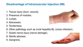
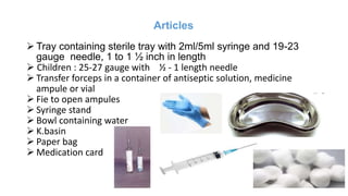
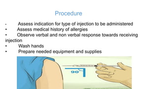
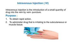
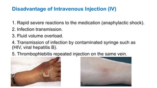
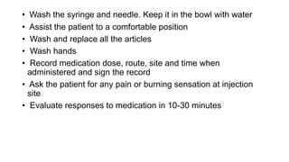
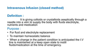
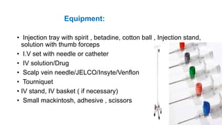
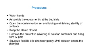
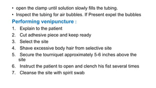
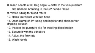
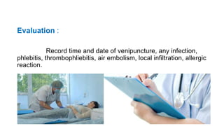
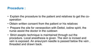
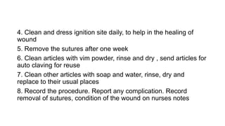
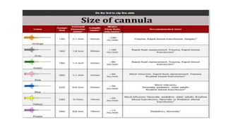
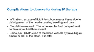
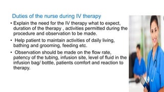
INTRAMUSCULAR
&
INTRAVENOUS INJECTION
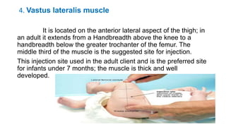
Prof.Saranya.R,M.sc(N),[Ph.D],
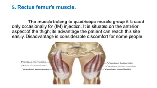
Associate Dean
Dhanalakshmik Srinivasan University
OBJECTIVES
At the end of the session the learners will be able to
• define Intramuscular injection
• enlist the sites of intramuscular injection
• describe the purpose of intramuscular injection
• explain about the procedure of intramuscular injection
• enumerate the advantages and disadvantages of IM injection
• define intravenous injection
• enlist the purpose of I.V injection
• explain the purpose of I.V injection
• describe the procedure of I.V injection
• enlist the advantages and disadvantages of I.V injection
• define intravenous infusion
• enlist the purpose of intravenous infusion
• list down the articles of intravenous infusion
• explain the procedure of intravenous infusion
• define venesection procedure
• explain the site of venesection procedure
• enlist the indication of venesection procedure
• describe the venesection procedure
• enumerate the after care
• list down the complication of venesection procedure
• describe the nurses responsibility
Intramuscular Injection( IM)
Definition :
Insertion of a needle into the muscle tissue to administer
medication.
1. Ventrogluteal muscle.
It lies over the gluteus minimums. Involves the gluteus
Medias and minimums it is situated deep and away from major
nerves and blood vessels and is a safe site for all clients. It's the
preferred injection site for adults and for children younger than 7
months. it preferred site for (IM) injection.
2. Dorsogluteal muscle
It is composed of the thick gluteal muscle of the
buttocks. The muscle is in the posterior superior iliac spine, the
injection site then lateral and superior site by positioning the
patient on prone position or side – lying position. The
Dorsogluteal site can be used for adult and children with well
develop gluteal muscle witch develop by walking
3. Deltoid muscle
 The deltoid muscle is in the edge of the acromion
process, which forms the base of a triangle in line with the midpoint
of the lateral aspect of the upper arm.
 The injection site is in the center of the triangle, about 2.5
to 5 cm (1to 2 inches) below the acromion process Should be use
this site only for small medication volumes (0.5 to 1.0ml) and when
other sites are inaccessible because of dressings or casts.
4. Vastus lateralis muscle
It is located on the anterior lateral aspect of the thigh; in
an adult it extends from a Handbreadth above the knee to a
handbreadth below the greater trochanter of the femur. The
middle third of the muscle is the suggested site for injection.
This injection site used in the adult client and is the preferred site
for infants under 7 months; the muscle is thick and well
developed.
5. Rectus femur's muscle.
The muscle belong to quadriceps muscle group it is used
only occasionally for (IM) injection. It is situated on the anterior
aspect of the thigh; its advantage the patient can reach this site
easily. Disadvantage is considerable discomfort for some people.
Purpose
• Provide the means of administration for medication that cannot
be given orally.
• To provide the needed effect when the patient is unconscious ,
unable to swallow due to neurological or surgical alteration
effecting the throat and mouth or when the patient is un co-
operative.
Advantage of Intramuscular Injection (IM)
1. The muscle is less sensitive to irritating and viscous drugs.
2. Adult client can safely tolerate as much as 4 ml of medication
in larger muscles such as the gluteus medius without discomfort
than subcutaneous tissue.
3. Safe method than other parenteral administration route.
4. Slow action of medication can be achieved by this route of
administration.
5. Some medication need to absorbed slowly and harm if given
intravenous such as oily hormone, long acting penicillin.
Disadvantage of Intramuscular Injection (IM)
1. Tissue injury (burn, wound).
2. Presence of nodules.
3. Lumps.
4. Abscesses.
5. Tenderness.
6. Other pathology such as (viral hepatitis B), (cross infection) .
7. Sciatic nerve injury (nerve damage).
8. Sterile abscess.
9. Gangrene.
Articles
 Tray containing sterile tray with 2ml/5ml syringe and 19-23
gauge needle, 1 to 1 ½ inch in length
 Children : 25-27 gauge with ½ - 1 length needle
 Transfer forceps in a container of antiseptic solution, medicine
ampule or vial
 Fie to open ampules
 Syringe stand
 Bowl containing water
 K.basin
 Paper bag
 Medication card
Procedure
• Assess indication for type of injection to be administered
• Assess medical history of allergies
• Observe verbal and non verbal response towards receiving
injection
• Wash hands
• Prepare needed equipment and supplies
• Check medication order
• Prepare correct medication dosage from ampule or vial ,
Check dosage carefully. Be sure air is expelled.
• Prepare an air lock by drawing 0.2cc air in syringe
• Keep the syringe over syringe stand after loading
• Identify patient by checking identification arm band and
asking patients name
• Explain procedure to client and proceed in calm confident manner
• Close room door or curtain
• Keep sheet or gown draped over body parts not
requiring exposure
• Select appropriate injection site
• Inspect skin surface over sites for bruises , inflammation
or edema. Note integrity and size of muscleand palpate
for tenderness
• Assist patient to comfortable position. Depending on the
site chosen, the patient lies flat, on his side or pone or sits
• Relocate site using anatomical land marks in the deltoid,
ventro gluteal, dorsogluteal, vastus lateralis or rectus
femoris regions
• Cleanse site with antiseptic swab. Apply the swab at the centre
of the site and rotate outward in a circular direction for about 5
cm
• Hold swab between 3rd and 4th fingers of non-dominant hand
• Remove needle cap from needle by pulling it straight off
• Hold syringe correctly between thumb and fore finger of
dominant hand hold as dart with palm down
• Administration of injection :
• Position non-dominant hand at proper anatomical landmarks
and spread skin tightly. Inject needle quickly at 90 degree angle
into muscle
• If patient muscle mass is small grasp body of muscle between
thumb and other fingers
• If medication is irritating use ‘z’ tract method . Here the nurse
pulls the overlying skin and subcutaneous tissues approx.. 2.5 to
3.5cm laterally to the side. Holding the skin taut with the non-
dominant hand the nurse injects the needle deep into the muscle
and after injecting the nurse release the skin.
• After the needle enters site of injection grasp lower end of
syringe barrel with non-dominant hand. More dominant hand to
end of plunger
• Avoid moving syringe while slowly pulling back on plunger to
aspirate drugs. If blood appears in the syringe remove needle
discard medication and syringe and repeat procedure.
• Inject medication slowly
• Withdraw needle while applying alcohol swab gently above
injection site
• Massage skin lightly
• Assist patient to comfortable position
• Flush out the syringe and needle and keep it in the bowl with
water
• Wash and replace all articles
• Wash hands
• Chart medication , dose route and site , time and date given in
medication sheet
• Ask patient for acute pain, burning, numbness or tingling at
injection site
• Evaluate responses to medication in 10-30 mins
Intravenous Injection ( IV)
Intravenous injection is the introduction of a small quantity of
drug into the vein by veni- puncture.
Purpose :
1. To obtain rapid action.
2. To administer drug that is irritating to the subcutaneous or
muscle tissue.
Advantage
1. When rapid effect is required.
2. Rout is appropriate when medications are too irritating to the
tissue when given by other routes.
3. When there are contraindications to give medication by other
rout such as abscesses on gluteal muscles occur.
4. When large volume infusion or medication are indicated.
5. When there are multi dose of drug administration for long
period.
6. Easy to perform venipuncture's by needle to administer
medication.
Disadvantage of Intravenous Injection (IV)
1. Rapid severe reactions to the medication (anaphylactic shock).
2. Infection transmission.
3. Fluid volume overload.
4. Transmission of infection by contaminated syringe such as
(HIV, viral hepatitis B).
5. Thrombophlebitis repeated injection on the same vein.
Procedure :
• Check physicians order for type of medication to be
administered, dosage and route
• Wash hands
• Prepare needed equipment and supplies:
Tray containing
 Sterile tray containing syringe & needle 18-21 gauge 1”-2”
needle length
 Bowl containing cotton swab
 Antiseptic solution
 Paper bag
 Medication card & k.basin
• Check the medication record
• prepare ordered medication as per direction for dilution. Load
medication in syringe and keep it over the syringe stand
• Identify patient by checking arm band or asking name
• Explain procedure to the patient
• Assess intravenous insertion site
• Assess patient to comfortable position. Ask patient to extend
his elbow and support it on a flat surface
• Talk with the patient about subject of interest.
• Apply tourniquet about 6-8 cm proximal to injection site
• Massage or stroke the vein distal to the knot and in the
direction of the venous flow
• Encourage the patient to clench and unclench the first rapidly
• Lightly tap the vein with antiseptic swab
• Hold swab between third and fourth finger of non dominant
hand
• Remove cap from needle by pulling it straight off
• For insertion of needle into the vein grasp the arm distally to
the point of entry of the needle. Place left thumb one inch below
the expected point of entry. Pull the site taut.
• Holding the needle at a 30 deg. Angle with the bevel up, pierce
the skin lateral to the vein, lower the angle of the needle so it
becomes parallel with the skin
• Follow the course of vein and pierce the side of vein
• When the back flow of blood occurs, insert the needle further
into the vein about ¾”-1”
• Release the tourniquet
• Inject the medication slowly
• Watch for swelling at and around the site of injection
• Withdraw the needle quickly after injection of medication
• Apply pressure over the injection site, do not massage
• Wash the syringe and needle. Keep it in the bowl with water
• Assist the patient to a comfortable position
• Wash and replace all the articles
• Wash hands
• Record medication dose, route, site and time when
administered and sign the record
• Ask the patient for any pain or burning sensation at injection
site
• Evaluate responses to medication in 10-30 minutes
Intravenous Infusion (closed method)
Definition :
It is giving colloids or crystalloids aseptically through a
needle into a vein to supply the body with fluids electrolyte,
nutrients and medication
Purpose :
• For fluid and electrolyte replacement
• To maintain homeostatic balance
• When a change in the patient condition is anticipated the I.V
line is maintained at a keep open state to instill
fluids/medication at the time of emergency.
Equipment:
• Injection tray with spirit , betadine, cotton ball , Injection stand,
solution with thumb forceps
• I.V set with needle or catheter
• IV solution/Drug
• Scalp vein needle/JELCO/Insyte/Venflon
• Tourniquet
• IV stand, IV basket ( if necessary)
• Small mackintosh, adhesive , scissors
Procedure:
• Wash hands
• Assemble the equipment's at the bed side
• Open the administration set and tubing maintaining sterility of
contents
• Keep the clamp closed
• Remove the protective covering of solution container and hang
from IV pole
• Squeeze flexible drip chamber gently. Until solution enters the
chamber
• open the clamp until solution slowly fills the tubing.
• Inspect the tubing for air bubbles. If Present expel the bubbles
Performing venipuncture :
1. Explain to the patient
2. Cut adhesive piece and keep ready
3. Select the site
4. Shave excessive body hair from selective site
5. Secure the tourniquet approximately 5-6 inches above the
site
6. Instruct the patient to open and clench his fist several times
7. Cleanse the site with spirit swab
8. Insert needle at 30 Deg angle ½ distal to the vein puncture
site Connect IV tubing to the S/V needle /Jelco
9. Watch tubing for blood return
10. Relax tourniquet with free hand
11. Open clamp on IV tubing and monitar drip chamber for
dripping solution
12. Inspect the puncture site for swelling discoloration
13. Secure it with the adhesive
14. Adjust the flow rate
15. Wash hands
Evaluation :
Record time and date of venipuncture, any infection,
phlebitis, thrombophliebitis, air embolism, local infiltration, allergic
reaction.
Venesection- Cut down procedure
Definition :
Whenever blood vessels become collapsed and invisible
or becomes difficult to trace a vein and to start infusion, the vein
will have to be exposed opened and blunt metal canula or fine
piece of polythene tubing is inserted into the vein to start infusion
it is called as ‘venesection’ or cut down
Site :
Usually saphenous vein over the ankle or cephalic vein over
the arm is selected.
Indications:
• To replace fluid into the circulation when a patient is in shock
and collapse
• To supply large amount of fluids rapidly i.e in cardiac arrest
• Before a major surgery where a fall in blood pressure is
anticipated
• To monitor central venous pressure
Articles :
Covered sterile tray :
• Sponge holding forceps-1
• B.P handle with knife-1
• Scissors, Small pointed-1
• Sterile rounded scissors – 1
• Mosquito forceps – 1
• Tooth forceps – 1
• Allis forceps -1
• Needle holder – 1
• Curved cutting needle – 2
• Annuerysm needle – 1 to pass the thread under the vein
• Venous section canula with stiller or polythene tubing
• 5ml syringe with needle no.20 and 22
• Cotton suture no.10
• Hole towel, towel clips
• Dressing material i.e. cotton swabs, guaze pieces and cotton
pads
• Gloves , gown and mask
Unsterile tray
• Cheatle forceps in a lotion jar
• kidney tray
• Mackintosh and draw sheet
• Tourniquet
• Adhesive tape with scissors
• spirit iodine
• Lignocaine 1 %
• IV infusion set , Screen
• Torch
• IV solution
Procedure :
• Explain the procedure to the patient and relatives to get the co-
operation
• Obtain written consent from the patient or his relatives
• Prepare the site for venesection with Dettol, iodine spirit, the
nurse assist the doctor in the cutdown
• Strict aseptic technique is maintained through out the
procedure. Local anesthesia is given. The skin is incised and
vein is exposed. An aneurysm needle is passed below the vein ,
threaded and drawn back.
• The distal end of the vein is tired and the proximal end is caught
between the thread, the vein is then cut partially between the
two ligatures. The canula is passed and the proximal ligature is
tied to keep the canula in place.
• IV drip is connected to the venesection needle. Skin is sutured
and bandaged
After care :
1. Observe the site to detect infiltration of fluid and
dislodgement of cannula
2. Restrict the movement of the patient
3. Make the patient comfortable and relaxed
4. Clean and dress ignition site daily, to help in the healing of
wound
5. Remove the sutures after one week
6. Clean articles with vim powder, rinse and dry , send articles for
auto claving for reuse
7. Clean other articles with soap and water, rinse, dry and
replace to their usual places
8. Record the procedure. Report any complication. Record
removal of sutures, condition of the wound on nurses notes
Complications to observe for during IV therapy
• Infiltration : escape of fluid into subcutaneous tissue due to
dislodgement of the needle causing swelling and pain .
• Circulation overload : The intravascular fluid compartment
contain more fluid than normal.
• Embolism : Obstruction of the blood vessels by travelling air
emboli or clot of the blood. It is fatal
Duties of the nurse during IV therapy
• Explain the need for the IV therapy what to expect,
duration of the therapy , activities permitted during the
procedure and observation to be made.
• Help patient to maintain activities of daily living,
bathing and grooming, feeding etc.
• Observation should be made on the flow rate,
patency of the tubing, infusion site, level of fluid in the
infusion bag/ bottle, patients comfort and reaction to
therapy.
Intramuscular & Intravenous Injection.pptx

Intramuscular & Intravenous Injection.pptx

  • 1.
  • 2.
    OBJECTIVES At the endof the session the learners will be able to • define Intramuscular injection • enlist the sites of intramuscular injection • describe the purpose of intramuscular injection • explain about the procedure of intramuscular injection • enumerate the advantages and disadvantages of IM injection
  • 3.
    • define intravenousinjection • enlist the purpose of I.V injection • explain the purpose of I.V injection • describe the procedure of I.V injection • enlist the advantages and disadvantages of I.V injection • define intravenous infusion • enlist the purpose of intravenous infusion • list down the articles of intravenous infusion • explain the procedure of intravenous infusion
  • 4.
    • define venesectionprocedure • explain the site of venesection procedure • enlist the indication of venesection procedure • describe the venesection procedure • enumerate the after care • list down the complication of venesection procedure • describe the nurses responsibility
  • 5.
    Intramuscular Injection( IM) Definition: Insertion of a needle into the muscle tissue to administer medication. 1. Ventrogluteal muscle. It lies over the gluteus minimums. Involves the gluteus Medias and minimums it is situated deep and away from major nerves and blood vessels and is a safe site for all clients. It's the preferred injection site for adults and for children younger than 7 months. it preferred site for (IM) injection.
  • 6.
    2. Dorsogluteal muscle Itis composed of the thick gluteal muscle of the buttocks. The muscle is in the posterior superior iliac spine, the injection site then lateral and superior site by positioning the patient on prone position or side – lying position. The Dorsogluteal site can be used for adult and children with well develop gluteal muscle witch develop by walking
  • 8.
    3. Deltoid muscle The deltoid muscle is in the edge of the acromion process, which forms the base of a triangle in line with the midpoint of the lateral aspect of the upper arm.  The injection site is in the center of the triangle, about 2.5 to 5 cm (1to 2 inches) below the acromion process Should be use this site only for small medication volumes (0.5 to 1.0ml) and when other sites are inaccessible because of dressings or casts.
  • 10.
    4. Vastus lateralismuscle It is located on the anterior lateral aspect of the thigh; in an adult it extends from a Handbreadth above the knee to a handbreadth below the greater trochanter of the femur. The middle third of the muscle is the suggested site for injection. This injection site used in the adult client and is the preferred site for infants under 7 months; the muscle is thick and well developed.
  • 11.
    5. Rectus femur'smuscle. The muscle belong to quadriceps muscle group it is used only occasionally for (IM) injection. It is situated on the anterior aspect of the thigh; its advantage the patient can reach this site easily. Disadvantage is considerable discomfort for some people.
  • 12.
    Purpose • Provide themeans of administration for medication that cannot be given orally. • To provide the needed effect when the patient is unconscious , unable to swallow due to neurological or surgical alteration effecting the throat and mouth or when the patient is un co- operative.
  • 13.
    Advantage of IntramuscularInjection (IM) 1. The muscle is less sensitive to irritating and viscous drugs. 2. Adult client can safely tolerate as much as 4 ml of medication in larger muscles such as the gluteus medius without discomfort than subcutaneous tissue. 3. Safe method than other parenteral administration route. 4. Slow action of medication can be achieved by this route of administration. 5. Some medication need to absorbed slowly and harm if given intravenous such as oily hormone, long acting penicillin.
  • 14.
    Disadvantage of IntramuscularInjection (IM) 1. Tissue injury (burn, wound). 2. Presence of nodules. 3. Lumps. 4. Abscesses. 5. Tenderness. 6. Other pathology such as (viral hepatitis B), (cross infection) . 7. Sciatic nerve injury (nerve damage). 8. Sterile abscess. 9. Gangrene.
  • 15.
    Articles  Tray containingsterile tray with 2ml/5ml syringe and 19-23 gauge needle, 1 to 1 ½ inch in length  Children : 25-27 gauge with ½ - 1 length needle  Transfer forceps in a container of antiseptic solution, medicine ampule or vial  Fie to open ampules  Syringe stand  Bowl containing water  K.basin  Paper bag  Medication card
  • 16.
    Procedure • Assess indicationfor type of injection to be administered • Assess medical history of allergies • Observe verbal and non verbal response towards receiving injection • Wash hands • Prepare needed equipment and supplies
  • 17.
    • Check medicationorder • Prepare correct medication dosage from ampule or vial , Check dosage carefully. Be sure air is expelled. • Prepare an air lock by drawing 0.2cc air in syringe • Keep the syringe over syringe stand after loading • Identify patient by checking identification arm band and asking patients name • Explain procedure to client and proceed in calm confident manner • Close room door or curtain
  • 18.
    • Keep sheetor gown draped over body parts not requiring exposure • Select appropriate injection site • Inspect skin surface over sites for bruises , inflammation or edema. Note integrity and size of muscleand palpate for tenderness • Assist patient to comfortable position. Depending on the site chosen, the patient lies flat, on his side or pone or sits • Relocate site using anatomical land marks in the deltoid, ventro gluteal, dorsogluteal, vastus lateralis or rectus femoris regions
  • 19.
    • Cleanse sitewith antiseptic swab. Apply the swab at the centre of the site and rotate outward in a circular direction for about 5 cm • Hold swab between 3rd and 4th fingers of non-dominant hand • Remove needle cap from needle by pulling it straight off • Hold syringe correctly between thumb and fore finger of dominant hand hold as dart with palm down • Administration of injection : • Position non-dominant hand at proper anatomical landmarks and spread skin tightly. Inject needle quickly at 90 degree angle into muscle
  • 20.
    • If patientmuscle mass is small grasp body of muscle between thumb and other fingers • If medication is irritating use ‘z’ tract method . Here the nurse pulls the overlying skin and subcutaneous tissues approx.. 2.5 to 3.5cm laterally to the side. Holding the skin taut with the non- dominant hand the nurse injects the needle deep into the muscle and after injecting the nurse release the skin.
  • 21.
    • After theneedle enters site of injection grasp lower end of syringe barrel with non-dominant hand. More dominant hand to end of plunger • Avoid moving syringe while slowly pulling back on plunger to aspirate drugs. If blood appears in the syringe remove needle discard medication and syringe and repeat procedure. • Inject medication slowly • Withdraw needle while applying alcohol swab gently above injection site • Massage skin lightly • Assist patient to comfortable position
  • 22.
    • Flush outthe syringe and needle and keep it in the bowl with water • Wash and replace all articles • Wash hands • Chart medication , dose route and site , time and date given in medication sheet • Ask patient for acute pain, burning, numbness or tingling at injection site • Evaluate responses to medication in 10-30 mins
  • 23.
    Intravenous Injection (IV) Intravenous injection is the introduction of a small quantity of drug into the vein by veni- puncture. Purpose : 1. To obtain rapid action. 2. To administer drug that is irritating to the subcutaneous or muscle tissue.
  • 24.
    Advantage 1. When rapideffect is required. 2. Rout is appropriate when medications are too irritating to the tissue when given by other routes. 3. When there are contraindications to give medication by other rout such as abscesses on gluteal muscles occur. 4. When large volume infusion or medication are indicated. 5. When there are multi dose of drug administration for long period. 6. Easy to perform venipuncture's by needle to administer medication.
  • 25.
    Disadvantage of IntravenousInjection (IV) 1. Rapid severe reactions to the medication (anaphylactic shock). 2. Infection transmission. 3. Fluid volume overload. 4. Transmission of infection by contaminated syringe such as (HIV, viral hepatitis B). 5. Thrombophlebitis repeated injection on the same vein.
  • 26.
    Procedure : • Checkphysicians order for type of medication to be administered, dosage and route • Wash hands • Prepare needed equipment and supplies: Tray containing  Sterile tray containing syringe & needle 18-21 gauge 1”-2” needle length  Bowl containing cotton swab  Antiseptic solution  Paper bag  Medication card & k.basin
  • 27.
    • Check themedication record • prepare ordered medication as per direction for dilution. Load medication in syringe and keep it over the syringe stand • Identify patient by checking arm band or asking name • Explain procedure to the patient • Assess intravenous insertion site • Assess patient to comfortable position. Ask patient to extend his elbow and support it on a flat surface • Talk with the patient about subject of interest.
  • 28.
    • Apply tourniquetabout 6-8 cm proximal to injection site • Massage or stroke the vein distal to the knot and in the direction of the venous flow • Encourage the patient to clench and unclench the first rapidly • Lightly tap the vein with antiseptic swab • Hold swab between third and fourth finger of non dominant hand • Remove cap from needle by pulling it straight off • For insertion of needle into the vein grasp the arm distally to the point of entry of the needle. Place left thumb one inch below the expected point of entry. Pull the site taut.
  • 29.
    • Holding theneedle at a 30 deg. Angle with the bevel up, pierce the skin lateral to the vein, lower the angle of the needle so it becomes parallel with the skin • Follow the course of vein and pierce the side of vein • When the back flow of blood occurs, insert the needle further into the vein about ¾”-1” • Release the tourniquet • Inject the medication slowly • Watch for swelling at and around the site of injection • Withdraw the needle quickly after injection of medication • Apply pressure over the injection site, do not massage
  • 30.
    • Wash thesyringe and needle. Keep it in the bowl with water • Assist the patient to a comfortable position • Wash and replace all the articles • Wash hands • Record medication dose, route, site and time when administered and sign the record • Ask the patient for any pain or burning sensation at injection site • Evaluate responses to medication in 10-30 minutes
  • 31.
    Intravenous Infusion (closedmethod) Definition : It is giving colloids or crystalloids aseptically through a needle into a vein to supply the body with fluids electrolyte, nutrients and medication Purpose : • For fluid and electrolyte replacement • To maintain homeostatic balance • When a change in the patient condition is anticipated the I.V line is maintained at a keep open state to instill fluids/medication at the time of emergency.
  • 32.
    Equipment: • Injection traywith spirit , betadine, cotton ball , Injection stand, solution with thumb forceps • I.V set with needle or catheter • IV solution/Drug • Scalp vein needle/JELCO/Insyte/Venflon • Tourniquet • IV stand, IV basket ( if necessary) • Small mackintosh, adhesive , scissors
  • 33.
    Procedure: • Wash hands •Assemble the equipment's at the bed side • Open the administration set and tubing maintaining sterility of contents • Keep the clamp closed • Remove the protective covering of solution container and hang from IV pole • Squeeze flexible drip chamber gently. Until solution enters the chamber
  • 34.
    • open theclamp until solution slowly fills the tubing. • Inspect the tubing for air bubbles. If Present expel the bubbles Performing venipuncture : 1. Explain to the patient 2. Cut adhesive piece and keep ready 3. Select the site 4. Shave excessive body hair from selective site 5. Secure the tourniquet approximately 5-6 inches above the site 6. Instruct the patient to open and clench his fist several times 7. Cleanse the site with spirit swab
  • 35.
    8. Insert needleat 30 Deg angle ½ distal to the vein puncture site Connect IV tubing to the S/V needle /Jelco 9. Watch tubing for blood return 10. Relax tourniquet with free hand 11. Open clamp on IV tubing and monitar drip chamber for dripping solution 12. Inspect the puncture site for swelling discoloration 13. Secure it with the adhesive 14. Adjust the flow rate 15. Wash hands
  • 36.
    Evaluation : Record timeand date of venipuncture, any infection, phlebitis, thrombophliebitis, air embolism, local infiltration, allergic reaction.
  • 37.
    Venesection- Cut downprocedure Definition : Whenever blood vessels become collapsed and invisible or becomes difficult to trace a vein and to start infusion, the vein will have to be exposed opened and blunt metal canula or fine piece of polythene tubing is inserted into the vein to start infusion it is called as ‘venesection’ or cut down Site : Usually saphenous vein over the ankle or cephalic vein over the arm is selected.
  • 38.
    Indications: • To replacefluid into the circulation when a patient is in shock and collapse • To supply large amount of fluids rapidly i.e in cardiac arrest • Before a major surgery where a fall in blood pressure is anticipated • To monitor central venous pressure
  • 39.
    Articles : Covered steriletray : • Sponge holding forceps-1 • B.P handle with knife-1 • Scissors, Small pointed-1 • Sterile rounded scissors – 1 • Mosquito forceps – 1 • Tooth forceps – 1 • Allis forceps -1
  • 40.
    • Needle holder– 1 • Curved cutting needle – 2 • Annuerysm needle – 1 to pass the thread under the vein • Venous section canula with stiller or polythene tubing • 5ml syringe with needle no.20 and 22 • Cotton suture no.10 • Hole towel, towel clips • Dressing material i.e. cotton swabs, guaze pieces and cotton pads • Gloves , gown and mask
  • 41.
    Unsterile tray • Cheatleforceps in a lotion jar • kidney tray • Mackintosh and draw sheet • Tourniquet • Adhesive tape with scissors • spirit iodine • Lignocaine 1 % • IV infusion set , Screen • Torch • IV solution
  • 42.
    Procedure : • Explainthe procedure to the patient and relatives to get the co- operation • Obtain written consent from the patient or his relatives • Prepare the site for venesection with Dettol, iodine spirit, the nurse assist the doctor in the cutdown • Strict aseptic technique is maintained through out the procedure. Local anesthesia is given. The skin is incised and vein is exposed. An aneurysm needle is passed below the vein , threaded and drawn back.
  • 43.
    • The distalend of the vein is tired and the proximal end is caught between the thread, the vein is then cut partially between the two ligatures. The canula is passed and the proximal ligature is tied to keep the canula in place. • IV drip is connected to the venesection needle. Skin is sutured and bandaged After care : 1. Observe the site to detect infiltration of fluid and dislodgement of cannula 2. Restrict the movement of the patient 3. Make the patient comfortable and relaxed
  • 44.
    4. Clean anddress ignition site daily, to help in the healing of wound 5. Remove the sutures after one week 6. Clean articles with vim powder, rinse and dry , send articles for auto claving for reuse 7. Clean other articles with soap and water, rinse, dry and replace to their usual places 8. Record the procedure. Report any complication. Record removal of sutures, condition of the wound on nurses notes
  • 46.
    Complications to observefor during IV therapy • Infiltration : escape of fluid into subcutaneous tissue due to dislodgement of the needle causing swelling and pain . • Circulation overload : The intravascular fluid compartment contain more fluid than normal. • Embolism : Obstruction of the blood vessels by travelling air emboli or clot of the blood. It is fatal
  • 47.
    Duties of thenurse during IV therapy • Explain the need for the IV therapy what to expect, duration of the therapy , activities permitted during the procedure and observation to be made. • Help patient to maintain activities of daily living, bathing and grooming, feeding etc. • Observation should be made on the flow rate, patency of the tubing, infusion site, level of fluid in the infusion bag/ bottle, patients comfort and reaction to therapy.