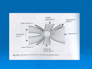
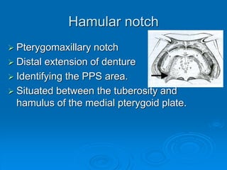
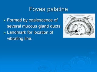
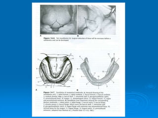
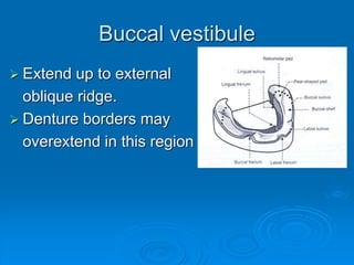
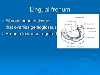
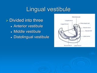
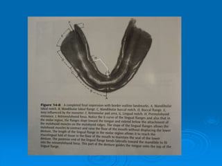
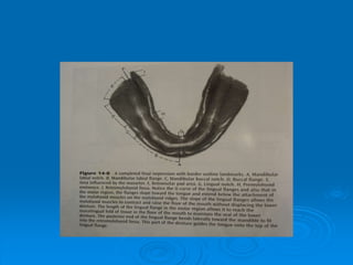
Anatomical landmarks
Introduction
 M.M Devan Dictum “Aim of a prosthodontist is
not only the meticulous replacement of what is
missing, but also perpetual preservation of
what is present”
 Complete denture must function in harmony
with the remaining natural tissues so for the
success, a through knowledge of the anatomy
is a must.
Maxillary landmarks
1. Labial frenum
2. Labial vestibule
3. Buccal frenum
4. Buccal vestibule
5. Distobuccal sulcus
6. Alveolar ridge
7. Tuberosity
8. Hamular notch
9. Hard palate (flat portions)
10. Fovea palatani
11. Mid palatine suture
12. Incissive papillae
13. Rugae
Labial frenum
 Single band of fibrous
connective tissue
 Properly relieved
Labial vestibule
 Extends from labial frenum
to buccal frenum.
 Proper lip support should be
provided.
Buccal frenum
 Consist of one or more
bands.
 Relieved
 Influenced by 3 muscles
 Orbicularis oris (forward)
 Buccinator (backward)
 Caninus (position)
Buccal vestibule
 Buccal frenum to hamular
notch
 Influenced by
 Buccinator
 Modiolus
 Coronid process of mandible
Alveolar ridge
 Ridge left after the extraction
of teeth.
 Mucosa is firmly attached to
the periosteum of the bone.
 Consist of dense collagenous fibers.
Maxillary tuberosity
 Posterior convexity
 Combination syndrome
Hamular notch
 Pterygomaxillary notch
 Distal extension of denture
 Identifying the PPS area.
 Situated between the tuberosity and
hamulus of the medial pterygoid plate.
Hard palate
 Flat areas as secondary
retentive areas.
 U shaped palate
 Flat palate (not good support)
 V shaped palate (least favorable)
Fovea palatine
 Formed by coalescence of
several mucous gland ducts.
 Landmark for location of
vibrating line.
Mid palatine suture
 Junction of palatine process
of maxilla.
 Covered by a thin mucosa
 Slightly elevated or raised
 Proper relief required.
Incisive papillae
 Pad of fiberous connective
tissue over incisive foramen
 Proper relief should be given.
 Aid in arrangement of anterior teeth and
jaw relation.
Rugae
 Resist the horizontal forces
against the denture.
 Should not be over
compressed.
Mandibular land marks
1. Labial frenum
2. Labial vestibule
3. Buccal frenum
4. Buccal vestibule
5. Buccal shelf area
6. Retromolar pad
7. Pear shaped pad
8. Lingual frenum
9. Lingual vestibule
Labial frenum
 Single narrow band
 Usually shorter and
wider than maxillary.
Labial vestibule
 Between labial and
buccal frenum
 Support to the lower lip.
Buccal frenum
 Single or more bands.
 More clearance required
Buccal vestibule
 Extend up to external
oblique ridge.
 Denture borders may
overextend in this region
Buccal shelf area
 It is the primary stress
bearing area.
 Consist of thick cortical
bone & dense connective
tissue
Retromolar pad area
 Forms the distal end of
the denture.
 Consist of loose
connective tissue
Pear shaped pad
 Aggregation of mucous
glands and loose,
connective tissue.
 Formed after residual
scar of third molar.
 Muscle attachments
 Posteriorly – temporalis tendons
 Laterally – buccinator
 Medially – pterygomandibular raphe and superior
constrictor of pharynx.
Lingual frenum
 Fibrous band of tissue
that overlies genioglossus
 Proper clearance required
Lingual vestibule
 Divided into three
 Anterior vestibule
 Middle vestibule
 Distolingual vestibule
Anterior
 Also called sublingual crescent area
 Structures present
 Genioglossus muscles
 Lingual frenum
 Anterior portion of sublingual gland
Middle
 Also called mylohyoid vestibule
 Is the largest portion of lingual vestibule
 Structures seen
 Mylohyoid muscle
 Sublingual gland
Posterior
 Also called lateral throat form or
retromylohyoid fossa
 Boundaries
 Anteriorly – mylohyoid muscle
 Laterally – pear shaped pad
 Posteriorly – retromylohyoid curtain
 Medially - tongue
Anatomic LandmarkT.PPT

Anatomic LandmarkT.PPT

  • 1.
  • 2.
    Introduction  M.M DevanDictum “Aim of a prosthodontist is not only the meticulous replacement of what is missing, but also perpetual preservation of what is present”  Complete denture must function in harmony with the remaining natural tissues so for the success, a through knowledge of the anatomy is a must.
  • 3.
    Maxillary landmarks 1. Labialfrenum 2. Labial vestibule 3. Buccal frenum 4. Buccal vestibule 5. Distobuccal sulcus 6. Alveolar ridge 7. Tuberosity 8. Hamular notch 9. Hard palate (flat portions) 10. Fovea palatani 11. Mid palatine suture 12. Incissive papillae 13. Rugae
  • 4.
    Labial frenum  Singleband of fibrous connective tissue  Properly relieved
  • 6.
    Labial vestibule  Extendsfrom labial frenum to buccal frenum.  Proper lip support should be provided.
  • 8.
    Buccal frenum  Consistof one or more bands.  Relieved  Influenced by 3 muscles  Orbicularis oris (forward)  Buccinator (backward)  Caninus (position)
  • 10.
    Buccal vestibule  Buccalfrenum to hamular notch  Influenced by  Buccinator  Modiolus  Coronid process of mandible
  • 11.
    Alveolar ridge  Ridgeleft after the extraction of teeth.  Mucosa is firmly attached to the periosteum of the bone.  Consist of dense collagenous fibers.
  • 13.
    Maxillary tuberosity  Posteriorconvexity  Combination syndrome
  • 15.
    Hamular notch  Pterygomaxillarynotch  Distal extension of denture  Identifying the PPS area.  Situated between the tuberosity and hamulus of the medial pterygoid plate.
  • 16.
    Hard palate  Flatareas as secondary retentive areas.  U shaped palate  Flat palate (not good support)  V shaped palate (least favorable)
  • 17.
    Fovea palatine  Formedby coalescence of several mucous gland ducts.  Landmark for location of vibrating line.
  • 19.
    Mid palatine suture Junction of palatine process of maxilla.  Covered by a thin mucosa  Slightly elevated or raised  Proper relief required.
  • 21.
    Incisive papillae  Padof fiberous connective tissue over incisive foramen  Proper relief should be given.  Aid in arrangement of anterior teeth and jaw relation.
  • 23.
    Rugae  Resist thehorizontal forces against the denture.  Should not be over compressed.
  • 26.
    Mandibular land marks 1.Labial frenum 2. Labial vestibule 3. Buccal frenum 4. Buccal vestibule 5. Buccal shelf area 6. Retromolar pad 7. Pear shaped pad 8. Lingual frenum 9. Lingual vestibule
  • 28.
    Labial frenum  Singlenarrow band  Usually shorter and wider than maxillary.
  • 30.
    Labial vestibule  Betweenlabial and buccal frenum  Support to the lower lip.
  • 31.
    Buccal frenum  Singleor more bands.  More clearance required
  • 33.
    Buccal vestibule  Extendup to external oblique ridge.  Denture borders may overextend in this region
  • 35.
    Buccal shelf area It is the primary stress bearing area.  Consist of thick cortical bone & dense connective tissue
  • 38.
    Retromolar pad area Forms the distal end of the denture.  Consist of loose connective tissue
  • 39.
    Pear shaped pad Aggregation of mucous glands and loose, connective tissue.  Formed after residual scar of third molar.  Muscle attachments  Posteriorly – temporalis tendons  Laterally – buccinator  Medially – pterygomandibular raphe and superior constrictor of pharynx.
  • 40.
    Lingual frenum  Fibrousband of tissue that overlies genioglossus  Proper clearance required
  • 41.
    Lingual vestibule  Dividedinto three  Anterior vestibule  Middle vestibule  Distolingual vestibule
  • 42.
    Anterior  Also calledsublingual crescent area  Structures present  Genioglossus muscles  Lingual frenum  Anterior portion of sublingual gland
  • 44.
    Middle  Also calledmylohyoid vestibule  Is the largest portion of lingual vestibule  Structures seen  Mylohyoid muscle  Sublingual gland
  • 46.
    Posterior  Also calledlateral throat form or retromylohyoid fossa  Boundaries  Anteriorly – mylohyoid muscle  Laterally – pear shaped pad  Posteriorly – retromylohyoid curtain  Medially - tongue