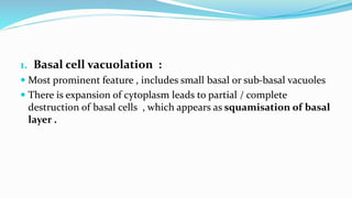
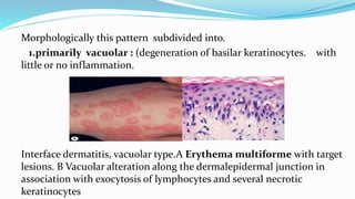
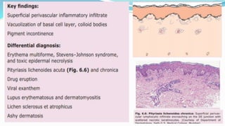
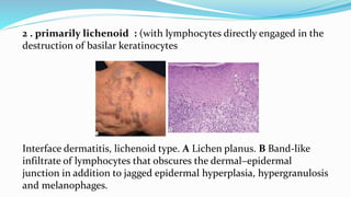
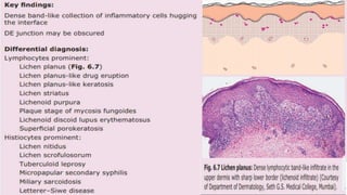
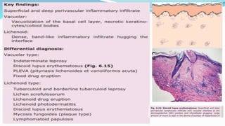
By
Dr.LAXMI PRASANNA
PG FIRST YEAR
REFERENCES :
IADVL
FITZPATRICKS
ROOKS
 Definition : Inflammation along the dermoepidermal junction associated
with Vacuolation characterizes interface dermatitis
 This common type of reaction leads to papules or plaques in some skin
diseases & bullae in others
 Interface dermatitis includes conditions in which the primary
pathology involves the "interface," i.e., the dermo-epidermal
junction. The components of this "interface" include the basal
layer of the epidermis, the dermo-epidermal junction, the
papillary dermis, and the adventitial dermis around the adnexal
struc
 Interface dermatitis includes conditions in
 Interface dermatitis include conditions in which primary pathology
involves interface i.e DEJ
 The components of interface includes basal layer of the epidermis ,
DEJ , Papillary dermis , adventitial dermis along the adnexal
structures
 Pathophysilogically apoptosis , individual cell necrosis of
keratinocytes is the most prominent feature of the primary interface
dermatitis
 T – cell mediated cytokine damage is major mechanism resulting in
disease
MORPHOLOGICAL CHANGES :
 Basal cell vacuolation
 Apoptotic keratinocytes / Colloid bodies / civatte
bodies
 Obscuring of DEJ
 Epidermal changes
 Papillary dermal changes
1. Basal cell vacuolation :
 Most prominent feature , includes small basal or sub-basal vacuoles
 There is expansion of cytoplasm leads to partial / complete
destruction of basal cells , which appears as squamisation of basal
layer .
APOPTOTIC BODIES :
 Small rounded eosinophilic hyaline anucleared structures &
slightky smaller than basal cells
 They are present individually or clumps in basal layer , papillary
dermis or spinous layer .
 Sometimes as whorls in acrosyringia & in s.c
OBSCURING OF DEJ :
 Lymphocytes are the major cells in interface dermatitis other cells
like eosinophils ,neutrophils , mast cells also seen .
 Accumulation of lymphocytes along basal layer obliterates the
distinction b/w epidermis & papillary dermis
 Inflammatory infiltrates may vary depending on disease & its stage
may be from sparse ( pauci inflammatory ) to dense band like (
lichenoid ) infiltrate
EPIDERMAL CHANGES :
 A thin flat atropic epidermis is typical for SLE
 Epidermal acanthosis with hypergranulosis & thick Compact
orthokeratotic stratum corneum is seen in LICHEN PLANUS
 Remarkable irregular hyperplasia in verrucuous discoid lupus
PAPILLARY DERMAL CHANGES :
 It undergoes expansion to accommodate inflammatory infiltrate
 The effect of long standing infiltrate results in fibrosis & sclerosis
 Dense infiltrate arranged in band like i.e lichenoid type
 2nd basal damage leads to incontinence of melanin into papillary
dermis
Morphologically this pattern subdivided into.
1.primarily vacuolar : (degeneration of basilar keratinocytes. with
little or no inflammation.
Interface dermatitis, vacuolar type.A Erythema multiforme with target
lesions. B Vacuolar alteration along the dermalepidermal junction in
association with exocytosis of lymphocytes and several necrotic
keratinocytes
2 . primarily lichenoid : (with lymphocytes directly engaged in the
destruction of basilar keratinocytes
Interface dermatitis, lichenoid type. A Lichen planus. B Band-like
infiltrate of lymphocytes that obscures the dermal–epidermal
junction in addition to jagged epidermal hyperplasia, hypergranulosis
and melanophages.
 frequently there is overlap between these two categories.
 even though an entity has lichenoid features histologically (e.g. fixed drug
eruption),
 That doesn’t mean it must resemble lichen planus clinically.
 Also, some degree of lichenoid inflammation can be associated with a
variety of benign and malignant neoplasms of the skin – e.g. solitary
lichenoid keratosis and melanoma, respectively.
THANK YOU…

interface dermatitis.pptx

  • 1.
    By Dr.LAXMI PRASANNA PG FIRSTYEAR REFERENCES : IADVL FITZPATRICKS ROOKS
  • 2.
     Definition :Inflammation along the dermoepidermal junction associated with Vacuolation characterizes interface dermatitis  This common type of reaction leads to papules or plaques in some skin diseases & bullae in others  Interface dermatitis includes conditions in which the primary pathology involves the "interface," i.e., the dermo-epidermal junction. The components of this "interface" include the basal layer of the epidermis, the dermo-epidermal junction, the papillary dermis, and the adventitial dermis around the adnexal struc  Interface dermatitis includes conditions in
  • 3.
     Interface dermatitisinclude conditions in which primary pathology involves interface i.e DEJ  The components of interface includes basal layer of the epidermis , DEJ , Papillary dermis , adventitial dermis along the adnexal structures  Pathophysilogically apoptosis , individual cell necrosis of keratinocytes is the most prominent feature of the primary interface dermatitis  T – cell mediated cytokine damage is major mechanism resulting in disease
  • 4.
    MORPHOLOGICAL CHANGES : Basal cell vacuolation  Apoptotic keratinocytes / Colloid bodies / civatte bodies  Obscuring of DEJ  Epidermal changes  Papillary dermal changes
  • 5.
    1. Basal cellvacuolation :  Most prominent feature , includes small basal or sub-basal vacuoles  There is expansion of cytoplasm leads to partial / complete destruction of basal cells , which appears as squamisation of basal layer .
  • 6.
    APOPTOTIC BODIES : Small rounded eosinophilic hyaline anucleared structures & slightky smaller than basal cells  They are present individually or clumps in basal layer , papillary dermis or spinous layer .  Sometimes as whorls in acrosyringia & in s.c
  • 7.
    OBSCURING OF DEJ:  Lymphocytes are the major cells in interface dermatitis other cells like eosinophils ,neutrophils , mast cells also seen .  Accumulation of lymphocytes along basal layer obliterates the distinction b/w epidermis & papillary dermis  Inflammatory infiltrates may vary depending on disease & its stage may be from sparse ( pauci inflammatory ) to dense band like ( lichenoid ) infiltrate
  • 8.
    EPIDERMAL CHANGES : A thin flat atropic epidermis is typical for SLE  Epidermal acanthosis with hypergranulosis & thick Compact orthokeratotic stratum corneum is seen in LICHEN PLANUS  Remarkable irregular hyperplasia in verrucuous discoid lupus
  • 9.
    PAPILLARY DERMAL CHANGES:  It undergoes expansion to accommodate inflammatory infiltrate  The effect of long standing infiltrate results in fibrosis & sclerosis  Dense infiltrate arranged in band like i.e lichenoid type  2nd basal damage leads to incontinence of melanin into papillary dermis
  • 10.
    Morphologically this patternsubdivided into. 1.primarily vacuolar : (degeneration of basilar keratinocytes. with little or no inflammation. Interface dermatitis, vacuolar type.A Erythema multiforme with target lesions. B Vacuolar alteration along the dermalepidermal junction in association with exocytosis of lymphocytes and several necrotic keratinocytes
  • 12.
    2 . primarilylichenoid : (with lymphocytes directly engaged in the destruction of basilar keratinocytes Interface dermatitis, lichenoid type. A Lichen planus. B Band-like infiltrate of lymphocytes that obscures the dermal–epidermal junction in addition to jagged epidermal hyperplasia, hypergranulosis and melanophages.
  • 14.
     frequently thereis overlap between these two categories.  even though an entity has lichenoid features histologically (e.g. fixed drug eruption),  That doesn’t mean it must resemble lichen planus clinically.  Also, some degree of lichenoid inflammation can be associated with a variety of benign and malignant neoplasms of the skin – e.g. solitary lichenoid keratosis and melanoma, respectively.
  • 16.