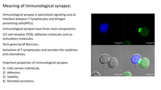
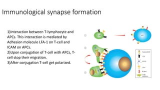
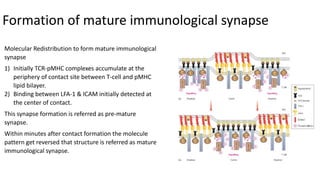
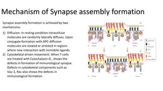
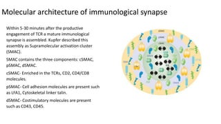
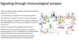
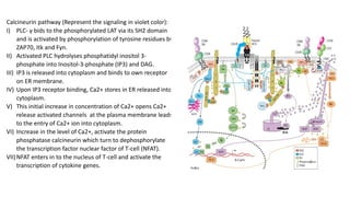
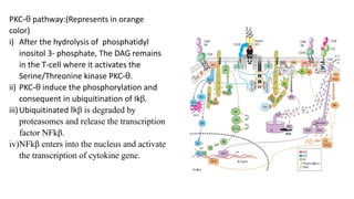
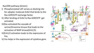
The immunological synapse is a specialized signaling structure formed at the interface between T lymphocytes and antigen presenting cells. It consists of three main components: T cell receptors, adhesion molecules, and co-stimulatory molecules. Formation of the mature immunological synapse involves molecular redistribution through diffusion and cytoskeletal movement over 5-30 minutes, resulting in a structure with central, peripheral, and distal supramolecular activation clusters that facilitate T cell signaling and activation. This signaling activates transcription factors that induce cytokine gene expression and T cell effector functions.
T-Cell Activation
• Concept of immune response
• T cell-mediated immune response
• B cell-mediated immune response
I. Concept of immune response
• A collective and coordinated response to the introduction of foreign substances in an individual mediated by the cells and molecules in the immune system.
II. T cell-mediated immune response
• Cell-mediated immunity is the arm of the adaptive immune response whose role is to combat infection of intracellular pathogens, such as intracellular bacteria (mycobacteria, listeria monocytogens), viruses, protozoa, etc.
Graduate level educational lectures on innate and adaptive immune signaling mechanisms in two parts. Part 1 focuses on Antigen Receptor Signaling with focus on TCR singaling & the Immunological Synapse. Part 2 focuses on Cytokine Receptor, Notch and Innate Immunoreceptor Signaling as well as Regulation of signal dynamics. This material is taught as part of Immunobiology (BIOM514) at the University of New Mexico School of Medicine.
T-Cell Activation
• Concept of immune response
• T cell-mediated immune response
• B cell-mediated immune response
I. Concept of immune response
• A collective and coordinated response to the introduction of foreign substances in an individual mediated by the cells and molecules in the immune system.
II. T cell-mediated immune response
• Cell-mediated immunity is the arm of the adaptive immune response whose role is to combat infection of intracellular pathogens, such as intracellular bacteria (mycobacteria, listeria monocytogens), viruses, protozoa, etc.
Richard's entangled aventures in wonderlandRichard Gill
Since the loophole-free Bell experiments of 2020 and the Nobel prizes in physics of 2022, critics of Bell's work have retreated to the fortress of super-determinism. Now, super-determinism is a derogatory word - it just means "determinism". Palmer, Hance and Hossenfelder argue that quantum mechanics and determinism are not incompatible, using a sophisticated mathematical construction based on a subtle thinning of allowed states and measurements in quantum mechanics, such that what is left appears to make Bell's argument fail, without altering the empirical predictions of quantum mechanics. I think however that it is a smoke screen, and the slogan "lost in math" comes to my mind. I will discuss some other recent disproofs of Bell's theorem using the language of causality based on causal graphs. Causal thinking is also central to law and justice. I will mention surprising connections to my work on serial killer nurse cases, in particular the Dutch case of Lucia de Berk and the current UK case of Lucy Letby.
The increased availability of biomedical data, particularly in the public domain, offers the opportunity to better understand human health and to develop effective therapeutics for a wide range of unmet medical needs. However, data scientists remain stymied by the fact that data remain hard to find and to productively reuse because data and their metadata i) are wholly inaccessible, ii) are in non-standard or incompatible representations, iii) do not conform to community standards, and iv) have unclear or highly restricted terms and conditions that preclude legitimate reuse. These limitations require a rethink on data can be made machine and AI-ready - the key motivation behind the FAIR Guiding Principles. Concurrently, while recent efforts have explored the use of deep learning to fuse disparate data into predictive models for a wide range of biomedical applications, these models often fail even when the correct answer is already known, and fail to explain individual predictions in terms that data scientists can appreciate. These limitations suggest that new methods to produce practical artificial intelligence are still needed.
In this talk, I will discuss our work in (1) building an integrative knowledge infrastructure to prepare FAIR and "AI-ready" data and services along with (2) neurosymbolic AI methods to improve the quality of predictions and to generate plausible explanations. Attention is given to standards, platforms, and methods to wrangle knowledge into simple, but effective semantic and latent representations, and to make these available into standards-compliant and discoverable interfaces that can be used in model building, validation, and explanation. Our work, and those of others in the field, creates a baseline for building trustworthy and easy to deploy AI models in biomedicine.
Bio
Dr. Michel Dumontier is the Distinguished Professor of Data Science at Maastricht University, founder and executive director of the Institute of Data Science, and co-founder of the FAIR (Findable, Accessible, Interoperable and Reusable) data principles. His research explores socio-technological approaches for responsible discovery science, which includes collaborative multi-modal knowledge graphs, privacy-preserving distributed data mining, and AI methods for drug discovery and personalized medicine. His work is supported through the Dutch National Research Agenda, the Netherlands Organisation for Scientific Research, Horizon Europe, the European Open Science Cloud, the US National Institutes of Health, and a Marie-Curie Innovative Training Network. He is the editor-in-chief for the journal Data Science and is internationally recognized for his contributions in bioinformatics, biomedical informatics, and semantic technologies including ontologies and linked data.
Earliest Galaxies in the JADES Origins Field: Luminosity Function and Cosmic ...Sérgio Sacani
We characterize the earliest galaxy population in the JADES Origins Field (JOF), the deepest
imaging field observed with JWST. We make use of the ancillary Hubble optical images (5 filters
spanning 0.4−0.9µm) and novel JWST images with 14 filters spanning 0.8−5µm, including 7 mediumband filters, and reaching total exposure times of up to 46 hours per filter. We combine all our data
at > 2.3µm to construct an ultradeep image, reaching as deep as ≈ 31.4 AB mag in the stack and
30.3-31.0 AB mag (5σ, r = 0.1” circular aperture) in individual filters. We measure photometric
redshifts and use robust selection criteria to identify a sample of eight galaxy candidates at redshifts
z = 11.5 − 15. These objects show compact half-light radii of R1/2 ∼ 50 − 200pc, stellar masses of
M⋆ ∼ 107−108M⊙, and star-formation rates of SFR ∼ 0.1−1 M⊙ yr−1
. Our search finds no candidates
at 15 < z < 20, placing upper limits at these redshifts. We develop a forward modeling approach to
infer the properties of the evolving luminosity function without binning in redshift or luminosity that
marginalizes over the photometric redshift uncertainty of our candidate galaxies and incorporates the
impact of non-detections. We find a z = 12 luminosity function in good agreement with prior results,
and that the luminosity function normalization and UV luminosity density decline by a factor of ∼ 2.5
from z = 12 to z = 14. We discuss the possible implications of our results in the context of theoretical
models for evolution of the dark matter halo mass function.
Introduction:
RNA interference (RNAi) or Post-Transcriptional Gene Silencing (PTGS) is an important biological process for modulating eukaryotic gene expression.
It is highly conserved process of posttranscriptional gene silencing by which double stranded RNA (dsRNA) causes sequence-specific degradation of mRNA sequences.
dsRNA-induced gene silencing (RNAi) is reported in a wide range of eukaryotes ranging from worms, insects, mammals and plants.
This process mediates resistance to both endogenous parasitic and exogenous pathogenic nucleic acids, and regulates the expression of protein-coding genes.
What are small ncRNAs?
micro RNA (miRNA)
short interfering RNA (siRNA)
Properties of small non-coding RNA:
Involved in silencing mRNA transcripts.
Called “small” because they are usually only about 21-24 nucleotides long.
Synthesized by first cutting up longer precursor sequences (like the 61nt one that Lee discovered).
Silence an mRNA by base pairing with some sequence on the mRNA.
Discovery of siRNA?
The first small RNA:
In 1993 Rosalind Lee (Victor Ambros lab) was studying a non- coding gene in C. elegans, lin-4, that was involved in silencing of another gene, lin-14, at the appropriate time in the
development of the worm C. elegans.
Two small transcripts of lin-4 (22nt and 61nt) were found to be complementary to a sequence in the 3' UTR of lin-14.
Because lin-4 encoded no protein, she deduced that it must be these transcripts that are causing the silencing by RNA-RNA interactions.
Types of RNAi ( non coding RNA)
MiRNA
Length (23-25 nt)
Trans acting
Binds with target MRNA in mismatch
Translation inhibition
Si RNA
Length 21 nt.
Cis acting
Bind with target Mrna in perfect complementary sequence
Piwi-RNA
Length ; 25 to 36 nt.
Expressed in Germ Cells
Regulates trnasposomes activity
MECHANISM OF RNAI:
First the double-stranded RNA teams up with a protein complex named Dicer, which cuts the long RNA into short pieces.
Then another protein complex called RISC (RNA-induced silencing complex) discards one of the two RNA strands.
The RISC-docked, single-stranded RNA then pairs with the homologous mRNA and destroys it.
THE RISC COMPLEX:
RISC is large(>500kD) RNA multi- protein Binding complex which triggers MRNA degradation in response to MRNA
Unwinding of double stranded Si RNA by ATP independent Helicase
Active component of RISC is Ago proteins( ENDONUCLEASE) which cleave target MRNA.
DICER: endonuclease (RNase Family III)
Argonaute: Central Component of the RNA-Induced Silencing Complex (RISC)
One strand of the dsRNA produced by Dicer is retained in the RISC complex in association with Argonaute
ARGONAUTE PROTEIN :
1.PAZ(PIWI/Argonaute/ Zwille)- Recognition of target MRNA
2.PIWI (p-element induced wimpy Testis)- breaks Phosphodiester bond of mRNA.)RNAse H activity.
MiRNA:
The Double-stranded RNAs are naturally produced in eukaryotic cells during development, and they have a key role in regulating gene expression .
A brief information about the SCOP protein database used in bioinformatics.
The Structural Classification of Proteins (SCOP) database is a comprehensive and authoritative resource for the structural and evolutionary relationships of proteins. It provides a detailed and curated classification of protein structures, grouping them into families, superfamilies, and folds based on their structural and sequence similarities.
(May 29th, 2024) Advancements in Intravital Microscopy- Insights for Preclini...Scintica Instrumentation
Intravital microscopy (IVM) is a powerful tool utilized to study cellular behavior over time and space in vivo. Much of our understanding of cell biology has been accomplished using various in vitro and ex vivo methods; however, these studies do not necessarily reflect the natural dynamics of biological processes. Unlike traditional cell culture or fixed tissue imaging, IVM allows for the ultra-fast high-resolution imaging of cellular processes over time and space and were studied in its natural environment. Real-time visualization of biological processes in the context of an intact organism helps maintain physiological relevance and provide insights into the progression of disease, response to treatments or developmental processes.
In this webinar we give an overview of advanced applications of the IVM system in preclinical research. IVIM technology is a provider of all-in-one intravital microscopy systems and solutions optimized for in vivo imaging of live animal models at sub-micron resolution. The system’s unique features and user-friendly software enables researchers to probe fast dynamic biological processes such as immune cell tracking, cell-cell interaction as well as vascularization and tumor metastasis with exceptional detail. This webinar will also give an overview of IVM being utilized in drug development, offering a view into the intricate interaction between drugs/nanoparticles and tissues in vivo and allows for the evaluation of therapeutic intervention in a variety of tissues and organs. This interdisciplinary collaboration continues to drive the advancements of novel therapeutic strategies.
2. Meaning of Immunological synapse
Immunological synapse formation
Formation of mature immunological synapse
Mechanism of Synapse assembly formation
Molecular architecture of immunological synapse
Signaling through immunological synapse
Conclusion
Reference
Overview
3. Immunological synapse is specialized signaling area at
interface between T-lymphocytes and Antigen
presenting cells(APCs).
Immunological synapse have three main components:
1)T-cell receptor (TCR), Adhesion molecules and co-
stimulatory molecules.
Term given by M Norcross .
Activation of T-lymphocytes and secretes the cytokines
and chemokines.
Meaning of Immunological synapse:
Important properties of immunological synapse:
1) Cells remain individuals.
2) Adhesion.
3) Stability.
4) Directed secretions.
4. Immunological synapse formation
1)Interaction between T-lymphocyte and
APCs. This interaction is mediated by
Adhesion molecule LFA-1 on T-cell and
ICAM on APCs.
2)Upon conjugation of T-cell with APCs, T-
cell stop their migration.
3)After conjugation T-cell get polarized.
5. Molecular Redistribution to form mature immunological
synapse
1) Initially TCR-pMHC complexes accumulate at the
periphery of contact site between T-cell and pMHC
lipid bilayer.
2) Binding between LFA-1 & ICAM initially detected at
the center of contact.
This synapse formation is referred as pre-mature
synapse.
Within minutes after contact formation the molecule
pattern get reversed that structure is referred as mature
immunological synapse.
Formation of mature immunological synapse
6. Synapse assembly formation is achieved by two
mechanisms:
1) Diffusion: In resting condition intracellular
molecules are randomly laterally diffuses. Upon
conjugate formation with APC-diffusion
molecules are slowed or arrested in regions
where new interaction with immobile ligands.
2) Cytoskeletal driven movement: When T-cells
are treated with Cytochalasin-D , shows the
defects in formation of immunological synapse.
Defects in cytoskeletal components such as
Vau-1, Rac also shows the defects in
immunological formation.
Mechanism of Synapse assembly formation
7. Within 5-30 minutes after the productive
engagement of TCR a mature immunological
synapse is assembled. Kupfer described this
assembly as Supramolecular activation cluster
(SMAC).
SMAC contains the three components: cSMAC,
pSMAC, dSMAC.
cSMAC- Enriched in the TCRs, CD2, CD4/CD8
molecules.
pSMAC- Cell adhesion molecules are present such
as LFA1, Cytoskeletal linker talin.
dSMAC- Costimulatory molecules are present
such as CD43, CD45.
Molecular architecture of immunological synapse
8. Signaling through immunological synapse
TCR is a multimeric protein complex is made up of six different
subunits α, β, γ, δ, ε and ζ.
α, β heterodimer is responsible for the Antigen recognition.
CD3 chains (γ, δ, ε) and ζ homodimer for signal transduction.
TCR when engaged with pMHC-II, the cytoplasmic domains of
the CD3 subunits and ζ homodimer get phosphorylated by p56-
Lck and Fyn.
ZAP70 is activated by PTK, phosphorylation of TCR.
ZAP70 activate the LAT(Linker activation of T-cell) and
phosphorylated LAT serves as docking site for multiple adaptor
molecules that help in the expression of cytokine gene.
Activation of LAT activates the four different intracellular
transduction pathway for expression of cytokine gene.
9. Calcineurin pathway (Represent the signaling in violet color):
I) PLC- γ bids to the phosphorylated LAT via its SH2 domain
and is activated by phosphorylation of tyrosine residues by
ZAP70, Itk and Fyn.
II) Activated PLC hydrolyses phosphatidyl inositol 3-
phosphate into Inositol-3-phosphate (IP3) and DAG.
III) IP3 is released into cytoplasm and binds to own receptor
on ER membrane.
IV) Upon IP3 receptor binding, Ca2+ stores in ER released into
cytoplasm.
V) This initial increase in concentration of Ca2+ opens Ca2+
release activated channels at the plasma membrane leads
to the entry of Ca2+ ion into cytoplasm.
VI) Increase in the level of Ca2+, activate the protein
phosphatase calcineurin which turn to dephosphorylate
the transcription factor nuclear factor of T-cell (NFAT).
VII)NFAT enters in to the nucleus of T-cell and activate the
transcription of cytokine genes.
10. PKC-θ pathway:(Represents in orange
color)
i) After the hydrolysis of phosphatidyl
inositol 3- phosphate, The DAG remains
in the T-cell where it activates the
Serine/Threonine kinase PKC-θ.
ii) PKC-θ induce the phosphorylation and
consequent in ubiquitination of Ikβ.
iii)Ubiquitinated Ikβ is degraded by
proteasomes and release the transcription
factor NFkβ.
iv)NFkβ enters into the nucleus and activate
the transcription of cytokine gene.
11. Ras/ERK pathway (Green):
I) Phosphorylated LAT serves as docking site
for adopter molecule Grb2 that binds to the
Ras-GDP/GTP exchange factor.
II) After binding of Grb2 to Ras-GDP/GTP get
activated.
III)Ras activated the cascade of
serine/threonine kinase that leads to the
activation of MAP-kinase(Erk1/2).
IV)Erk1/2 activation leads to the expression of
Fos.
V) Fos helps in the expression of cytokine gene.
12. Conclusion
Immunological synapse has no specification in function
but is simply the manifestation of signal transmission
between APCs and T-cell.
IS play role in the activation of T-cell by which activated T-
cells shows the effect of cellular mode of immunity as well
as humoral immunity.