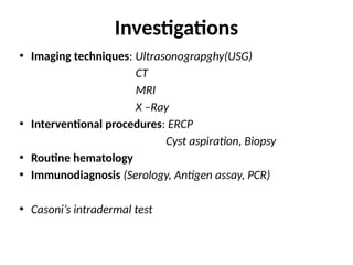
Dr. Zahid Iqbal Mir, MBBS, MS (General Surgery), DNB (General Surgery) has done his bachelors and masters in General Surgery from the prestigious Govt Medical College Jammu and DNB in General Surgery from NBEMS New Delhi. He is a passionate surgeon, earlier practising at Government Medical College, Jammu as Registrar in Department of General Surgery. Nowadays working as Senior Resident in Department of General Surgery, Government Medical College & Hospital, Sector 32, Chandigarh and a rising name in field of surgery.
He is an enthusiastic, enigmatic and dedicated teacher as well. He is not just a resolute learner, but also an awe inspiring guiding light for his juniors, which makes him the most loveable and respected senior.
4. Ultrasonography
• Diagnostic investigation of choice
• Sensitivity for evaluation is 90-95%
• It can detect cyst, localise the cyst and can be
used for aspiration purposes
• Advantage – easy availability, affordability,
diagnostic sensitivity, no radiation exposure, non
invasive.
5. Pathognomic ultrasound diagnostic features
✓Unmistakable daughter cysts ‘rosettes’
within the cyst cavity
✓Detachment of the membrane of the cyst
‘double-contoured membrane’ – floating
membrane ‘water lily sign’
✓Agglomeration of daughter cysts in the
dependent portion of a cyst – hydatid sand
✓Calcification of the cyst wall.
6. A well-defined cystic lesion measuring 8.0 x 6.5 x 7.4 cm in segment 7 and 8 with low-
level internal echoes. The cyst shows a detached, irregular laminated membrane
inside it which appears to float within the contents of the cyst.
7. USG in hydatid disease-
✓ Screening in endemic areas and family members in
infested families
✓ Staging and treatment planning
✓ First-line diagnostics
✓ Interventional, nonoperative treatment procedures
✓ intraoperative US
✓ Treatment monitoring and postoperative follow-up
✓ Workup of patients presenting with jaundice
( daughter cysts vs stones )
8. HASSEN GHARBI’S classification of hydatid cyst (1981)
Type I – Pure fluid collection
Type II – Fluid collection with split wall
Type III – Fluid collection with septa
Type IV – Heterogenous appearance
Type V – Solid cyst with calcified wall
• Type I, II, V – diagnostic for liver hydatid disease.
• In endemic area type I &V - cystic hydatid disease; type IV – diagnostic problem
9. WHO – IWGE USG CLASSIFICATION
✓ Group 1 : Active group – Cyst larger than 2 cm and
often fertile.
✓ Group 2 : Transition group – Cyst starting to
degenerate because of host resistance or
treatment ; may contain viable protoscolices.
✓ Group 3 : Inactive group – Degenerated partially or
totally calcified cyst ; unlikely to contain viable
protoscolices.
10. WHO
IWGE
USG FEATURES STATU
S
REMARKS GHARBI
S
CL • Signs not
pathognomic
• Unilocular
• No cyst wall
Active • Early stage
• Not fertile
• D/D
necessary
-
CE1 • Unilocular
• Anechoic cystic
lesion
with double line sign
Active • Usually fertile I
CE2 • Multiseptated
“rosette like”
“honeycomb” cyst
Active • Usually fertile III
11. WHO
IWGE
USG FEATURES STATUS REMARKS GHARBI
S
CE3
A
• Cyst with detached
membrane
“water lilly sign”
- floating membrane
• Less round
( decreased intracystic
pressure)
Transitional II
CE3
B
• Cyst with daughter cyst
in solid matrix
Transitional -
CE4 • Cyst with heterogenous
hypoechoic or
hyperechoic contents
• No daughter cyst
Inactive Usually
no living
protoscoleces
✓ D/D
necessary
IV
12.
13. How IOUS affects operative
strategy?
✓ Mapping
✓ Multiple cysts
✓ When atrophy / hypertrophy complex
distorts the anatomy
✓ In reoperations for recurrence
✓ Localization and management of
small, nonpalpable cysts deep in liver
parenchyma
✓ Examination of the EHBR
✓ Search for exogenous vesiculation,
especially if nonresectional surgery is
performed.
✓ Detection of CBC
14. When to suspect CBC on USG ?
✓Dead cyst (heterogeneous US pattern of
the cyst)
✓Hydatid material in the biliary tree
✓Sign of "lost continuity" of the bile duct
adjacent to the cyst wall
✓Gas in the cyst.
15. Computed tomography
• Most accurate in determining number, position, cyst
characteristics and extent of intra-abdominal disease.
• Discontinuity of the cyst wall in the vicinity of bile ducts
is highly suggestive of CBC.
• Demonstrates exogenous vesiculation - cause of
recurrence
• Characteristically show “Cart wheel” – multivesicular
rossete like pattern.
• Detecting transdiaphragmatic extension and for
assessing the thoracic component
• Used for monitoring lesions during therapy and to detect
reccurences.
17. CT of a 50 year old woman showing hepatic hydatid cyst
and the serpentine membrane within the cyst
18. MRI & MRCP
• High quality images of liver and abdominal hydatidosis
• MRI & MRCP indicated in
✓ Subdiaphragmatic site
✓ Disseminated disease
✓ Extra abdominal location
✓ Complicated, symptomatic cysts , CBC
✓ Jaundiced patients
• Skeletal , vertebral disease and in cardiac cysts
• MR angiography –large cyst , in close proximity to major
vessels or when the patient has been previously operated
several times for hydatid disease and has distorted liver
anatomy
20. MRI showing large fluid cyst within the liver containing
multiple septations
21. XRAY
• Limited value
• Endemic area- Elevation of rt.
hemidiaphragm is suggestive
• Plain X-ray may demonstrate
speckled calcification within a
cyst but only in later stages.
22. ERCP
• Little value in asymptomatic patients - avoided.
• Indications for endoscopic papillotomy in the
preoperative period are
✓ Hydatid material in the CBD
✓ Cholangitis regardless of whether a CBC is
detected
✓ Biliary fistula lasting longer than 3 weeks
✓ High output biliary fistula (>1,000 mL / 24 hours)
✓ Jaundice
✓ Short stricture obstructing the papilla
25. Immunological tests
✓IHA
✓Indirect immunoflourescence
✓Latex agglutination
✓immunoelectrophoresis
✓RIA
✓ELISA
✓ELIEDA
✓IHA
✓Indirect immunoflourescence
✓Latex agglutination
✓immunoelectrophoresis
✓RIA
✓ELISA
✓ELIEDA
✓Detection of precipitation
line (arc 5)
✓Identification of IgG
✓Immunoblot.
✓PCR
✓Detection of precipitation
line (arc 5)
✓Identification of IgG
✓Immunoblot.
✓PCR
• Useful for primary diagnosis and for follow-up after treatment
• Antibody detection is more sensitive than antigen detection
for diagnosis of E. granulosus
26.
27. ALOGRITHM FOR SEROLOGY
Primary test -ive , asymptomatic, imaging (cystic
echinococcosis ) – Advanced imaging, wait and
watch, repeat serology
Imaging -ive — no further investigation
Symptomatic, imaging (cystic echinococcosis ) – no
further investigation, manage
Primary test +ive, imaging (+/-), Symptoms (+/-) —
Needs Secondary tests
28. CYST ASPIRATION & BIOPSY
✓ Rarely done — in absence of a + serologic test
— presence of protoscolices, hooklets, or
hydatid membranes
✓ Risk of anaphylaxis and secondary spread of
the infection
✓ Ryan trichrome blue stain ,modified
babxy stain & Ziehl-Neelsen stain
✓ under US or CT guidance; with administration
of albendazole / praziquantel
Wet mount of contents of hydatid cyst of
liver - scolex of Echinococcus.
30. Casoni’s intradermal test
• Type 1 hypersensitivity skin test.
• Antigen in hydatid fluid is collected from
animal or human cyst and is stertilised.
• Injected (0.2ml) intradermally in one
arm and saline as control in other arm.
• wheal (5cm) in diameter with multiple
pseudopodia like projections within half
hour and fades in an hour.
➢ abandoned now due to nonspecificity.
32. ESTABLISHING THE DIAGNOSIS
• Clinical criteria:
✓ slowly growing or static cystic mass (imaging)
✓ anaphylactic reaction (leaking or ruptured cyst)
✓ incidental finding of a cyst in asymptomatic carriers or
detected by screening techniques.
• Diagnostic criteria:
✓ typical organ lesion (imaging)
✓ Specific serum antibodies (serology)
✓ histopathology or parasitology (protoscoleces or hooklets)
✓ pathognomonic macroscopic morphology