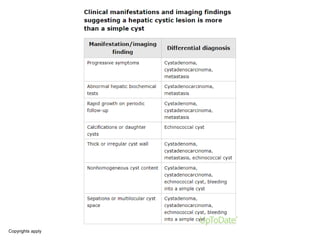
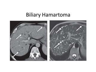
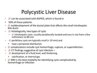
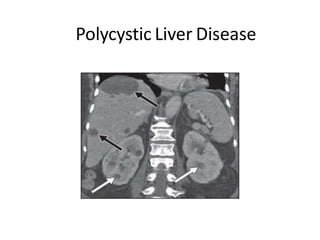
Ciste jetre, dijagnostika i liječenje
Dinko Ladić
Zavod za gastroenterologiju i
hepatologiju
KBC OSIJEK
OBJECTIVES
O review the appearance of different cystic
hepatic lesions on imaging
O propose a practical algorithm for
approaching the diagnosis
O establish a definitive diagnosis or provide
a reasonable differential diagnosis
Hepatic cystic lesions
O differential diagnoses range from benign
to malignant and potentially lethal
conditions
O key radiologic features in combination with
reviewing the clinical data important
O key imaging features
O solid component
O number and morphology
Copyrights apply
Copyrights apply
Copyrights apply
Classification
Developmental Inflammatory Neoplastic Trauma-
related
Simple cyst Pyogenic
abscess
Biliary
cystadenoma
spectrum
Biloma
Biliary
hamartoma
Amebic abscess Cystic HCC Seroma and
hematoma
Caroli disease Hydatid cyst Cystic metastasis
Polycystic liver
disease
Fungal
microabscess
Undifferentiated
embryonal
carcinoma
Ciliated foregut
duplication cyst
Intrahepatic
pseudocyst
Copyrights apply
Hepatic Cysts
O most commonly encountered hepatic lesion
O deranged development of the biliary tree (i.e., a
hamartoma of biliary origin or so-called “von
Meyenburg complex”
O no communication with the biliary tree
O ultrasound features
O well-marginated , anechoic round to ovoid
structure with an imperceptible wall
O enhancement of the posterior wall and increased
through-transmission
O CT and MRI,
O simple cysts have attenuation (0–15 HU) and
signal intensity (T1 hypointensity, T2
hyperintensity) similar to water
O Simple cysts do not show enhancement; can rarely
become complex
Hepatic Cysts
Biliary Hamartoma (von Meyenburg Complex)
O dilated small bile ducts surrounded by fibrous
stroma
O ductal plate malformation
O lack of communication with biliary system
O multiple, small (< 15 mm), round or irregular
scattered cysts with predilection for the
subcapsular region
O ultrasound findings
O Variable- might appear anechoic, hypoechoic,
or hyperechoic,sometimes comet-tai artefacts
O CT and MRI -simple cystic appearance
Biliary Hamartoma
Copyrights apply
Copyrights apply
Caroli Disease
O saccular dilatation of large intrahepatic bile ducts,
O associated with other diseases along the spectrum
of ductal plate malformations (e.g., biliary
hamartomas, polycystic liver disease, or hepatic
fibrosis), polycystic kidney disease, or renal tubular
ectasia
O Caroli disease and hepatic fibrosis is designated
as Caroli syndrome
O Caroli disease is classified as type V in the revised
Todani classification of biliary cysts
O become symptomatic by the age of 30 years
O Complications include recurrent cholangitis and
abscess, stone formation, cholangiocarcinoma,
and the development of secondary biliary cirrhosis
Caroli Disease
O multiple intrahepatic cysts of varying sizes
that communicate with the biliary system
O diffuse or localized to one segment or one
lobe, usually the left lobe
O On CT and MRI
O lesions are cystic and usually have a
central enhancing component, the “central
dot” sign, which is the portal radicle
O communication with the biliary system can
be further confirmed on cholangiography
Caroli Disease
Polycystic Liver Disease
• O can be associated with ADPKD, which is found in
• 50% of these patients
• O maldevelopment of the ductal plate that affects the small intrahepatic
bile ducts
• O Histologically, two types of cysts
• O intrahepatic cysts -usually peripherally located and vary in size from a few
millimeters to 80 mm
• O peribiliary cysts are typically small (< 10 mm) and
• have a periportal distribution
• O complications include cyst hemorrhage, rupture, or superinfection.
• O CT findings suggestive of cyst infection –
• O development of a fluid level, wall thickening,
• calcification, or internal gas
• O MRI is the best modality for identifying cysts complicated by
hemorrhage or infection
Polycystic Liver Disease
Ciliated Hepatic Foregut Duplication
Cyst
O rare congenital cystic lesion thought to arise from the
embryonic foregut- many similarities with a bronchogenic
cyst
O solitary lesion that typically measures less than 3
O most commonly located in the subcapsular aspect of segment
IV
O few cases of malignant transformation to squamous cell
carcinoma have been reported
O symptomatic, enlarging, larger than 4 cm, or containing
atypical features (e.g., solid components, thick septations)
should be resected
O ultrasound: anechoic or hypoechoic
O The cyst content ranges from clear serous fluid to mucous
fluid of different viscosities- - CT attenuation and T1 signal
intensity vary
O high signal intensity on T2-weighted imaging, no
enhancement on MRI
Ciliated Hepatic Foregut
Duplication Cyst
Pyogenic Liver
Abscess
O risk factors
O diabetes,
O gastrointestinal tract cancers,
O diverticulitis, cholangitis, cholecystitis
O recent hepatobiliary surgery or trauma
O more likely to form in the right lobe
O ultrasound- an anechoic mass with
iso- to hypoattenuating compared with background liver on the
unenhanced phase
peripheral rim of enhancement on administration of IV contrast
O well-defined or indistinct borders
O increased through transmission
O may contain echogenic debris or gas
O with Klebsiella, more likely to be solid
O CT-
O
O
O MRI
O
O
the central portion of the lesion will show low signal intensity on T1-
weighted imaging and high signal intensity on T2-weighted imaging
a peripheral halo of hyperintensity indicating edema may be seen on T2-
weighted imaging.
Copyrights apply
Amebic Liver Abscess
O amebic and pyogenic liver abscesses are virtually
indistinguishable on imaging
O diagnosis on the basis of clinical and serologic findings
O Extrahepatic disease, such as a right pleural effusion, a
pericardial effusion, or intraperitoneal rupture may suggest an
amebic abscess
O typically solitary most often in the posterior segment
O ultrasound-
O hypoechoic round or oval lesions located close to the liver
capsule
O show low-level internal echoes and posterior acoustic
enhancement
O CT-
O slightly higher attenuation than water may have smooth or
nodular borders
O thick (3–15 mm) wall that typically enhances
O MRI
O the central portion cystic,
O “rind” exhibits variable intensities on T1- and T2-weighted
imaging
Amebic Liver Abscess
Copyrights apply
Copyrights apply
Hydatid Cyst
O symptoms include pain; biliary obstruction;
superinfection; and, rarely, cyst rupture, which can
lead to anaphylactic reaction
O On imaging, the lesions present as unilocular or
multilocular cysts
O Four different radiographic appearances
O simple cyst with no internal architecture
O cyst with daughter cysts and a matrix
O calcified cyst
O complicated cyst
O The classic type is a cyst containing multiple
peripheral daughter cysts
O content of the daughter cysts is different from that of
the mother cyst- daughter cysts are usually
hypodense on CT and have a slightly different signal
intensity than the mother cyst on MR
Copyrights apply
Fungal
Microabscesses
• O typically seen in the immunocompromised population
• O small lesions(< 2 cm) and disseminated throughout the
• liver and the spleen, sometimes renal
• O Ultrasound appearance
• O “bull’s eye”: a round hyperechoic lesion with an outer hypoechoic ring
• O “wheel within a wheel”- adding a central hypoechoic dot to the bull’s eye
• O CT
• O triphasic liver imaging is most sensitive
• O most lesions being detectable on the arterial phase as uniformly
hypoattenuating
• O MRI
• O most conspicuous on the T2
• O Mimics of hepatosplenic fungal infection include granulomatous diseases
(e.g., sarcoidosis) and rarely aseptic abscesses
Fungal Microabscesses
Intrahepatic pseudocyst
O extremely rare condition- usually as a complication
of acute alcoholic pancreatitis
O affect young and middle-age men
O high propensity for the right lobe
O ancillary findings of pancreatitis is key
O spread of pancreatic enzymes and lesser sac fluid
along the hepatogastric and hepatoduodenal
ligaments or the portal triad into the liver
parenchyma intrahepatic tissue damage and
necrosis
O can spontaneously resolve or can progress to
become a pseudocyst with a fibrous capsule
O On imaging, the lesions manifest as a simple fluid
collection with an enhancing thin peripheral capsule
Biliary Cystadenoma (BCA) and Cystadenocarcinoma
(BCAC)
O more common in women: mean age at presentation is 45
years for BCA and almost 55 years for BCAC-
O the female predominance is much more pronounced in BCA
(female/male ratio- 9:1)
O arise from ectopic rests of embryonic bile ducts or aberrant
ducts- majority intrahepatic
O BCAC is usually a result of malignant transformation of BCA
(risk as high as 20%) but can also arise de novo
O imaging findings overlap
O multilocular with enhancing walls,
O fine septations, and variable calcification
O can be as large as 30 cm
O biliary ductal dilatation in the left lobe common
O Enhancing mural nodules are more common in BCAC than
BCA
O can mimic that of pyogenic abscess, amebic abscess, or
cystic metastasis
Copyrights apply
Copyrights apply
Cystic Hepatocellular Carcinoma
O Classically HCC is a hypervascular mass with
rapid washout on the portal venous phase and
an enhancing peripheral capsule
O Very rarely, may manifest as a predominantly
cystic mass with enhancing septa- an irregular
multilocular hypoattenuating lesion with a
peripheral rim of enhancement
O hypoattenuating central portion is necrosis and
the peripheral enhancing septa contain
malignant cells
O liquefactive necrosis after locoregional
treatment is a more common cause for the
cystic morphology
Cystic Hepatocellular Carcinoma
Cystic Liver Metastases
• O 10% of focal liver lesions in patients with a known primary are found to
be metastatic disease-
• O neuroendocrine tumors,
• O gastrointestinal stromal tumor (GIST),
• O lung adenocarcinoma, colorectal carcinoma,
• O transitional cell carcinoma, adenoid cystic carcinoma,
• O ovarian carcinoma, choriocarcinoma, sarcoma,
• O Cause of cystic appearance
• O high mucinous content
• O rapid growth of the tumor with hemorrhage, necrosis, or cystic
degeneration
• O cystic degeneration after chemotherapy
• O hypoattenuating to background liver on CT ,usually irregular peripheral
rim of enhancement
Cystic Liver Metastases
Undifferentiated Embryonal Sarcoma
(UES)
O highly malignant hepatic neoplasm: the pediatric age
group (typical age at presentation, 6–10 years)
O mesenchymal origin with sarcomatous features
O large (> 10 cm) solitary lesion commonly in the right lobe
O ultrasound- solid iso to hyperechoic with small anechoic
areas corresponding to areas of necrosis or cystic
degeneration
O CT
O unenhanced CT-cystic with near-water attenuation: high
water content of its myxoid stroma
O Contrast-enhanced CT can show different degrees of
enhancement
O MR
O cystic on unenhanced T1- and T2-weighted sequences
O heterogeneous enhancement post contrast -better seen in
the late portal venous phase
Undifferentiated Embryonal Sarcoma
O Post trauma or iatrogenic injury ,collection of
O bile-biloma,
O lymph- seroma
O blood- hematoma
O Inflammatory response leads to pseudocapsule
formation
O On imaging,
O seromas and bilomas appear as a simple fluid collection
that may or may not show a thin rim of enhancement
O Hematomas, on the other hand, have different density
and intensity based on the age of the blood products.
O GRE T2-weighted sequence—the most sensitive
method for detecting blood products
Mimics
• O Pseudoaneurysm
• O cystic on ultrasound and on unenhanced CT
• O vascular nature established on color and
• spectral Doppler imaging.
• O Contrast-enhanced CT and MRI show enhancement similar
to the blood pool.
• O Focal Steatosis
• O nodular steatosis rarely can mimic a cystic lesion on
unenhanced CT
• O MRI with the use of chemical-shift gradient- echo imaging
Mimics
O Peribiliary Cyst
O obstruction of the neck of the periductal glands
O high association with cirrhosis, portal
hypertension, and AD polycystic disease
O lesions usually increase in size and number as
cirrhosis and portal hypertension progress
O located along the portal tracts in the hilum and
adjacent to the large intrahepatic ducts
O discrete, clustered, or confluent
O The confluent type can mimic biliary ductal
dilatation
O Ultrasound can depict the thin septa between the
cysts to differentiate them fromprimary sclerosing
cholangitis
Peribiliary Cyst
Hvala Vam na pažnji

ciste1.pptx

  • 1.
    Ciste jetre, dijagnostikai liječenje Dinko Ladić Zavod za gastroenterologiju i hepatologiju KBC OSIJEK
  • 2.
    OBJECTIVES O review theappearance of different cystic hepatic lesions on imaging O propose a practical algorithm for approaching the diagnosis O establish a definitive diagnosis or provide a reasonable differential diagnosis
  • 3.
    Hepatic cystic lesions Odifferential diagnoses range from benign to malignant and potentially lethal conditions O key radiologic features in combination with reviewing the clinical data important O key imaging features O solid component O number and morphology
  • 4.
  • 5.
  • 6.
  • 7.
    Classification Developmental Inflammatory NeoplasticTrauma- related Simple cyst Pyogenic abscess Biliary cystadenoma spectrum Biloma Biliary hamartoma Amebic abscess Cystic HCC Seroma and hematoma Caroli disease Hydatid cyst Cystic metastasis Polycystic liver disease Fungal microabscess Undifferentiated embryonal carcinoma Ciliated foregut duplication cyst Intrahepatic pseudocyst
  • 8.
  • 9.
    Hepatic Cysts O mostcommonly encountered hepatic lesion O deranged development of the biliary tree (i.e., a hamartoma of biliary origin or so-called “von Meyenburg complex” O no communication with the biliary tree O ultrasound features O well-marginated , anechoic round to ovoid structure with an imperceptible wall O enhancement of the posterior wall and increased through-transmission O CT and MRI, O simple cysts have attenuation (0–15 HU) and signal intensity (T1 hypointensity, T2 hyperintensity) similar to water O Simple cysts do not show enhancement; can rarely become complex
  • 10.
  • 11.
    Biliary Hamartoma (vonMeyenburg Complex) O dilated small bile ducts surrounded by fibrous stroma O ductal plate malformation O lack of communication with biliary system O multiple, small (< 15 mm), round or irregular scattered cysts with predilection for the subcapsular region O ultrasound findings O Variable- might appear anechoic, hypoechoic, or hyperechoic,sometimes comet-tai artefacts O CT and MRI -simple cystic appearance
  • 12.
  • 13.
  • 14.
  • 15.
    Caroli Disease O sacculardilatation of large intrahepatic bile ducts, O associated with other diseases along the spectrum of ductal plate malformations (e.g., biliary hamartomas, polycystic liver disease, or hepatic fibrosis), polycystic kidney disease, or renal tubular ectasia O Caroli disease and hepatic fibrosis is designated as Caroli syndrome O Caroli disease is classified as type V in the revised Todani classification of biliary cysts O become symptomatic by the age of 30 years O Complications include recurrent cholangitis and abscess, stone formation, cholangiocarcinoma, and the development of secondary biliary cirrhosis
  • 16.
    Caroli Disease O multipleintrahepatic cysts of varying sizes that communicate with the biliary system O diffuse or localized to one segment or one lobe, usually the left lobe O On CT and MRI O lesions are cystic and usually have a central enhancing component, the “central dot” sign, which is the portal radicle O communication with the biliary system can be further confirmed on cholangiography
  • 17.
  • 18.
    Polycystic Liver Disease •O can be associated with ADPKD, which is found in • 50% of these patients • O maldevelopment of the ductal plate that affects the small intrahepatic bile ducts • O Histologically, two types of cysts • O intrahepatic cysts -usually peripherally located and vary in size from a few millimeters to 80 mm • O peribiliary cysts are typically small (< 10 mm) and • have a periportal distribution • O complications include cyst hemorrhage, rupture, or superinfection. • O CT findings suggestive of cyst infection – • O development of a fluid level, wall thickening, • calcification, or internal gas • O MRI is the best modality for identifying cysts complicated by hemorrhage or infection
  • 19.
  • 20.
    Ciliated Hepatic ForegutDuplication Cyst O rare congenital cystic lesion thought to arise from the embryonic foregut- many similarities with a bronchogenic cyst O solitary lesion that typically measures less than 3 O most commonly located in the subcapsular aspect of segment IV O few cases of malignant transformation to squamous cell carcinoma have been reported O symptomatic, enlarging, larger than 4 cm, or containing atypical features (e.g., solid components, thick septations) should be resected O ultrasound: anechoic or hypoechoic O The cyst content ranges from clear serous fluid to mucous fluid of different viscosities- - CT attenuation and T1 signal intensity vary O high signal intensity on T2-weighted imaging, no enhancement on MRI
  • 21.
  • 22.
    Pyogenic Liver Abscess O riskfactors O diabetes, O gastrointestinal tract cancers, O diverticulitis, cholangitis, cholecystitis O recent hepatobiliary surgery or trauma O more likely to form in the right lobe O ultrasound- an anechoic mass with iso- to hypoattenuating compared with background liver on the unenhanced phase peripheral rim of enhancement on administration of IV contrast O well-defined or indistinct borders O increased through transmission O may contain echogenic debris or gas O with Klebsiella, more likely to be solid O CT- O O O MRI O O the central portion of the lesion will show low signal intensity on T1- weighted imaging and high signal intensity on T2-weighted imaging a peripheral halo of hyperintensity indicating edema may be seen on T2- weighted imaging.
  • 23.
  • 24.
    Amebic Liver Abscess Oamebic and pyogenic liver abscesses are virtually indistinguishable on imaging O diagnosis on the basis of clinical and serologic findings O Extrahepatic disease, such as a right pleural effusion, a pericardial effusion, or intraperitoneal rupture may suggest an amebic abscess O typically solitary most often in the posterior segment O ultrasound- O hypoechoic round or oval lesions located close to the liver capsule O show low-level internal echoes and posterior acoustic enhancement O CT- O slightly higher attenuation than water may have smooth or nodular borders O thick (3–15 mm) wall that typically enhances O MRI O the central portion cystic, O “rind” exhibits variable intensities on T1- and T2-weighted imaging
  • 25.
  • 26.
  • 27.
  • 28.
    Hydatid Cyst O symptomsinclude pain; biliary obstruction; superinfection; and, rarely, cyst rupture, which can lead to anaphylactic reaction O On imaging, the lesions present as unilocular or multilocular cysts O Four different radiographic appearances O simple cyst with no internal architecture O cyst with daughter cysts and a matrix O calcified cyst O complicated cyst O The classic type is a cyst containing multiple peripheral daughter cysts O content of the daughter cysts is different from that of the mother cyst- daughter cysts are usually hypodense on CT and have a slightly different signal intensity than the mother cyst on MR
  • 29.
  • 30.
    Fungal Microabscesses • O typicallyseen in the immunocompromised population • O small lesions(< 2 cm) and disseminated throughout the • liver and the spleen, sometimes renal • O Ultrasound appearance • O “bull’s eye”: a round hyperechoic lesion with an outer hypoechoic ring • O “wheel within a wheel”- adding a central hypoechoic dot to the bull’s eye • O CT • O triphasic liver imaging is most sensitive • O most lesions being detectable on the arterial phase as uniformly hypoattenuating • O MRI • O most conspicuous on the T2 • O Mimics of hepatosplenic fungal infection include granulomatous diseases (e.g., sarcoidosis) and rarely aseptic abscesses
  • 31.
  • 32.
    Intrahepatic pseudocyst O extremelyrare condition- usually as a complication of acute alcoholic pancreatitis O affect young and middle-age men O high propensity for the right lobe O ancillary findings of pancreatitis is key O spread of pancreatic enzymes and lesser sac fluid along the hepatogastric and hepatoduodenal ligaments or the portal triad into the liver parenchyma intrahepatic tissue damage and necrosis O can spontaneously resolve or can progress to become a pseudocyst with a fibrous capsule O On imaging, the lesions manifest as a simple fluid collection with an enhancing thin peripheral capsule
  • 33.
    Biliary Cystadenoma (BCA)and Cystadenocarcinoma (BCAC) O more common in women: mean age at presentation is 45 years for BCA and almost 55 years for BCAC- O the female predominance is much more pronounced in BCA (female/male ratio- 9:1) O arise from ectopic rests of embryonic bile ducts or aberrant ducts- majority intrahepatic O BCAC is usually a result of malignant transformation of BCA (risk as high as 20%) but can also arise de novo O imaging findings overlap O multilocular with enhancing walls, O fine septations, and variable calcification O can be as large as 30 cm O biliary ductal dilatation in the left lobe common O Enhancing mural nodules are more common in BCAC than BCA O can mimic that of pyogenic abscess, amebic abscess, or cystic metastasis
  • 34.
  • 35.
  • 36.
    Cystic Hepatocellular Carcinoma OClassically HCC is a hypervascular mass with rapid washout on the portal venous phase and an enhancing peripheral capsule O Very rarely, may manifest as a predominantly cystic mass with enhancing septa- an irregular multilocular hypoattenuating lesion with a peripheral rim of enhancement O hypoattenuating central portion is necrosis and the peripheral enhancing septa contain malignant cells O liquefactive necrosis after locoregional treatment is a more common cause for the cystic morphology
  • 37.
  • 38.
    Cystic Liver Metastases •O 10% of focal liver lesions in patients with a known primary are found to be metastatic disease- • O neuroendocrine tumors, • O gastrointestinal stromal tumor (GIST), • O lung adenocarcinoma, colorectal carcinoma, • O transitional cell carcinoma, adenoid cystic carcinoma, • O ovarian carcinoma, choriocarcinoma, sarcoma, • O Cause of cystic appearance • O high mucinous content • O rapid growth of the tumor with hemorrhage, necrosis, or cystic degeneration • O cystic degeneration after chemotherapy • O hypoattenuating to background liver on CT ,usually irregular peripheral rim of enhancement
  • 39.
  • 40.
    Undifferentiated Embryonal Sarcoma (UES) Ohighly malignant hepatic neoplasm: the pediatric age group (typical age at presentation, 6–10 years) O mesenchymal origin with sarcomatous features O large (> 10 cm) solitary lesion commonly in the right lobe O ultrasound- solid iso to hyperechoic with small anechoic areas corresponding to areas of necrosis or cystic degeneration O CT O unenhanced CT-cystic with near-water attenuation: high water content of its myxoid stroma O Contrast-enhanced CT can show different degrees of enhancement O MR O cystic on unenhanced T1- and T2-weighted sequences O heterogeneous enhancement post contrast -better seen in the late portal venous phase
  • 41.
  • 42.
  • 43.
    O Post traumaor iatrogenic injury ,collection of O bile-biloma, O lymph- seroma O blood- hematoma O Inflammatory response leads to pseudocapsule formation O On imaging, O seromas and bilomas appear as a simple fluid collection that may or may not show a thin rim of enhancement O Hematomas, on the other hand, have different density and intensity based on the age of the blood products. O GRE T2-weighted sequence—the most sensitive method for detecting blood products
  • 45.
    Mimics • O Pseudoaneurysm •O cystic on ultrasound and on unenhanced CT • O vascular nature established on color and • spectral Doppler imaging. • O Contrast-enhanced CT and MRI show enhancement similar to the blood pool. • O Focal Steatosis • O nodular steatosis rarely can mimic a cystic lesion on unenhanced CT • O MRI with the use of chemical-shift gradient- echo imaging
  • 46.
    Mimics O Peribiliary Cyst Oobstruction of the neck of the periductal glands O high association with cirrhosis, portal hypertension, and AD polycystic disease O lesions usually increase in size and number as cirrhosis and portal hypertension progress O located along the portal tracts in the hilum and adjacent to the large intrahepatic ducts O discrete, clustered, or confluent O The confluent type can mimic biliary ductal dilatation O Ultrasound can depict the thin septa between the cysts to differentiate them fromprimary sclerosing cholangitis
  • 47.
  • 48.
    Hvala Vam napažnji