![[JX Huxley][JX Huxley]
‘‘Self-multiplication of living substance’.Self-multiplication of living substance’.
[Krogman][Krogman]
‘‘Increase in size, change in proportion & progressiveIncrease in size, change in proportion & progressive
complexity’.complexity’.
[Meredith][Meredith]
‘‘Entire series of sequential anatomic & physiologicalEntire series of sequential anatomic & physiological
changes taking place from the beginning of prenatalchanges taking place from the beginning of prenatal
life to senility’.life to senility’.
[Moyers][Moyers]
‘‘Quantitative aspect of biologic development per unitQuantitative aspect of biologic development per unit
of time’.of time’.
[Moss][Moss]
‘‘Change in any morphological parameter which isChange in any morphological parameter which is
measurable’.measurable’.
www.indiandentalacademy.comwww.indiandentalacademy.com](https://image.slidesharecdn.com/humangrowthdevelopment-160510095456/85/Human-growth-development-2-320.jpg)
![ Thorough background in craniofacialThorough background in craniofacial
growth & development is necessary forgrowth & development is necessary for
every dentist. Even for those who neverevery dentist. Even for those who never
work with children, it is difficult towork with children, it is difficult to
comprehend conditions observed incomprehend conditions observed in
adults without understanding theadults without understanding the
developmental processes that produceddevelopmental processes that produced
these problems.these problems.
[William R.[William R.
Proffit]Proffit]
www.indiandentalacademy.comwww.indiandentalacademy.com](https://image.slidesharecdn.com/humangrowthdevelopment-160510095456/85/Human-growth-development-3-320.jpg)

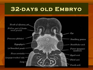
![ During first 2months we call the developingDuring first 2months we call the developing
individualindividual An EmbryoAn Embryo
From third month until birthFrom third month until birth FetusFetus
From birth to 1yr of ageFrom birth to 1yr of age An InfantAn Infant
A young child who is just beginning to walkA young child who is just beginning to walk
ToddlerToddler
Period shows onset of puberty. Period markPeriod shows onset of puberty. Period mark
the secondary sex characters [12-15y(G) 13-the secondary sex characters [12-15y(G) 13-
16y(B)]16y(B)] TeenagerTeenager
Developing from child to an adult (13-19y)Developing from child to an adult (13-19y)
AdolescentAdolescent
Body attained full growth & maturity of anBody attained full growth & maturity of an
organism (18-21y)organism (18-21y) AdultAdultwww.indiandentalacademy.comwww.indiandentalacademy.com](https://image.slidesharecdn.com/humangrowthdevelopment-160510095456/85/Human-growth-development-5-320.jpg)


![Formation of EmbryonicFormation of Embryonic
DiscDisc
Embryobalsts form the embryonic Spherical discEmbryobalsts form the embryonic Spherical disc
which forms three germ layers.which forms three germ layers.
1]1] EndodermEndoderm (Hypoblast-Cuboidal cells)—Inside (1(Hypoblast-Cuboidal cells)—Inside (1stst
germ layer to be formed)germ layer to be formed)
2]2] EctodermEctoderm (Epiblast-Columnar cell)—Outside (2(Epiblast-Columnar cell)—Outside (2ndnd
germ layer)germ layer)
3]3] MesodermMesoderm ––In the middleIn the middle
www.indiandentalacademy.comwww.indiandentalacademy.com](https://image.slidesharecdn.com/humangrowthdevelopment-160510095456/85/Human-growth-development-34-320.jpg)


![Neural PlateNeural Plate
Notochord functions as the primaryNotochord functions as the primary
inductor in the early embryoinductor in the early embryo
The developing notochord induces theThe developing notochord induces the
overlying embryonic ectoderm tooverlying embryonic ectoderm to
thicken and form thethicken and form the “neural plate”“neural plate”
[Primordium of central nervous system][Primordium of central nervous system]
www.indiandentalacademy.comwww.indiandentalacademy.com](https://image.slidesharecdn.com/humangrowthdevelopment-160510095456/85/Human-growth-development-49-320.jpg)


![Intra-embryonic mesoderm subdivided intoIntra-embryonic mesoderm subdivided into
three parts—three parts—
1]1] Paraxial mesodermParaxial mesoderm
2]2] Lateral mesodermLateral mesoderm
3]3] Intermediate mesodermIntermediate mesoderm
In 1638, 1In 1638, 1stst
-human dissection was done in America.-human dissection was done in America.
www.indiandentalacademy.comwww.indiandentalacademy.com](https://image.slidesharecdn.com/humangrowthdevelopment-160510095456/85/Human-growth-development-52-320.jpg)
![[1] Paraxial[1] Paraxial
MesodermMesoderm
OOn either side of the notochord. It is organized inton either side of the notochord. It is organized into
the segments called “somitomeres”. These arethe segments called “somitomeres”. These are
arranged into somite. Each somite give rise to:-arranged into somite. Each somite give rise to:-
Sclerotome –cartilage & bone of the axial &Sclerotome –cartilage & bone of the axial &
paraxial skeleton (remember neural crest cellsparaxial skeleton (remember neural crest cells
give rise to cartilage & bone of the skull & face).give rise to cartilage & bone of the skull & face).
Myotome –segmental muscle component.Myotome –segmental muscle component.
Dermatome—Segmental skin component.Dermatome—Segmental skin component.
Each myotome & dermatome has its own muscleEach myotome & dermatome has its own muscle
& nerve component.& nerve component.
www.indiandentalacademy.comwww.indiandentalacademy.com](https://image.slidesharecdn.com/humangrowthdevelopment-160510095456/85/Human-growth-development-53-320.jpg)
![[2] Lateral Mesoderm[2] Lateral Mesoderm
LLateral side of the notochord. Differentiateateral side of the notochord. Differentiate
into excretory unit of urinary system &into excretory unit of urinary system &
Gonads.Gonads.
www.indiandentalacademy.comwww.indiandentalacademy.com](https://image.slidesharecdn.com/humangrowthdevelopment-160510095456/85/Human-growth-development-54-320.jpg)
![[3] Inter-mediate[3] Inter-mediate
MesodermMesoderm
BBetween Paraxial & Lateral mesoderms.etween Paraxial & Lateral mesoderms.
Split into partial & visceral layers.Split into partial & visceral layers.
In the head region, cranial to somites,In the head region, cranial to somites,
somitomeres give origin to thesomitomeres give origin to the
mesenchyme.mesenchyme.
Striated muscle of the tongue is derivedStriated muscle of the tongue is derived
from the occipital myotome.from the occipital myotome.
www.indiandentalacademy.comwww.indiandentalacademy.com](https://image.slidesharecdn.com/humangrowthdevelopment-160510095456/85/Human-growth-development-55-320.jpg)


![ Initially separate centres of cartilageInitially separate centres of cartilage
formation in the cranial base, fuseformation in the cranial base, fuse
together into a single irregular & greatlytogether into a single irregular & greatly
perforated cranial base. [Earlyperforated cranial base. [Early
development of the various nerve &development of the various nerve &
vessels from & to the brain results invessels from & to the brain results in
numerous perforations (foramina)].numerous perforations (foramina)].
The ossifying chondro-cranial meets theThe ossifying chondro-cranial meets the
ossifying desmocranium (cranial vault)ossifying desmocranium (cranial vault)
to formto form neurocranium.neurocranium.
www.indiandentalacademy.comwww.indiandentalacademy.com](https://image.slidesharecdn.com/humangrowthdevelopment-160510095456/85/Human-growth-development-75-320.jpg)


![Nasal PitsNasal Pits
Ectoderm overlying the fronto-nasalEctoderm overlying the fronto-nasal
process shows bilateral localizedprocess shows bilateral localized
thickenings above the stomodeum whichthickenings above the stomodeum which
soon sink & form thesoon sink & form the nasal pitsnasal pits. The. The
formation of these nasal pits divides theformation of these nasal pits divides the
fronto-nasal process into two parts.fronto-nasal process into two parts.
1]1] Medial nasal processMedial nasal process
2]2] Lateral nasal process.Lateral nasal process.
www.indiandentalacademy.comwww.indiandentalacademy.com](https://image.slidesharecdn.com/humangrowthdevelopment-160510095456/85/Human-growth-development-99-320.jpg)


![Secondary GrowthSecondary Growth
CartilagesCartilages
Further growth of the mandibleFurther growth of the mandible
until birth is influenceduntil birth is influenced
strongly by the appearance ofstrongly by the appearance of
three secondary growth cartilages.three secondary growth cartilages.
[Endochondral Bone Formation][Endochondral Bone Formation]
www.indiandentalacademy.comwww.indiandentalacademy.com](https://image.slidesharecdn.com/humangrowthdevelopment-160510095456/85/Human-growth-development-135-320.jpg)
![1] Condylar1] Condylar
CartilageCartilage
Appears during 12th week of developmentAppears during 12th week of development
& rapidly formed a carrot-shaped mass that& rapidly formed a carrot-shaped mass that
occupies most of the developing ramus. Thisoccupies most of the developing ramus. This
mass of cartilage is converted quickly tomass of cartilage is converted quickly to
bone by endochondral ossification so that atbone by endochondral ossification so that at
20weeks only a thin layer of cartilage is20weeks only a thin layer of cartilage is
remains in the condylar head. This remnantremains in the condylar head. This remnant
of cartilage persists until the end of secondof cartilage persists until the end of second
decade of life, providing the mechanism fordecade of life, providing the mechanism for
growth of mandible, in the same way as thegrowth of mandible, in the same way as the
epiphyseal cartilage does in the limbs.epiphyseal cartilage does in the limbs.www.indiandentalacademy.comwww.indiandentalacademy.com](https://image.slidesharecdn.com/humangrowthdevelopment-160510095456/85/Human-growth-development-136-320.jpg)
![2] Coronoid2] Coronoid
CartilageCartilage
Appears about 4th month of development,Appears about 4th month of development,
surrounding the anterior border & top ofsurrounding the anterior border & top of
the coronoid process. Coronoid cartilagethe coronoid process. Coronoid cartilage
is a transient growth cartilage is believedis a transient growth cartilage is believed
to grow as a response to the developingto grow as a response to the developing
temporalis muscle & disappears longtemporalis muscle & disappears long
before birth.before birth.
www.indiandentalacademy.comwww.indiandentalacademy.com](https://image.slidesharecdn.com/humangrowthdevelopment-160510095456/85/Human-growth-development-137-320.jpg)
![3] Symphyseal3] Symphyseal
CartilageCartilage
In the mental region, on either side of theIn the mental region, on either side of the
symphysis, two cartilages, appear in thesymphysis, two cartilages, appear in the
connective tissue b/w the two ends ofconnective tissue b/w the two ends of
Meckel’s cartilage but are entirelyMeckel’s cartilage but are entirely
independent of it. They are obliteratedindependent of it. They are obliterated
within the 1st year after birth.within the 1st year after birth.
www.indiandentalacademy.comwww.indiandentalacademy.com](https://image.slidesharecdn.com/humangrowthdevelopment-160510095456/85/Human-growth-development-138-320.jpg)


The document provides an extensive overview of human growth and development, from prenatal stages through adulthood. It outlines the sequential changes during various developmental phases—embryo, fetus, infant, toddler, adolescent, and adult—highlighting the significance of understanding these processes for dental professionals. Key concepts such as embryology, teratogens, and cellular division processes are discussed, emphasizing their relevance in clinical practice and treatment approaches.