Download as PDF, PPTX
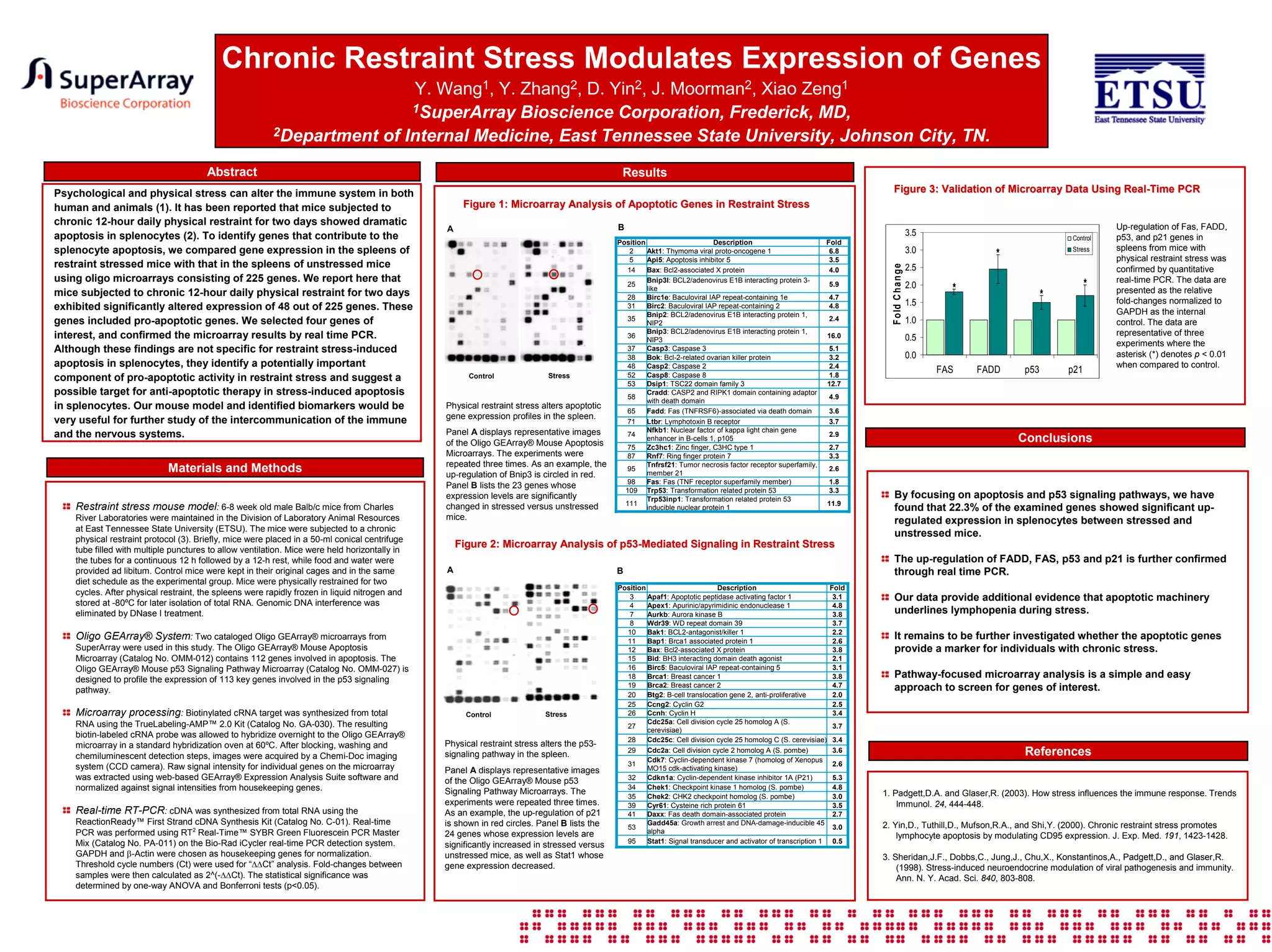

This study examined how chronic physical restraint stress alters gene expression in mouse spleens. Mice were subjected to restraint stress for 12 hours per day for two days. Microarray analysis found that 48 out of 225 apoptosis- and p53 signaling-related genes showed significant expression changes in stressed mice compared to unstressed controls. Four genes (Fas, FADD, p53, p21) were validated with real-time PCR. The results provide evidence that apoptotic pathways contribute to stress-induced lymphopenia and identify potential stress response biomarkers.


