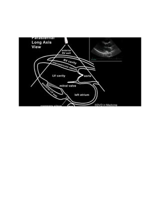
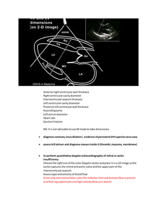
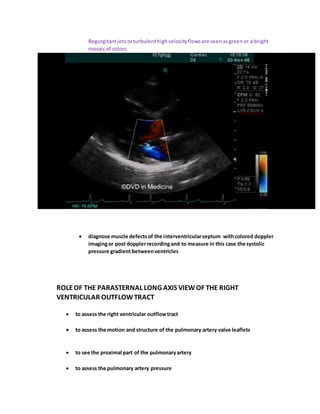
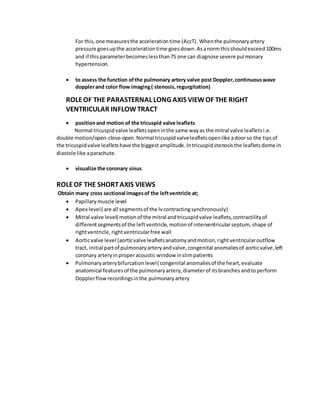
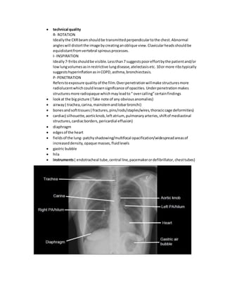
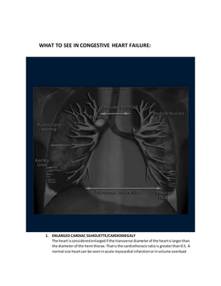
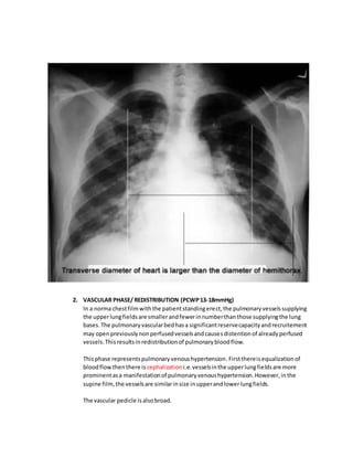
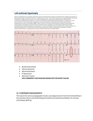
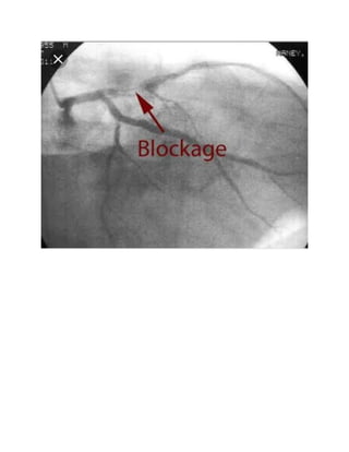
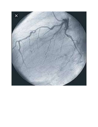
TOPIC: HEART FAILURE RADIOLOGY
DEFINITION
Heart failure isaclinical syndrome representing aconstellationof signsandsymptomsresultingfrom
the inabilityof the hearttopumpbloodforwardat a sufficientrate tomeetthe metabolicdemandsof
the body( forwardfailure) orthe abilitytodoso onlyif the cardiac fillingpressuresare abnormallyhigh
( backwardfailure) orboth.
CLASSIFICATION OF HEARTFAILURE
Variousclassificationsystemshave beenusedtoclassifyheartfailure.Thesesystemsdivide heartfailure
intovariousgroupsbasedon;
a) Onsetof heart failure
b) Ejectionfraction
c) Side of the heart affected
d) Cardiac output
e) Pathophysiologicmechanism
THIS THEREFORE GIVESUS:
1) Acute heartfailure vs. chronicheartfailure vs.acute onchronic heartfailure
2) Heart failure withreducedejectionfraction vs. heartfailurewithpreservedejectionfraction vs.
heartfailure midrange ejectionfraction
3) Leftsidedheartfailure vs. rightsidedheartfailurevs. biventricularheartfailure
4) Highoutputfailure vs.lowoutputheartfailure
5) Forwardfailure vs. backwardfailure
6) Compensated vs. decompensatedheartfailure
ETIOLOGY OF HEARTFAILURE
Differenttypesof heartfailurehave differentetiologies.Nonetheless,there isconsiderable overlap
betweenthe etiologiesof the differenttypes.
1) HEART FAILURE WITH REDUCED EJECTION FRACTION
 Ischemicheartdisease ( myocardial infarction,chronicmyocardialischemia)
 Pressure overload/increasedafterload( uncontrolledsystemicorpulmonary
hypertension,valve stenosis,chroniclungdiseases,pulmonarythromboembolism,
bronchiectasis,coarctationof the aorta)
 Volume overload/increasedpreload(thyrotoxicosis,severeanemia,arteriovenous
shunts,valvularinsufficiency,beriberi,hypoxiasecondarytolungdiseases)
 Cardiomyopathy
 Chaga’sdisease
 Congenital heartdisease ( atrial septal defects,ventricularseptal defect,patentductus
arteriosus)
 Cardiac arrhythmias
 Myocarditis
 Metabolicdisorders
 Drugs ( eitherrecreational ortherapeutic) withcardiacside effectse.gdoxorubicin
2) HEART FAILURE WITH PRESERVED EJECTION FRACTION
 Ischemicheartdisease ( myocardial infarction,chronicmyocardialinfarction)
 Pressure overload
 Myocardial fibrosis
 Infiltrativedisorders
 Cardiomyopathy( restrictive cardiomyopathy,hypertrophiccardiomyopathy)
 Massive pathological ventricularhypertrophy
3) HIGH OUTPUT FAILURE
 Anemia
 Thyrotoxicosis
 Beriberi
 Pregnancy
 Carcinoidsyndrome
 Albrightsyndrome
 Myxedema
 Systemicarteriovenousfistulas
4) ACUTE HEART FAILURE
 Acute myocardial infarction
 Cardiac arrhythmias
 Sepsis
 Drugs e.gcocaine,calciumchannel blockers
 Acute valvularregurgitation
 Myocarditis
PATHOPHYSIOLOGY
INTRODUCTION
Bloodpressure isa functionof cardiacoutputand total peripheral resistance.Cardiacoutputisitselfa
functionof stroke volume andheartrate.Stroke volume isinturndeterminedbyanumberof cardiac
and noncardiacfactorsincluding;
 Myocardial contractility
 Sodiumandwaterhomeostasis
 Mineralocorticoids
 Atrial natriureticpeptides
BLOOD PRESSURE = CARDIACOUTPUT X TOTAL PERIPHERALRESISTANCE
STROKE VOLUME XHEART RATE HUMORAL + NEURAL FACTORS+ LOCALFACTORS
The cardiovascularsystemattemptstocompensate forthe hemodynamicburdenobservedinheart
failure bythe following mechanisms;
1. Structural myocardial changes( hypertrophyanddilation)
2. Neurohumoral mechanisms( RAAS,ADRENERGICNERVOUSSYSTEM)
3. Frank- starlingmechanism
CLINICAL FEATURES
1. Exertional dyspnea
2. Dyspneaat rest
3. Fatigue
4. Orthopnea
5. Nocturnal cough
6. Paroxysmal nocturnal dyspnea
7. Acute pulmonaryedema
8. Cheyne strokesrespiration
INVESTIGATIONS FOR HEART FAILURE
NON IMAGING MODALITIES
 Complete bloodcount
 Renal panel
 Liverfunctiontests
 Iron studies
 Fastingbloodglucose/oral glucose tolerance test
 Lipidreview
 Thyroidfunctiontests(TSH)
 B type natriureticpeptide
 N type pro b type natriureticpeptide
IMAGING MODALITIES
1) ECHOCARDIOGRAPHY
TYPES
 2D TRANSTHORACICECHO
 DOPPLER ECHO
 3D TRANSTHORACICECHO
 TRANSESOPHAGEAL
VIEWS
 parasternal longaxisviewof the leftventricularinflow andoutflow tract
 parasternal longaxisviewof the right ventricularoutflow tract
 parasternal longaxisviewof the rightventricularinflow tract
 parasternal shortaxisviewsof the leftventricle
 the segmental approachtoanalyzingregional ventricularfunction (shortaxisviews)
 apical images
 subcostal views
POSITION OF THE PATIENT
 IDEAL: patientliesinthe left(lateral) decubitusposition
 OTHER: Supine
INDICATIONS
 diagnosis,management andfollow upof patientswithsuspectedorknownheart
disease
THE PARASTERNAL LONG AXIS VIEW
In this view, one is able to visualize the;
 anterior right ventricular wall
 the right ventricular cavity
 the interventricular septum
 the left ventricular cavity
 the mitral valve leaflets
 the aortic valve leaflets
 the proximal part of the aorta
 the posterior left ventricular wall
 the left atrium
ROLE OF THE PARASTERNAL LONG AXIS VIEW OF THE LEFT VENTRICLE
 Diagnose aortic root pathology ( dilation,dissectionand atherosclerosisofthe aorta)
 Diagnose aortic valve pathology ( anomalies,calcinosis,vegetations, thickening)
One can see the aortic valve leafletsbetterwhentheyare thickenedorina verythin
personwhenthere isa properacousticwindow)
 Diagnose subvalvular leftventricular outflowtract obstruction ( membranous or
muscular stenosis)
 To assess leftventricularsystolic function
Thisis done byassessingthe wall motion.Normallythe leftventricularwall must
contract and thickenuniformlyinall segmentsandopposite wallsmustapproachone
another.Onthe parasternal longaxisview the shape of the leftventricleresemblesan
equilateral triangle.The tipof thistriangle isformedbythe leftventricularapex andthe
base is a straightline thatconnectsbasal endsof the opposite walls.Duringsystole the
wallsof the triangle thicken uniformlyandthe tipof thistriangle approachesthe base
and the opposite dotsonthe wallsof the triangle approachtheiraxisatequal distances
therefore the ventricularwallsuniformlyapproacheachotheranduniformlythicken.
On the parasternal longaxisview of the leftventricle,one observescontractionof the
interventricularseptumandthe posteriorleftventricularwall.The parasternal longaxis
viewdoesnotshowthe apex inmostpatientstherefore one cannotassessits
shortening.
 visualize the coronary sinus
 to determine the amplitude of motionand thicknessofthe interventricularseptum
and posterior leftventricularwall
 to see membranous defectsofthe interventricularseptum
 to confirm mitral aortic fibrouscontinuance (important in the diagnosis ofsome
congenital heart diseases)
 to diagnose structural changesand dysfunctionsofthe mitral valve and its supporting
structures (cord, papillarymuscle)
Duringdiastole the mitral valve openstwice;earlydiastoleandlate diastole.Amplitude
of the late diastolicopeningislowerthanthe earlydiastolicopening.Normal mitral
valvesopenlike adoor.Inmitral stenosisthe anteriormitral leafletballoonsindiastole
like the canopy of a parachute.
 To measure dimensions/take various measurements
Anteriorrightventricularwall thickness
Rightventricularcavitydiameter
Interventricularseptumthickness
Leftventricularcavitydiameter
Posteriorleftventricularwall thickness
Ascendingaorta
Leftatriumdiameter
Heart rate
Ejectionfraction
NB: It isnot advisable touse M mode to take dimensions
 diagnose coronary sinusdilation( evidence ofpersistentleftsuperiorvena cava
 assessleftatrium and diagnose massesinside it (thrombi,myxoma, membrane)
 to perform quantitative dopplerechocardiography of mitral or aortic
insufficiency
Choose the rightsize of the color Dopplersectorandplace it ina 2d image sothe
sectorcapturesthe mitral and aortic valve andthe upperpart of the
interventricularseptum.
Assesstype andvelocityof bloodflow
If one only sees red and blue colorthis indicates thatonly laminar flow is present
and thatregurgitantjetsand high velocity flowsare absent
Regurgitantjetsorturbulenthighvelocityflowsare seenasgreenor a bright
mosaicof colors
 diagnose muscle defectsof the interventricularseptum withcolored doppler
imagingor post dopplerrecordingand to measure in this case the systolic
pressure gradient betweenventricles
ROLEOF THE PARASTERNAL LONG AXIS VIEWOF THE RIGHT
VENTRICULAR OUTFLOWTRACT
 to assess the right ventricular outflowtract
 to assess the motion and structure of the pulmonary artery valve leaflets
 to see the proximal part of the pulmonaryartery
 to assess the pulmonary artery pressure
For this,one measuresthe accelerationtime (AccT). Whenthe pulmonaryartery
pressure goesupthe accelerationtime goesdown.Asanormthisshouldexceed100ms
and if thisparameterbecomeslessthan75 one can diagnose severe pulmonary
hypertension.
 to assess the function ofthe pulmonary artery valve post Doppler,continuouswave
dopplerand color flow imaging( stenosis,regurgitation)
ROLEOF THE PARASTERNAL LONG AXIS VIEWOF THE RIGHT
VENTRICULAR INFLOWTRACT
 positionand motion of the tricuspid valve leaflets
Normal tricuspidvalve leafletsopeninthe same wayas the mitral valve leaflets i.e.
double motion/open-close-open.Normal tricuspidvalveleafletsopenlike adoorso the tipsof
the tricuspidvalve leafletshave the biggest amplitude.In tricuspidstenosisthe leafletsdome in
diastole like aparachute.
 visualize the coronary sinus
ROLEOF THE SHORTAXIS VIEWS
Obtain many cross sectional imagesof the leftventricle at;
 Papillarymuscle level
 Apex level( are all segmentsof the lvcontractingsynchronously)
 Mitral valve level( motionof the mitral andtricuspidvalve leaflets,contractilityof
differentsegmentsof the leftventricle,motionof interventricularseptum,shape of
rightventricle, rightventricularfree wall
 Aorticvalve level (aorticvalve leafletsanatomyandmotion,rightventricularoutflow
tract, initial partof pulmonaryarteryandvalve,congenital anomaliesof aorticvalve,left
coronary arteryinproperacoustic window inslimpatients
 Pulmonaryarterybifurcationlevel( congenital anomaliesof the heart,evaluate
anatomical featuresof the pulmonaryartery,diameterof itsbranches andtoperform
Dopplerflowrecordingsinthe pulmonary artery
ADVANTAGES OF ECHOCARDIOGRAPHY:
 noninvasive
 no side effects
 easyavailability
2) RADIOGRAPHY
When reading a chest x- ray the following areas should be included:
 patientsname ,hospital numberanddate of birth
 date the image wastaken
 whywas ittaken
 type offilm
PA- patientisusuallystandingwithanteriorchestagainstthe x ray plate.Xray beam
originatesfrom5-6 feetbehindthe patient.The beampenetratesfromposteriorto
anteriorchest.
AP- patientisusuallyinbedandleaningwithbackagainstthe x ray palte.Xray beam
originatesfrom2-4 feetinfrontof the patient.The beampenetratesfromanteriorto
posteriorchest.
Lateral
 positionof the patient( upright,lying)
 technical quality
R- ROTATION
Ideallythe CXRbeamshouldbe transmittedperpendiculartothe chest.Abnormal
angleswill distortthe image bycreatinganoblique view.Clavicularheadsshouldbe
equidistantfromvertebral spinousprocesses.
I- INSPIRATION
Ideally7-9ribsshouldbe visible.Lessthan7 suggestspooreffortbythe patientand/or
lowlungvolumesasinrestrictive lungdisease,atelectasisetc. 10or more ribstypically
suggestshyperinflationasinCOPD,asthma,bronchiectasis.
P- PENETRATION
Referstoexposure quality of the film.Overpenetrationwillmake structuresmore
radiolucentwhichcouldlessensignificance of opacities. Underpenetration makes
structuresmore radiopaque whichmayleadto“ overcalling”certainfindings
 lookat the big picture ( Take note of any obviousanomalies)
 airway ( trachea,carina,mainstemandlobarbronchi)
 bonesandsofttissues ( fractures,pins/rods/staples/wires,thoraciccage deformities)
 cardiac( silhouette,aorticknob,left atrium, pulmonary arteries,shiftof mediastinal
structures,cardiacborders,pericardial effusion)
 diaphragm
 edgesof the heart
 fieldsof the lung- patchyshadowing/multifocal opacification/widespreadareasof
increaseddensity, opaque masses,fluidlevels
 gastric bubble
 hila
 instruments( endotracheal tube,central line,pacemakerordefibrillator, chesttubes)
WHAT TO SEE IN CONGESTIVE HEART FAILURE:
1. ENLARGED CARDIAC SILHOUETTE/CARDIOMEGALY
The heart isconsideredenlargedif the transverse diameterof the heartislargerthan
the diameterof the hemi thorax.Thatis the cardiothoracicratiois greaterthan0.5. A
normal size heartcan be seeninacute myocardial infarctionorinvolume overload
2. VASCULAR PHASE/ REDISTRIBUTION (PCWP13-18mmHg)
In a norma chestfilmwiththe patientstandingerect,the pulmonaryvesselssupplying
the upperlungfieldsare smallerandfewerinnumberthanthose supplyingthe lung
bases.The pulmonaryvascularbedhasa significantreservecapacityandrecruitement
may openpreviouslynonperfusedvesselsandcausesdistentionof alreadyperfused
vessels.Thisresultsinredistributionof pulmonarybloodflow.
Thisphase representspulmonaryvenoushypertension. Firstthereisequalizationof
bloodflowthenthere is cephalization i.e.vesselsinthe upperlungfieldsare more
prominentasa manifestationof pulmonaryvenoushypertension.However,inthe
supine film, the vesselsare similarinsize inupperandlowerlungfields.
The vascular pedicle isalsobroad.
There isalso increased(>1) arteryto bronchusratio. Normallythe vesselsinthe upper
lobesare smallerthanthe accompanyingbronchuswitharatio of 0.85. At the level of
the hilumtheyare equal andin the lowerlobesthe arteriesare largerwitharatioof
1.35. whenthere isredistributionof pulmonarybloodflowthere will be anincreased
arteryto bronchusratio inthe upperand middle lobes.Thisisbestvisible inthe
perihilarregion.
Enlargedpulmonaryveinswithperivascularfluidcollectionleadstofull hazyhilumand
vessels.
3. INTERSTITIAL PHASE (PCWP18-25 mmHg)
KerleyBlines(interstitial septal thickening) are seenas1-2 cm longhorizontal linesin
the base of the lungsclose to the chestwall. They are the resultof interstitial edema
and lymphaticdrainage.
There isalso peribronchialcuffing andperihilarhaze( lossof definitionof vessels).This
occurs whenfluidleaksintothe peribronchovascularinterstitium
4. ALVEOLAR PHASE
Thisstage is characterizedbycontinuedfluidleakageintothe interstitiumwhichcannot
be compensatedbylymphaticdrainage.Thiseventuallyleadstofluidleakageintothe
alveoli ( alveolaredema) andleakageintothe pleuralspace.The distributionof the
alveolaredemacanbe influencedby;
 Gravity
 Obstructive lungdisease
Alveolaredemaisseenasconsolidationespeciallylowerlobesandhilarregion-
Air bronchogram
Cottonwool appearance
PLEURAL EFFUSION
BLUNTING OF COSTOPHRENICANGLES
5. ENLARGED MEDIASTINAL VEINS- INCREASED CENTRAL SYSTEMIC VENOUSVOLUME
3). ECG
Major ECG findingsare;
 Atrial fibrillations
 Previousmyocardial infarction
 Leftventricularhypertrophy
 Bundle branchblock
 Leftaxisdeviation
 Abnormal Qwaves
 ST depression
 Abnormal T waves
LESS COMMONLYUSED IMAGING MODALITIES FOR HEART FAILURE
4). CORONARYANGIOGRAPHY
The reasonsfor coronaryangiographyinclude,assessingpresence of restrictiontobloodflow in
the coronary arteries andestablishingwhichpatientsare potential candidates forcoronary
arterybypassgrafting.
Heart failure radiology

Heart failure radiology

  • 1.
    TOPIC: HEART FAILURERADIOLOGY DEFINITION Heart failure isaclinical syndrome representing aconstellationof signsandsymptomsresultingfrom the inabilityof the hearttopumpbloodforwardat a sufficientrate tomeetthe metabolicdemandsof the body( forwardfailure) orthe abilitytodoso onlyif the cardiac fillingpressuresare abnormallyhigh ( backwardfailure) orboth. CLASSIFICATION OF HEARTFAILURE Variousclassificationsystemshave beenusedtoclassifyheartfailure.Thesesystemsdivide heartfailure intovariousgroupsbasedon; a) Onsetof heart failure b) Ejectionfraction c) Side of the heart affected d) Cardiac output e) Pathophysiologicmechanism THIS THEREFORE GIVESUS: 1) Acute heartfailure vs. chronicheartfailure vs.acute onchronic heartfailure 2) Heart failure withreducedejectionfraction vs. heartfailurewithpreservedejectionfraction vs. heartfailure midrange ejectionfraction 3) Leftsidedheartfailure vs. rightsidedheartfailurevs. biventricularheartfailure 4) Highoutputfailure vs.lowoutputheartfailure 5) Forwardfailure vs. backwardfailure 6) Compensated vs. decompensatedheartfailure ETIOLOGY OF HEARTFAILURE Differenttypesof heartfailurehave differentetiologies.Nonetheless,there isconsiderable overlap betweenthe etiologiesof the differenttypes. 1) HEART FAILURE WITH REDUCED EJECTION FRACTION  Ischemicheartdisease ( myocardial infarction,chronicmyocardialischemia)  Pressure overload/increasedafterload( uncontrolledsystemicorpulmonary hypertension,valve stenosis,chroniclungdiseases,pulmonarythromboembolism, bronchiectasis,coarctationof the aorta)
  • 2.
     Volume overload/increasedpreload(thyrotoxicosis,severeanemia,arteriovenous shunts,valvularinsufficiency,beriberi,hypoxiasecondarytolungdiseases) Cardiomyopathy  Chaga’sdisease  Congenital heartdisease ( atrial septal defects,ventricularseptal defect,patentductus arteriosus)  Cardiac arrhythmias  Myocarditis  Metabolicdisorders  Drugs ( eitherrecreational ortherapeutic) withcardiacside effectse.gdoxorubicin 2) HEART FAILURE WITH PRESERVED EJECTION FRACTION  Ischemicheartdisease ( myocardial infarction,chronicmyocardialinfarction)  Pressure overload  Myocardial fibrosis  Infiltrativedisorders  Cardiomyopathy( restrictive cardiomyopathy,hypertrophiccardiomyopathy)  Massive pathological ventricularhypertrophy 3) HIGH OUTPUT FAILURE  Anemia  Thyrotoxicosis  Beriberi  Pregnancy  Carcinoidsyndrome  Albrightsyndrome  Myxedema  Systemicarteriovenousfistulas 4) ACUTE HEART FAILURE  Acute myocardial infarction  Cardiac arrhythmias  Sepsis  Drugs e.gcocaine,calciumchannel blockers  Acute valvularregurgitation  Myocarditis PATHOPHYSIOLOGY INTRODUCTION Bloodpressure isa functionof cardiacoutputand total peripheral resistance.Cardiacoutputisitselfa functionof stroke volume andheartrate.Stroke volume isinturndeterminedbyanumberof cardiac and noncardiacfactorsincluding;  Myocardial contractility  Sodiumandwaterhomeostasis  Mineralocorticoids
  • 3.
     Atrial natriureticpeptides BLOODPRESSURE = CARDIACOUTPUT X TOTAL PERIPHERALRESISTANCE STROKE VOLUME XHEART RATE HUMORAL + NEURAL FACTORS+ LOCALFACTORS The cardiovascularsystemattemptstocompensate forthe hemodynamicburdenobservedinheart failure bythe following mechanisms; 1. Structural myocardial changes( hypertrophyanddilation) 2. Neurohumoral mechanisms( RAAS,ADRENERGICNERVOUSSYSTEM) 3. Frank- starlingmechanism CLINICAL FEATURES 1. Exertional dyspnea 2. Dyspneaat rest 3. Fatigue 4. Orthopnea 5. Nocturnal cough 6. Paroxysmal nocturnal dyspnea 7. Acute pulmonaryedema 8. Cheyne strokesrespiration INVESTIGATIONS FOR HEART FAILURE NON IMAGING MODALITIES  Complete bloodcount  Renal panel  Liverfunctiontests  Iron studies  Fastingbloodglucose/oral glucose tolerance test  Lipidreview  Thyroidfunctiontests(TSH)  B type natriureticpeptide  N type pro b type natriureticpeptide IMAGING MODALITIES
  • 4.
    1) ECHOCARDIOGRAPHY TYPES  2DTRANSTHORACICECHO  DOPPLER ECHO  3D TRANSTHORACICECHO  TRANSESOPHAGEAL VIEWS  parasternal longaxisviewof the leftventricularinflow andoutflow tract  parasternal longaxisviewof the right ventricularoutflow tract  parasternal longaxisviewof the rightventricularinflow tract  parasternal shortaxisviewsof the leftventricle  the segmental approachtoanalyzingregional ventricularfunction (shortaxisviews)  apical images  subcostal views POSITION OF THE PATIENT  IDEAL: patientliesinthe left(lateral) decubitusposition  OTHER: Supine INDICATIONS  diagnosis,management andfollow upof patientswithsuspectedorknownheart disease THE PARASTERNAL LONG AXIS VIEW In this view, one is able to visualize the;  anterior right ventricular wall  the right ventricular cavity  the interventricular septum  the left ventricular cavity  the mitral valve leaflets  the aortic valve leaflets  the proximal part of the aorta  the posterior left ventricular wall  the left atrium
  • 6.
    ROLE OF THEPARASTERNAL LONG AXIS VIEW OF THE LEFT VENTRICLE  Diagnose aortic root pathology ( dilation,dissectionand atherosclerosisofthe aorta)
  • 7.
     Diagnose aorticvalve pathology ( anomalies,calcinosis,vegetations, thickening) One can see the aortic valve leafletsbetterwhentheyare thickenedorina verythin personwhenthere isa properacousticwindow)  Diagnose subvalvular leftventricular outflowtract obstruction ( membranous or muscular stenosis)  To assess leftventricularsystolic function Thisis done byassessingthe wall motion.Normallythe leftventricularwall must contract and thickenuniformlyinall segmentsandopposite wallsmustapproachone another.Onthe parasternal longaxisview the shape of the leftventricleresemblesan equilateral triangle.The tipof thistriangle isformedbythe leftventricularapex andthe base is a straightline thatconnectsbasal endsof the opposite walls.Duringsystole the wallsof the triangle thicken uniformlyandthe tipof thistriangle approachesthe base and the opposite dotsonthe wallsof the triangle approachtheiraxisatequal distances therefore the ventricularwallsuniformlyapproacheachotheranduniformlythicken. On the parasternal longaxisview of the leftventricle,one observescontractionof the interventricularseptumandthe posteriorleftventricularwall.The parasternal longaxis viewdoesnotshowthe apex inmostpatientstherefore one cannotassessits shortening.
  • 8.
     visualize thecoronary sinus  to determine the amplitude of motionand thicknessofthe interventricularseptum and posterior leftventricularwall  to see membranous defectsofthe interventricularseptum  to confirm mitral aortic fibrouscontinuance (important in the diagnosis ofsome congenital heart diseases)  to diagnose structural changesand dysfunctionsofthe mitral valve and its supporting structures (cord, papillarymuscle) Duringdiastole the mitral valve openstwice;earlydiastoleandlate diastole.Amplitude of the late diastolicopeningislowerthanthe earlydiastolicopening.Normal mitral valvesopenlike adoor.Inmitral stenosisthe anteriormitral leafletballoonsindiastole like the canopy of a parachute.  To measure dimensions/take various measurements
  • 9.
    Anteriorrightventricularwall thickness Rightventricularcavitydiameter Interventricularseptumthickness Leftventricularcavitydiameter Posteriorleftventricularwall thickness Ascendingaorta Leftatriumdiameter Heartrate Ejectionfraction NB: It isnot advisable touse M mode to take dimensions  diagnose coronary sinusdilation( evidence ofpersistentleftsuperiorvena cava  assessleftatrium and diagnose massesinside it (thrombi,myxoma, membrane)  to perform quantitative dopplerechocardiography of mitral or aortic insufficiency Choose the rightsize of the color Dopplersectorandplace it ina 2d image sothe sectorcapturesthe mitral and aortic valve andthe upperpart of the interventricularseptum. Assesstype andvelocityof bloodflow If one only sees red and blue colorthis indicates thatonly laminar flow is present and thatregurgitantjetsand high velocity flowsare absent
  • 10.
    Regurgitantjetsorturbulenthighvelocityflowsare seenasgreenor abright mosaicof colors  diagnose muscle defectsof the interventricularseptum withcolored doppler imagingor post dopplerrecordingand to measure in this case the systolic pressure gradient betweenventricles ROLEOF THE PARASTERNAL LONG AXIS VIEWOF THE RIGHT VENTRICULAR OUTFLOWTRACT  to assess the right ventricular outflowtract  to assess the motion and structure of the pulmonary artery valve leaflets  to see the proximal part of the pulmonaryartery  to assess the pulmonary artery pressure
  • 11.
    For this,one measurestheaccelerationtime (AccT). Whenthe pulmonaryartery pressure goesupthe accelerationtime goesdown.Asanormthisshouldexceed100ms and if thisparameterbecomeslessthan75 one can diagnose severe pulmonary hypertension.  to assess the function ofthe pulmonary artery valve post Doppler,continuouswave dopplerand color flow imaging( stenosis,regurgitation) ROLEOF THE PARASTERNAL LONG AXIS VIEWOF THE RIGHT VENTRICULAR INFLOWTRACT  positionand motion of the tricuspid valve leaflets Normal tricuspidvalve leafletsopeninthe same wayas the mitral valve leaflets i.e. double motion/open-close-open.Normal tricuspidvalveleafletsopenlike adoorso the tipsof the tricuspidvalve leafletshave the biggest amplitude.In tricuspidstenosisthe leafletsdome in diastole like aparachute.  visualize the coronary sinus ROLEOF THE SHORTAXIS VIEWS Obtain many cross sectional imagesof the leftventricle at;  Papillarymuscle level  Apex level( are all segmentsof the lvcontractingsynchronously)  Mitral valve level( motionof the mitral andtricuspidvalve leaflets,contractilityof differentsegmentsof the leftventricle,motionof interventricularseptum,shape of rightventricle, rightventricularfree wall  Aorticvalve level (aorticvalve leafletsanatomyandmotion,rightventricularoutflow tract, initial partof pulmonaryarteryandvalve,congenital anomaliesof aorticvalve,left coronary arteryinproperacoustic window inslimpatients  Pulmonaryarterybifurcationlevel( congenital anomaliesof the heart,evaluate anatomical featuresof the pulmonaryartery,diameterof itsbranches andtoperform Dopplerflowrecordingsinthe pulmonary artery
  • 12.
    ADVANTAGES OF ECHOCARDIOGRAPHY: noninvasive  no side effects  easyavailability 2) RADIOGRAPHY When reading a chest x- ray the following areas should be included:  patientsname ,hospital numberanddate of birth  date the image wastaken  whywas ittaken  type offilm PA- patientisusuallystandingwithanteriorchestagainstthe x ray plate.Xray beam originatesfrom5-6 feetbehindthe patient.The beampenetratesfromposteriorto anteriorchest. AP- patientisusuallyinbedandleaningwithbackagainstthe x ray palte.Xray beam originatesfrom2-4 feetinfrontof the patient.The beampenetratesfromanteriorto posteriorchest. Lateral  positionof the patient( upright,lying)
  • 13.
     technical quality R-ROTATION Ideallythe CXRbeamshouldbe transmittedperpendiculartothe chest.Abnormal angleswill distortthe image bycreatinganoblique view.Clavicularheadsshouldbe equidistantfromvertebral spinousprocesses. I- INSPIRATION Ideally7-9ribsshouldbe visible.Lessthan7 suggestspooreffortbythe patientand/or lowlungvolumesasinrestrictive lungdisease,atelectasisetc. 10or more ribstypically suggestshyperinflationasinCOPD,asthma,bronchiectasis. P- PENETRATION Referstoexposure quality of the film.Overpenetrationwillmake structuresmore radiolucentwhichcouldlessensignificance of opacities. Underpenetration makes structuresmore radiopaque whichmayleadto“ overcalling”certainfindings  lookat the big picture ( Take note of any obviousanomalies)  airway ( trachea,carina,mainstemandlobarbronchi)  bonesandsofttissues ( fractures,pins/rods/staples/wires,thoraciccage deformities)  cardiac( silhouette,aorticknob,left atrium, pulmonary arteries,shiftof mediastinal structures,cardiacborders,pericardial effusion)  diaphragm  edgesof the heart  fieldsof the lung- patchyshadowing/multifocal opacification/widespreadareasof increaseddensity, opaque masses,fluidlevels  gastric bubble  hila  instruments( endotracheal tube,central line,pacemakerordefibrillator, chesttubes)
  • 14.
    WHAT TO SEEIN CONGESTIVE HEART FAILURE: 1. ENLARGED CARDIAC SILHOUETTE/CARDIOMEGALY The heart isconsideredenlargedif the transverse diameterof the heartislargerthan the diameterof the hemi thorax.Thatis the cardiothoracicratiois greaterthan0.5. A normal size heartcan be seeninacute myocardial infarctionorinvolume overload
  • 15.
    2. VASCULAR PHASE/REDISTRIBUTION (PCWP13-18mmHg) In a norma chestfilmwiththe patientstandingerect,the pulmonaryvesselssupplying the upperlungfieldsare smallerandfewerinnumberthanthose supplyingthe lung bases.The pulmonaryvascularbedhasa significantreservecapacityandrecruitement may openpreviouslynonperfusedvesselsandcausesdistentionof alreadyperfused vessels.Thisresultsinredistributionof pulmonarybloodflow. Thisphase representspulmonaryvenoushypertension. Firstthereisequalizationof bloodflowthenthere is cephalization i.e.vesselsinthe upperlungfieldsare more prominentasa manifestationof pulmonaryvenoushypertension.However,inthe supine film, the vesselsare similarinsize inupperandlowerlungfields. The vascular pedicle isalsobroad.
  • 16.
    There isalso increased(>1)arteryto bronchusratio. Normallythe vesselsinthe upper lobesare smallerthanthe accompanyingbronchuswitharatio of 0.85. At the level of the hilumtheyare equal andin the lowerlobesthe arteriesare largerwitharatioof 1.35. whenthere isredistributionof pulmonarybloodflowthere will be anincreased arteryto bronchusratio inthe upperand middle lobes.Thisisbestvisible inthe perihilarregion. Enlargedpulmonaryveinswithperivascularfluidcollectionleadstofull hazyhilumand vessels.
  • 18.
    3. INTERSTITIAL PHASE(PCWP18-25 mmHg) KerleyBlines(interstitial septal thickening) are seenas1-2 cm longhorizontal linesin the base of the lungsclose to the chestwall. They are the resultof interstitial edema and lymphaticdrainage.
  • 19.
    There isalso peribronchialcuffingandperihilarhaze( lossof definitionof vessels).This occurs whenfluidleaksintothe peribronchovascularinterstitium
  • 21.
    4. ALVEOLAR PHASE Thisstageis characterizedbycontinuedfluidleakageintothe interstitiumwhichcannot be compensatedbylymphaticdrainage.Thiseventuallyleadstofluidleakageintothe alveoli ( alveolaredema) andleakageintothe pleuralspace.The distributionof the alveolaredemacanbe influencedby;  Gravity  Obstructive lungdisease Alveolaredemaisseenasconsolidationespeciallylowerlobesandhilarregion- Air bronchogram Cottonwool appearance
  • 23.
    PLEURAL EFFUSION BLUNTING OFCOSTOPHRENICANGLES 5. ENLARGED MEDIASTINAL VEINS- INCREASED CENTRAL SYSTEMIC VENOUSVOLUME 3). ECG Major ECG findingsare;  Atrial fibrillations
  • 25.
     Previousmyocardial infarction Leftventricularhypertrophy
  • 26.
     Bundle branchblock Leftaxisdeviation  Abnormal Qwaves  ST depression  Abnormal T waves LESS COMMONLYUSED IMAGING MODALITIES FOR HEART FAILURE 4). CORONARYANGIOGRAPHY The reasonsfor coronaryangiographyinclude,assessingpresence of restrictiontobloodflow in the coronary arteries andestablishingwhichpatientsare potential candidates forcoronary arterybypassgrafting.