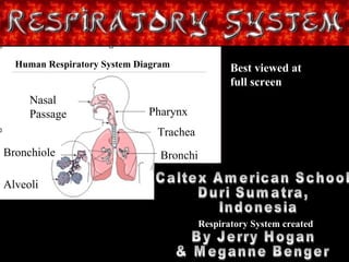
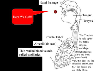
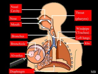
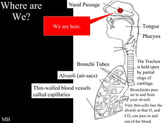
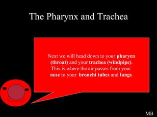
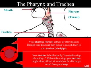
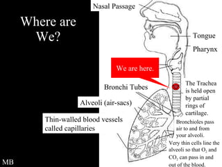
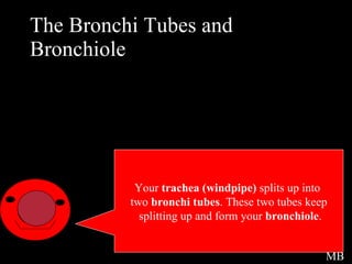
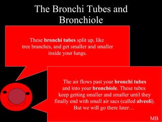
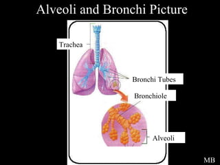
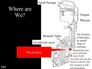
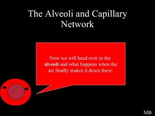
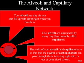
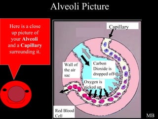
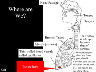
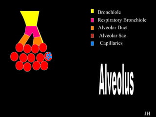
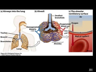
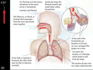
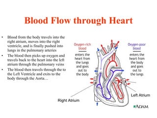
Welcome to... The Show of the Century Recline Your Chair,  Put Your Feet Up and Enjoy... Learning About the Respiratory and Circulatory Systems of the Human Body Best viewed at full screen and high resolution
Title Page Caltex American School  Duri Sumatra, Indonesia By Jerry Hogan & Meganne Benger Nasal Passage Bronchiole Alveoli Pharynx Trachea Bronchi Human Respiratory System Diagram Best viewed at full screen Respiratory System created
Oxygen Cell Hi I am O 2  ,you can call  me oxygen, and I will be your guide today.  I advise you keep all feet  and hands inside the ride at all times. JH
Respiratory Intro You may be asking, what is the Respiratory system? Well, the Respiratory system is the system that helps you breath in and out, so oxygen (0 2 ) can be pumped through your body and carbon dioxide (CO 2 ) can be removed from the blood stream. You must remember that the Respiratory system is made up of many different organs.  JH
Where are we? Here We Go!!! JH Nasal Passage Bronchi Tubes Alveoli (air-sacs) Thin-walled blood vessels called capillaries Very thin cells line the alveoli so that O 2  and CO 2  can pass in and out of the blood. Bronchioles pass air to and from your alveoli. The Trachea is held open by partial rings of cartilage. Tongue Pharynx
Picture Intro Here is a overview picture of the  Respiratory System . Just go to the next slide to see it. MB
Respiratory Overview  Picture MB Nasal Cavity Nose Mouth Bronchus Bronchiole Alveolus Diaphragm Throat (pharynx) Windpipe (Trachea) Left lungs Ribs
Welcome The Respiratory System Now we will begin our tour.  Welcome to… MB
The Nose and Mouth This is where it all begins.  This is where the  oxygen  first  enters your body and also where  Carbon Dioxide  leaves. MB
The Nose and Mouth When the air comes into your  nose  it gets filtered by tiny hairs and it is moistened by the mucus that is in your nose.  Your  sinuses  also help out with your  Respiratory System . They help to moisten and heat the air that you breath. Air can also get into your body through your mouth/oral cavity  but air is not filtered as much when it enters in through your  mouth .  MB
Nose and Mouth Picture Nasal Cavity Nostril Oral Cavity Pharynx Here is a picture of your  nasal  and  oral cavity . MB
Where are We? We are here. MB Nasal Passage Bronchi Tubes Alveoli (air-sacs) Thin-walled blood vessels called capillaries Very thin cells line the alveoli so that O 2  and CO 2  can pass in and out of the blood. Bronchioles pass air to and from your alveoli. The Trachea is held open by partial rings of cartilage. Tongue Pharynx
The Pharynx and Trachea Next we will head down to your  pharynx (throat)  and your  trachea (windpipe) . This is where the air passes from your  nose  to your  bronchi tubes  and  lungs . MB
The Pharynx and Trachea Your  pharynx (throat)  gathers air after it passes  through your  nose  and then the air is passed down to  your  trachea (windpipe) .   Your  trachea  is held open by “incomplete rings of cartilage.” Without these rings your  trachea  might close off and air would not be able to get  to and from your  lungs . MB Pharynx (Throat) Mouth Trachea
Where are We? We are here. MB Nasal Passage Bronchi Tubes Alveoli (air-sacs) Thin-walled blood vessels called capillaries Very thin cells line the alveoli so that O 2  and CO 2  can pass in and out of the blood. Bronchioles pass air to and from your alveoli. The Trachea is held open by partial rings of cartilage. Tongue Pharynx
The Bronchi Tubes and Bronchiole  Intro Your  trachea (windpipe)  splits up into  two  bronchi tubes . These two tubes keep splitting up and form your  bronchiole . MB
The Bronchi Tubes and Bronchiole These  bronchi tubes  split up, like  tree branches, and get smaller and smaller inside your lungs. The air flows past your  bronchi tubes and into your  bronchiole . These tubes  keep getting smaller and smaller until they finally end with small air sacs (called  alveoli ). But we will go there later…  MB
Alveoli and Bronchi Picture Trachea Bronchi Tubes Bronchiole Alveoli MB
Where are We? We are here. MB Nasal Passage Bronchi Tubes Alveoli (air-sacs) Thin-walled blood vessels called capillaries Very thin cells line the alveoli so that O 2  and CO 2  can pass in and out of the blood. Bronchioles pass air to and from your alveoli. The Trachea is held open by partial rings of cartilage. Tongue Pharynx
The Alveoli and Capillary Network Now we will head over to the  alveoli  and what happens when the air finally makes it down there. MB
The Alveoli and Capillary Network Your  alveoli  are tiny air sacs that fill up with air/oxygen when you  breath in. Your  alveoli  are surrounded by many tiny blood vessels called capillaries .  The walls of your  alveoli  (and  capillaries ) are so thin that the  oxygen  or  carbon dioxide  can  pass through them, traveling right into, or out of your blood stream. MB
Alveoli Picture Here is a close up picture of your  Alveoli   and a  Capillary surrounding it. MB Capillary Red Blood Cell Oxygen is picked up Carbon Dioxide is dropped off Wall of the air sac
Where are We? We are here. MB Nasal Passage Bronchi Tubes Alveoli (air-sacs) Thin-walled blood vessels called capillaries Very thin cells line the alveoli so that O 2  and CO 2  can pass in and out of the blood. Bronchioles pass air to and from your alveoli. The Trachea is held open by partial rings of cartilage. Tongue Pharynx
Alveolus Bronchiole Respiratory Bronchiole Alveolar Duct Alveolar Sac Capillaries Alveolus JH
Looking at the Alveoli Lets take a closer  look shall we. JH
Chemicals JH Red blood cell carrying  Carbon dioxide Chemical change is taking place in cell Red blood cell carrying oxygen Alveolus Contiguous Basal Laminae (Membrane) Capillary
Diffusion Oxygen Carbon Dioxide Alveolus Contiguous Basal Laminae (Membrane*) Capillary JH * A specialized  thin layer of skin that oxygen and carbon dioxide can pass through. Oxygen diffuses through the membrane into the blood stream. Carbon Dioxide diffuses through the membrane and enters the alveolus.
Cool pictures JH
Intro to Diaphragm Now we will look at the  Diaphragm . You might be wondering, what does the  Diaphragm  do? The  Diaphragm  is an important factor in breathing.  JH
Diagram of Diaphragm JH
Diaphragm Experiment Here is an experiment that you can try. JH
Experiment Instructions 1 st  you need a bottle that you can sacrifice to  cut up.  2 nd  you cut the bottom of the bottle and put a big balloon on the bottom. 3 rd  get a rubber cork ( make sure it blocks the hole)and put a hole through it ( top to bottom). Insert a thin tube into the cork and place a balloon on the bottom of the tube. 4 th  make sure the thing is airtight. JH
Respiratory Overview Review CO 2 JH The Pharynx, or throat, is located where passages from the nose and mouth came together. Air Passing over the mucus membrane of the nasal cavity is moistened, warmed, and filtered   Inside the lungs the Bronchi branch into small tubes called bronchioles At the end of the bronchioles are bunches of alveoli, air sacs, arranged like grapes on a stem Air enters the trachea, or wind pipe which leads to and from the lungs The trachea divides into two tubes called bronchi If one lobe is injured or diseased, the other lobes may be able to function normally
Fun Facts *  At rest, the body takes in and breathes out about 10 liters of air each minute. * The right lung is slightly larger than the left. * The highest recorded "sneeze speed" is 165 km per hour. * The surface area of the lungs is roughly the same size as a tennis court. * The capillaries in the lungs would extend 1,600 kilometers if placed end to end. * We lose half a liter of water a day through breathing. This is the water vapor we see when we breathe onto glass. * A person at rest usually breathes between 12 and 15 times a minute. * The breathing rate is faster in children and women than in men.
Key Words Respiratory System - The group of organs in your body that are responsible for taking in Oxygen and breathing out the Carbon Dioxide which is the waste product of cellular respiration. Oxygen -The gas that your body needs to work and function. Carbon Dioxide - The waste product (gas) that is produced through respiration of people and animals. Nose/Nasal Cavity - Where Oxygen first enters your body. Tiny hairs help filter the air and air is moistened and heated by your nose. Your Nose leads into your Nasal Cavity. Mouth/Oral Cavity - Oxygen/air can also enter through your Mouth but it is not filtered. Your Mouth opens up into your Oral Cavity. Sinus - A cavity in the bones of your skull that helps moisten and heat the air that you breath. Pharynx/Throat - Gathers air from your Nasal and Oral Cavities and passes it to your Trachea. Trachea/Windpipe - A tube like pathway that connects your throat to your Bronchi Tubes and lungs. Air passes through it when it travels from the Pharynx to the Bronchi Tubes.
Key Words Cont. Bronchi Tubes - Each tube (one per lung) splits up into many smaller tubes called Bronchiole, like branches on a tree. Bronchiole - Keep splitting up until they reach your Alveoli. Respiratory Bronchiole - The air-tubes that are actually connected to the Alveoli. Alveolar Duct - The final tube, which is part of the Alveoli, that leads to the air-sacs. Alveolar Sac - Where the chemical change takes place and where blood  cells pick up oxygen and drop off carbon dioxide. Alveoli - Tiny air-sacs at the end of your Alveolar Duct. They fill up with Oxygen and are surrounded by Capillaries. Capillaries - Tiny blood streams (around one cell wide) that surround your Alveoli. They take Oxygen out of our Lungs and replace it with Carbon Dioxide, which you later breath out. Diaphragm-  The muscle membrane that helps you breath in and out by changing the pressure in your chest cavity.
Works Cited For more information please visit: http://yucky.kids.discovery.com/flash/body/pg000138.html -Why do you need to breathe? And basic info on parts of the Respiratory system   http://www.lung.ca/children/grades7_12/respiratory/index.html -An overview of the parts of Respiratory System http://www4.tpgi.com.au/users/amcgann/body/respiratory.html -A basic look at the Respiratory System http://www4.tpgi.com.au/users/amcgann/body/respiratory_facts.html -Fun Facts http://users.pandora.be/educypedia/education/respiratory.htm -Very detailed info and some animation-Has many other body systems too http://www. bioedonline .org/slides/slide01.cfm?tk=5&pg=2S -Web slides with a little info and good pictures www.geocities.com/medinotes/nasal_cavity.htm -The Nose and Nasal Cavity Human anatomy coloring book
Works Cited Cont. Where we got some of our pictures: 	 http://www.bbc.co.uk/schools/gcsebitesize/img/pe01021.gif 	 http://www.sirinet.net/~jgjohnso/respiratorylungs.gif http://academic.pg.cc.md.us/~aimholtz/AandP/206_ONLINE/Resp/Images/respstructures.jpg http://www. cancersa .org.au/files/1/2/17/226/ airwaysfullylabelled .jpg http://www. researchmatters . harvard .edu/photos/645.jpg http://www4.tpgi.com.au/users/amcgann/body/respiratory.html http://www.sirinet.net/~jgjohnso/humanrespiratory8.jpg
Prepare Your Cranium For The Further Insertion of  Knowledge
Title Page The Circulatory System Hannah Redlich and Joe Zalan  Caltex American School Duri, Indonesia Circulatory System Created by
Introduction The Circulatory System is the main cooling and transportation system for the human body  The body has about 5 liters of blood continuously traveling through it by way of the Circulatory System  In the Circulatory System, the heart, lungs, and blood vessels have to work together The Circulatory System has three different parts: pulmonary circulation (lungs), coronary circulation (heart), and systemic circulation, (the rest of the system’s processes).
The Heart This organ is what pumps oxygen rich blood, nutrients, hormones, and the other things your body needs to maintain your health, to your organs and tissues. The pulmonary veins you see on the right side of the diagram come from your lungs, where the blood cells collect oxygen. It’s then pumped out to the rest of the body through the Aorta (Top).  All of the blue sections show blood cells carrying waste, (C0 2 ) moving back to the lungs (where the C0 2  will be replaced by oxygen) through the Pulmonary Artery (Top, blue) Pulmonary Veins Pulmonary Artery (Inferior Vena Cava) From the Body (Aortic Artery) To the body Whenever the blood is pumped from one section of the heart another a valve closes behind it preventing the blood from moving backwards. By The Way… Valves: ( tricuspid valve semilunar (pulmonary) valve, bicuspid (mitral) valve, and the semilunar (aortic) valve (Superior Vena Cava) From the Body
Blood Flow through Heart Blood from the body travels into the right atrium, moves into the right ventricle, and is finally pushed into lungs in the pulmonary arteries The blood then picks up oxygen and travels back to the heart into the left atrium through the pulmonary veins The blood then travels through the to the Left Ventricle and exits to the body through the Aorta…  Right Atrium Left Atrium
Blood Flow to Arms Oxygen rich blood leaves the heart and travels through arteries In the capillaries the oxygen and food is given to the body’s cells The blood finally travels back through veins to the heart to pick up oxygen ARTERIES- FROM HEART CAPILLARIES VEINS- TO HEART
Path to the Exchange A red blood cell then travels from the heart through arteries that eventually branch into the body’s vast system of capillaries (microscopic blood vessels which connect arteries and veins), they eventually lead to… Pulmonary Vein Aorta Brachial Artery Renal Artery Redial Artery Ulnar Artery Iliac Artery
The Exchange When the itty bitty teeny tiny red blood cells pass the desired tissue they………………………………. TRANSACT The oxygen the blood cells are carrying is given to the body’s tissue. And the CO 2  (waste) from the tissue is given to the same blood cell to be exhaled. Tissue Tissue Technically the Hemoglobin in the blood (a substance full of iron) attracts oxygen from the lungs. The red blood cell then carries it to the desired tissue. Because this tissue has a high CO 2  count the hemoglobin lets go of its oxygen and collects the carbon dioxide. You see the hemoglobin has an affinity for whichever gas has a greater count. Because the tissue has a large amount of built up waste (CO 2 ) the hemoglobin attracts it and then replaces it with oxygen, and vise versa in the lungs. How It Works… Oxy-Rich Blood Cell Oxy-Poor  Blood Cell Now lets travel to the legs!!!
Blood Flow to Legs Approximately 500 ml of blood moves from the heart and lungs down to the legs when a person stands up after lying down The oxygen rich blood cells then travel through the capillaries where yet another… !FUN FACT!
Gas Exchange Occurs, The oxygen and CO 2  are exchanged…in the cells Oxygen Rich Oxygen Rich Oxygen Poor Oxygen Poor Tissue Don’t forget that the Hemoglobin in the blood cells let go of the cell’s oxygen because of the large CO 2  (waste) count in the tissue. Now lets go back to the heart!!!
Circulation back to Heart Capillaries carry the blood to… Venules that connect to veins and the… Veins (wide blood vessels) carries the  oxygen-poor blood back to the heart. To upper body From upper body  To lung To lung From lung Left Atrium Left Ventricle To lower body From lower  body Right Ventricle Right Atrium From lung
Conclusion  As you have learned  (Hopefully )  the Circulatory System is one of the most important systems in the human body… It is the  only  reason you’re still alive today… and you can attribute the cooling down, feeding of and protection of your body to it. So the next time you bust open your leg skateboarding you can thank your Circulatory System for patching you up.
Works Cited For further information please visit: http://www.carolguze.com/images/organsystems/circulatory2.jpg   -circulation picture http://users.rcn.com/jkimball.ma.ultranet/BiologyPages/C/Circulation2.html   - how circulatory system works http://www.medical-art-service.de/assets/images/3_KA_704. jpg   - Heart and Leg Pictures http://www.ama-assn.org/ama1/pub/upload/images/446/circulationgeneral.gif   -circulation picture http://eduserv.hscer.washington.edu/hubio553/atlas/232.html   -arm picture http://adam.about.com/encyclopedia/19387.html   -heart picture http://www.tmc.edu/thi/anatomy1.html  -detailed views of the Cardiovascular System http://www.tmc.edu/thi/leg.jpg  -complex leg picture http://www.med.umich.edu/1libr/wha/circ.gif-   diagram of the circulatory system http://images.google.co.id/imgres?imgurl=http://www4.tpgi.com.au/users/amcgann/body/circulatory/body_circulation.jpg&imgrefurl=http://www4.tpgi.com.au/users/amcgann/body/circulatory.html&h=369&w=300&sz=23&tbnid=rSdZ_CMJpBYJ:&tbnh=117&tbnw=95&start=123&prev=/images%3Fq%3Dcirculatory%2Bsystem%26start%3D120%26hl%3Den%26lr%3D%26ie%3DUTF-8%26sa%3D   -picture of heart valves http:// eduserv . hscer . washington . edu /hubio553/atlas/232.html   -basic picture of arteries
So Take a Deep Breath and   Go Home The End The End!

heart and lungs

  • 1.
    Welcome to... TheShow of the Century Recline Your Chair, Put Your Feet Up and Enjoy... Learning About the Respiratory and Circulatory Systems of the Human Body Best viewed at full screen and high resolution
  • 2.
    Title Page CaltexAmerican School Duri Sumatra, Indonesia By Jerry Hogan & Meganne Benger Nasal Passage Bronchiole Alveoli Pharynx Trachea Bronchi Human Respiratory System Diagram Best viewed at full screen Respiratory System created
  • 3.
    Oxygen Cell HiI am O 2 ,you can call me oxygen, and I will be your guide today. I advise you keep all feet and hands inside the ride at all times. JH
  • 4.
    Respiratory Intro Youmay be asking, what is the Respiratory system? Well, the Respiratory system is the system that helps you breath in and out, so oxygen (0 2 ) can be pumped through your body and carbon dioxide (CO 2 ) can be removed from the blood stream. You must remember that the Respiratory system is made up of many different organs. JH
  • 5.
    Where are we?Here We Go!!! JH Nasal Passage Bronchi Tubes Alveoli (air-sacs) Thin-walled blood vessels called capillaries Very thin cells line the alveoli so that O 2 and CO 2 can pass in and out of the blood. Bronchioles pass air to and from your alveoli. The Trachea is held open by partial rings of cartilage. Tongue Pharynx
  • 6.
    Picture Intro Hereis a overview picture of the Respiratory System . Just go to the next slide to see it. MB
  • 7.
    Respiratory Overview Picture MB Nasal Cavity Nose Mouth Bronchus Bronchiole Alveolus Diaphragm Throat (pharynx) Windpipe (Trachea) Left lungs Ribs
  • 8.
    Welcome The RespiratorySystem Now we will begin our tour. Welcome to… MB
  • 9.
    The Nose andMouth This is where it all begins. This is where the oxygen first enters your body and also where Carbon Dioxide leaves. MB
  • 10.
    The Nose andMouth When the air comes into your nose it gets filtered by tiny hairs and it is moistened by the mucus that is in your nose. Your sinuses also help out with your Respiratory System . They help to moisten and heat the air that you breath. Air can also get into your body through your mouth/oral cavity but air is not filtered as much when it enters in through your mouth . MB
  • 11.
    Nose and MouthPicture Nasal Cavity Nostril Oral Cavity Pharynx Here is a picture of your nasal and oral cavity . MB
  • 12.
    Where are We?We are here. MB Nasal Passage Bronchi Tubes Alveoli (air-sacs) Thin-walled blood vessels called capillaries Very thin cells line the alveoli so that O 2 and CO 2 can pass in and out of the blood. Bronchioles pass air to and from your alveoli. The Trachea is held open by partial rings of cartilage. Tongue Pharynx
  • 13.
    The Pharynx andTrachea Next we will head down to your pharynx (throat) and your trachea (windpipe) . This is where the air passes from your nose to your bronchi tubes and lungs . MB
  • 14.
    The Pharynx andTrachea Your pharynx (throat) gathers air after it passes through your nose and then the air is passed down to your trachea (windpipe) . Your trachea is held open by “incomplete rings of cartilage.” Without these rings your trachea might close off and air would not be able to get to and from your lungs . MB Pharynx (Throat) Mouth Trachea
  • 15.
    Where are We?We are here. MB Nasal Passage Bronchi Tubes Alveoli (air-sacs) Thin-walled blood vessels called capillaries Very thin cells line the alveoli so that O 2 and CO 2 can pass in and out of the blood. Bronchioles pass air to and from your alveoli. The Trachea is held open by partial rings of cartilage. Tongue Pharynx
  • 16.
    The Bronchi Tubesand Bronchiole Intro Your trachea (windpipe) splits up into two bronchi tubes . These two tubes keep splitting up and form your bronchiole . MB
  • 17.
    The Bronchi Tubesand Bronchiole These bronchi tubes split up, like tree branches, and get smaller and smaller inside your lungs. The air flows past your bronchi tubes and into your bronchiole . These tubes keep getting smaller and smaller until they finally end with small air sacs (called alveoli ). But we will go there later… MB
  • 18.
    Alveoli and BronchiPicture Trachea Bronchi Tubes Bronchiole Alveoli MB
  • 19.
    Where are We?We are here. MB Nasal Passage Bronchi Tubes Alveoli (air-sacs) Thin-walled blood vessels called capillaries Very thin cells line the alveoli so that O 2 and CO 2 can pass in and out of the blood. Bronchioles pass air to and from your alveoli. The Trachea is held open by partial rings of cartilage. Tongue Pharynx
  • 20.
    The Alveoli andCapillary Network Now we will head over to the alveoli and what happens when the air finally makes it down there. MB
  • 21.
    The Alveoli andCapillary Network Your alveoli are tiny air sacs that fill up with air/oxygen when you breath in. Your alveoli are surrounded by many tiny blood vessels called capillaries . The walls of your alveoli (and capillaries ) are so thin that the oxygen or carbon dioxide can pass through them, traveling right into, or out of your blood stream. MB
  • 22.
    Alveoli Picture Hereis a close up picture of your Alveoli and a Capillary surrounding it. MB Capillary Red Blood Cell Oxygen is picked up Carbon Dioxide is dropped off Wall of the air sac
  • 23.
    Where are We?We are here. MB Nasal Passage Bronchi Tubes Alveoli (air-sacs) Thin-walled blood vessels called capillaries Very thin cells line the alveoli so that O 2 and CO 2 can pass in and out of the blood. Bronchioles pass air to and from your alveoli. The Trachea is held open by partial rings of cartilage. Tongue Pharynx
  • 24.
    Alveolus Bronchiole RespiratoryBronchiole Alveolar Duct Alveolar Sac Capillaries Alveolus JH
  • 25.
    Looking at theAlveoli Lets take a closer look shall we. JH
  • 26.
    Chemicals JH Redblood cell carrying Carbon dioxide Chemical change is taking place in cell Red blood cell carrying oxygen Alveolus Contiguous Basal Laminae (Membrane) Capillary
  • 27.
    Diffusion Oxygen CarbonDioxide Alveolus Contiguous Basal Laminae (Membrane*) Capillary JH * A specialized thin layer of skin that oxygen and carbon dioxide can pass through. Oxygen diffuses through the membrane into the blood stream. Carbon Dioxide diffuses through the membrane and enters the alveolus.
  • 28.
  • 29.
    Intro to DiaphragmNow we will look at the Diaphragm . You might be wondering, what does the Diaphragm do? The Diaphragm is an important factor in breathing. JH
  • 30.
  • 31.
    Diaphragm Experiment Hereis an experiment that you can try. JH
  • 32.
    Experiment Instructions 1st you need a bottle that you can sacrifice to cut up. 2 nd you cut the bottom of the bottle and put a big balloon on the bottom. 3 rd get a rubber cork ( make sure it blocks the hole)and put a hole through it ( top to bottom). Insert a thin tube into the cork and place a balloon on the bottom of the tube. 4 th make sure the thing is airtight. JH
  • 33.
    Respiratory Overview ReviewCO 2 JH The Pharynx, or throat, is located where passages from the nose and mouth came together. Air Passing over the mucus membrane of the nasal cavity is moistened, warmed, and filtered Inside the lungs the Bronchi branch into small tubes called bronchioles At the end of the bronchioles are bunches of alveoli, air sacs, arranged like grapes on a stem Air enters the trachea, or wind pipe which leads to and from the lungs The trachea divides into two tubes called bronchi If one lobe is injured or diseased, the other lobes may be able to function normally
  • 34.
    Fun Facts * At rest, the body takes in and breathes out about 10 liters of air each minute. * The right lung is slightly larger than the left. * The highest recorded "sneeze speed" is 165 km per hour. * The surface area of the lungs is roughly the same size as a tennis court. * The capillaries in the lungs would extend 1,600 kilometers if placed end to end. * We lose half a liter of water a day through breathing. This is the water vapor we see when we breathe onto glass. * A person at rest usually breathes between 12 and 15 times a minute. * The breathing rate is faster in children and women than in men.
  • 35.
    Key Words RespiratorySystem - The group of organs in your body that are responsible for taking in Oxygen and breathing out the Carbon Dioxide which is the waste product of cellular respiration. Oxygen -The gas that your body needs to work and function. Carbon Dioxide - The waste product (gas) that is produced through respiration of people and animals. Nose/Nasal Cavity - Where Oxygen first enters your body. Tiny hairs help filter the air and air is moistened and heated by your nose. Your Nose leads into your Nasal Cavity. Mouth/Oral Cavity - Oxygen/air can also enter through your Mouth but it is not filtered. Your Mouth opens up into your Oral Cavity. Sinus - A cavity in the bones of your skull that helps moisten and heat the air that you breath. Pharynx/Throat - Gathers air from your Nasal and Oral Cavities and passes it to your Trachea. Trachea/Windpipe - A tube like pathway that connects your throat to your Bronchi Tubes and lungs. Air passes through it when it travels from the Pharynx to the Bronchi Tubes.
  • 36.
    Key Words Cont.Bronchi Tubes - Each tube (one per lung) splits up into many smaller tubes called Bronchiole, like branches on a tree. Bronchiole - Keep splitting up until they reach your Alveoli. Respiratory Bronchiole - The air-tubes that are actually connected to the Alveoli. Alveolar Duct - The final tube, which is part of the Alveoli, that leads to the air-sacs. Alveolar Sac - Where the chemical change takes place and where blood cells pick up oxygen and drop off carbon dioxide. Alveoli - Tiny air-sacs at the end of your Alveolar Duct. They fill up with Oxygen and are surrounded by Capillaries. Capillaries - Tiny blood streams (around one cell wide) that surround your Alveoli. They take Oxygen out of our Lungs and replace it with Carbon Dioxide, which you later breath out. Diaphragm- The muscle membrane that helps you breath in and out by changing the pressure in your chest cavity.
  • 37.
    Works Cited Formore information please visit: http://yucky.kids.discovery.com/flash/body/pg000138.html -Why do you need to breathe? And basic info on parts of the Respiratory system http://www.lung.ca/children/grades7_12/respiratory/index.html -An overview of the parts of Respiratory System http://www4.tpgi.com.au/users/amcgann/body/respiratory.html -A basic look at the Respiratory System http://www4.tpgi.com.au/users/amcgann/body/respiratory_facts.html -Fun Facts http://users.pandora.be/educypedia/education/respiratory.htm -Very detailed info and some animation-Has many other body systems too http://www. bioedonline .org/slides/slide01.cfm?tk=5&pg=2S -Web slides with a little info and good pictures www.geocities.com/medinotes/nasal_cavity.htm -The Nose and Nasal Cavity Human anatomy coloring book
  • 38.
    Works Cited Cont.Where we got some of our pictures: http://www.bbc.co.uk/schools/gcsebitesize/img/pe01021.gif http://www.sirinet.net/~jgjohnso/respiratorylungs.gif http://academic.pg.cc.md.us/~aimholtz/AandP/206_ONLINE/Resp/Images/respstructures.jpg http://www. cancersa .org.au/files/1/2/17/226/ airwaysfullylabelled .jpg http://www. researchmatters . harvard .edu/photos/645.jpg http://www4.tpgi.com.au/users/amcgann/body/respiratory.html http://www.sirinet.net/~jgjohnso/humanrespiratory8.jpg
  • 39.
    Prepare Your CraniumFor The Further Insertion of Knowledge
  • 40.
    Title Page TheCirculatory System Hannah Redlich and Joe Zalan Caltex American School Duri, Indonesia Circulatory System Created by
  • 41.
    Introduction The CirculatorySystem is the main cooling and transportation system for the human body The body has about 5 liters of blood continuously traveling through it by way of the Circulatory System In the Circulatory System, the heart, lungs, and blood vessels have to work together The Circulatory System has three different parts: pulmonary circulation (lungs), coronary circulation (heart), and systemic circulation, (the rest of the system’s processes).
  • 42.
    The Heart Thisorgan is what pumps oxygen rich blood, nutrients, hormones, and the other things your body needs to maintain your health, to your organs and tissues. The pulmonary veins you see on the right side of the diagram come from your lungs, where the blood cells collect oxygen. It’s then pumped out to the rest of the body through the Aorta (Top). All of the blue sections show blood cells carrying waste, (C0 2 ) moving back to the lungs (where the C0 2 will be replaced by oxygen) through the Pulmonary Artery (Top, blue) Pulmonary Veins Pulmonary Artery (Inferior Vena Cava) From the Body (Aortic Artery) To the body Whenever the blood is pumped from one section of the heart another a valve closes behind it preventing the blood from moving backwards. By The Way… Valves: ( tricuspid valve semilunar (pulmonary) valve, bicuspid (mitral) valve, and the semilunar (aortic) valve (Superior Vena Cava) From the Body
  • 43.
    Blood Flow throughHeart Blood from the body travels into the right atrium, moves into the right ventricle, and is finally pushed into lungs in the pulmonary arteries The blood then picks up oxygen and travels back to the heart into the left atrium through the pulmonary veins The blood then travels through the to the Left Ventricle and exits to the body through the Aorta… Right Atrium Left Atrium
  • 44.
    Blood Flow toArms Oxygen rich blood leaves the heart and travels through arteries In the capillaries the oxygen and food is given to the body’s cells The blood finally travels back through veins to the heart to pick up oxygen ARTERIES- FROM HEART CAPILLARIES VEINS- TO HEART
  • 45.
    Path to theExchange A red blood cell then travels from the heart through arteries that eventually branch into the body’s vast system of capillaries (microscopic blood vessels which connect arteries and veins), they eventually lead to… Pulmonary Vein Aorta Brachial Artery Renal Artery Redial Artery Ulnar Artery Iliac Artery
  • 46.
    The Exchange Whenthe itty bitty teeny tiny red blood cells pass the desired tissue they………………………………. TRANSACT The oxygen the blood cells are carrying is given to the body’s tissue. And the CO 2 (waste) from the tissue is given to the same blood cell to be exhaled. Tissue Tissue Technically the Hemoglobin in the blood (a substance full of iron) attracts oxygen from the lungs. The red blood cell then carries it to the desired tissue. Because this tissue has a high CO 2 count the hemoglobin lets go of its oxygen and collects the carbon dioxide. You see the hemoglobin has an affinity for whichever gas has a greater count. Because the tissue has a large amount of built up waste (CO 2 ) the hemoglobin attracts it and then replaces it with oxygen, and vise versa in the lungs. How It Works… Oxy-Rich Blood Cell Oxy-Poor Blood Cell Now lets travel to the legs!!!
  • 47.
    Blood Flow toLegs Approximately 500 ml of blood moves from the heart and lungs down to the legs when a person stands up after lying down The oxygen rich blood cells then travel through the capillaries where yet another… !FUN FACT!
  • 48.
    Gas Exchange Occurs,The oxygen and CO 2 are exchanged…in the cells Oxygen Rich Oxygen Rich Oxygen Poor Oxygen Poor Tissue Don’t forget that the Hemoglobin in the blood cells let go of the cell’s oxygen because of the large CO 2 (waste) count in the tissue. Now lets go back to the heart!!!
  • 49.
    Circulation back toHeart Capillaries carry the blood to… Venules that connect to veins and the… Veins (wide blood vessels) carries the oxygen-poor blood back to the heart. To upper body From upper body To lung To lung From lung Left Atrium Left Ventricle To lower body From lower body Right Ventricle Right Atrium From lung
  • 50.
    Conclusion Asyou have learned (Hopefully ) the Circulatory System is one of the most important systems in the human body… It is the only reason you’re still alive today… and you can attribute the cooling down, feeding of and protection of your body to it. So the next time you bust open your leg skateboarding you can thank your Circulatory System for patching you up.
  • 51.
    Works Cited Forfurther information please visit: http://www.carolguze.com/images/organsystems/circulatory2.jpg -circulation picture http://users.rcn.com/jkimball.ma.ultranet/BiologyPages/C/Circulation2.html - how circulatory system works http://www.medical-art-service.de/assets/images/3_KA_704. jpg - Heart and Leg Pictures http://www.ama-assn.org/ama1/pub/upload/images/446/circulationgeneral.gif -circulation picture http://eduserv.hscer.washington.edu/hubio553/atlas/232.html -arm picture http://adam.about.com/encyclopedia/19387.html -heart picture http://www.tmc.edu/thi/anatomy1.html -detailed views of the Cardiovascular System http://www.tmc.edu/thi/leg.jpg -complex leg picture http://www.med.umich.edu/1libr/wha/circ.gif- diagram of the circulatory system http://images.google.co.id/imgres?imgurl=http://www4.tpgi.com.au/users/amcgann/body/circulatory/body_circulation.jpg&imgrefurl=http://www4.tpgi.com.au/users/amcgann/body/circulatory.html&h=369&w=300&sz=23&tbnid=rSdZ_CMJpBYJ:&tbnh=117&tbnw=95&start=123&prev=/images%3Fq%3Dcirculatory%2Bsystem%26start%3D120%26hl%3Den%26lr%3D%26ie%3DUTF-8%26sa%3D -picture of heart valves http:// eduserv . hscer . washington . edu /hubio553/atlas/232.html -basic picture of arteries
  • 52.
    So Take aDeep Breath and Go Home The End The End!