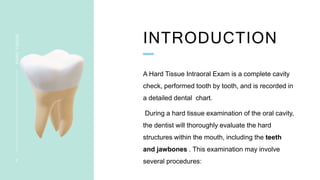
HARD TISSUE
EXAMINATION
ANISHMA KRISHNAN
INTRODUCTION
HARD
TISSUE
2
A Hard Tissue Intraoral Exam is a complete cavity
check, performed tooth by tooth, and is recorded in
a detailed dental chart.
During a hard tissue examination of the oral cavity,
the dentist will thoroughly evaluate the hard
structures within the mouth, including the teeth
and jawbones . This examination may involve
several procedures:
 Visual Examination: The dentist will visually inspect the oral
cavity, looking for any abnormalities or irregularities. They will check for
signs of decay, such as cavities or dark spots on the teeth, as well as
any fractures, chips, or wear on the tooth surfaces.
 Palpation: The dentist may gently press on the jawbones and
surrounding tissues to feel for any abnormalities, such as swelling,
tenderness, or irregularities in bone structure. This can help detect any
underlying issues, such as jawbone infections or tumors.
 Percussion; done by gently tapping the occlusal or incisal surfaces
of the suspected tooth and adjacent tooth using the end of mirror
handle , to check for involvement of periapex and periodontium
3
HARD
TISSUE
 Dental Probing: The dentist may use a dental probe, to measure
the depth of the pockets around each tooth. Deep pockets can
indicate gingival disease or periodontal diseases.
 Dental Radiographs : X-rays provide a detailed view of the teeth
and surrounding structures that may not be visible to the naked eye. X-
rays can reveal cavities, tooth root infections, impacted teeth, jawbone
abnormalities, and other hard tissue problems.
4
HARD
TISSUE
 Bite Evaluation: The dentist will assess how the upper and lower
teeth come together when biting and chewing. This evaluation helps
identify any issues with the bite alignment, such as malocclusions or
problems with the TMJ.
 Occlusal Analysis: The dentist may use articulating paper or bite
registration materials, to evaluate the contact between the teeth in
different jaw positions. This assessment helps identify areas of uneven
pressure or premature contacts that may lead to bite problems or tooth
damage.
Transillumination: In some cases, a transillumination device
may be used to examine the teeth. This involves shining a light through
the tooth to detect cracks, fractures, or other structural abnormalities
that may not be visible otherwise.
5
HARD
TISSUE
.
6
HARD
TISSUE
CONTENT
HARD
TISSUE
EXAMINATION
7
12. Examination of maxilla & mandible
I)TEETH PRESENT
3 TOOTH NUMBERING SYSTEMS ARE GENERALLY USED
- UNIVERSAL (ADOPTED BY ADA)
- THE FDI SYSTEM FEDERATION DENTARIA INTERNATIONAL
- THE ZSIGMONDY-PALMER SYSTEM
UNIVERSAL SYSTEM
HARD
TISSUE
9
Numbering of permanent
teeth: 1-32
Numbering of deciduous
tooth; A-T
Here each tooth is assigned
a number
THE FDI SYSTEM
PRESENTATION
TITLE
1 0
It is a two number
system
First digit indicate ; Quadrant
Second digit indicate ; tooth
in that quadrant
Quadrants ; permanent dentition 1-4
Quadrants ; deciduous dentition 5-8
ZSIGMONDY-PALMER
SYSTEM
1 1
HARD
TISSUE
• Quadrants identified by horizontal and vertical line
• Tooth identified by numbers / alphabets assigned
2)TEETH MISSING
3)DENTAL CARIES
It is a irreversible microbial disease of calcified tissues of teeth
characterized by demineralization of inorganic and destruction of
organic substance of teeth which lead to cavitation
TYPES OF DENTAL CARIES
HARD
TISSUE
1 4
PIT AND
FISSURE
CARIES
Occlusal surface of
molars and premolars
Deep narrow pits and
fissuresfavor the
retention of foodand
debris along with
microbes result in
caries
SOOMTH
SURFACE
CARIES
Can developon the
proximal surface of the
tooth
On the surface on the
tooth
ROOT SURFACE
CARIES
Caries on cementum
Usuallyoccur in older
age group with
significant gingival
recession and exposed
root surface
CERVICAL
CARIES
Seen on the buccal ,lingual
and labial surfaces
It is a crescent shaped
cavity which occur in the
proximal surface as a
roughened chalky area
that gradually become
excavated
HARD
TISSUE
1 5
NURSING
BOTTEL CARIES
Occurs in deciduous
dentition
Most commonly occur
in maxillary incisors
followedmy molars
Uppertooth decay
RADIATION
CARIES
Caries encircling the neck
of the tooth
Brown discoloration of the
tooth
Spot depression which
spread on tooth surface
ARESSTED
CARIES
They do not show the
tendency to progress
RAMPANT
CARIES
Sudden , rapid and
uncontrollable destruction
of multiple primary tooth
1 6
HARD
TISSUE
TENDERNESS/PERCUSSION
TEST
HARD
TISSUE
1 7
VERTICAL PERCUSSION HORIZONTAL
PERCUSSION
pressure is transferred to apical
periodontal tissue
Positive in periapical pathology
pressure is transferred to lateral
periodontal tissue
Positive in periodontium associated
problems
donebygentlytappingtheocclusalorincisalsurfacesof thesuspectedtoothandadjacenttooth
usingtheendofmirrorhandle
4)TOOTH MOBILITY
It is the movement of the teeth in its socket resulting from an applied force
Normally all healthy teeth have a physiological tooth mobility
EXAMINATION;
Applying firm pressure with either two metal instruments or
one metal instrument and gloved finger
SYMPTOMS;
There can be diurnal variations seen
morning-mobility is found to be more- due to lack of chewing or deglutition during
the sleep time
Mobility decreases as the chewing starts
Causes of tooth mobility
trauma from occlusion
periodontitis
Endo-Perio lesion
pathologies like cyst , tumour, osteomyelitis, fracture
After periodontal surgery
Mobility is increased in females after pregnancy, use of contraceptives,
menstruation
1 9
HARD
TISSUE
MILLERS CLASSIFICATION OF
TOOTH MOBILITY
2 0
HARD
TISSUE
5)OCCLUSION
 MOLAR RELATION
 CANINE RELATION
 OVERJET AND OVERBITE
 CROSSBITE
 OPENBITE
 DEEPBITE
ANGLE’S CLASS 1 MOLAR RELATION
2 2
HARD
TISSUE
Mesiobuccally
groove of maxillary
first permanent
molar occludes in
the buccal groove of
mandibular first
permanent molar
ANGLE’S CLASS II MOLAR RELATION
2 3
HARD
TISSUE
mandibular arch distal
to the normal in its
relationship to
maxillary arch
Distobuccal cusp of
maxillary first
permanent premolar
occludes in the
buccal groove of
mandibular first
ANGLE’S CLASS III MOLAR RELATION
2 4
HARD
TISSUE
mandibular arch in
mesial in normal
relation to maxillary
arch
Mesiobuccal cusp of
maxillary first
permanent molar
occludes in the
interdental space
between mandibular
first and second
molar
CANINE
RELATION
2 5
HARD
TISSUE
OVERJET &OVERBITE
OVERBITE OVERJET
2 6
HARD
TISSUE
The condition where the teeth on your upper jaw rest at an
outward angle , causing them to extend far in front of the teeth on
your lower
Overjet refers to the horizontal
distance between the upper and
lower front teeth when the jaws are
closed
Overbite refers to the vertical overlap
between the upper and lower front teeth
when the jaws are closed. It is the
amount by which the upper front teeth
vertically cover the lower front teeth.
6)WASTING DISEASE
It is defined as any gradual loss of tooth substance
characterized by formation of polished surface, without
regards to the possible mechanism of the loss
Types
 ATTRITION
 ABRASION
 EROSION
 ABFRACTION
TYPES OF WASTING
DISEASES
2 8
HARD
TISSUE
ABRACTION
It is the pathological
wearing away of tooth
substance through
some abnormal
mechanical process
V shaped groove and
the cervical third of
tooth with some
gingival recession
ATTRISION
It is the physiological
wearing away of tooth
as a result of a tooth to
tooth contact, as in
mastication
Small polishedfacet on
the cusp tip or
flattening of the incisal
edge
EROSION
It is the irreversible
lossof dental hard
tissue by a chemical
process that doesnot
involve bacteria
smooth shiny and flat
facets surface with
exposeddentin &loss
ofocclusalmorphology
ABFRACTION
lossof tooth structure
that result from
repeated tooth flexure
caused by occlusal
stress Wedge shaped
defects with sharp
margins and sharp
internal angles
7)DENTAL CALCULUS
Dental calculus consist of mineralized bacterial plaque that
forms on the surface of the natural teeth and prosthesis
TYPES ;
SUPRAGINGIVAL CALCULUS
SUBGINGIVAL CALCULUS
SUPRAGINGIVAL
CALCULUS
calculus present on the clinical
crown coronal to the margin of the
gingiva and visible in the oral
cavity
Appearance-
white chalky  creamy – yellow or
gray
may be stained by tobacco or
food
SUBGINGIVAL
CALCULUS
calculus present on the clinical
crown apical to the margin of the
gingiva , usually in periodontal
pockets
Not visible on oral examination
Appearance- light to dark brown ,
dark green, or black stains
3 0
HARD
TISSUE
8)TOOTH FRACTURE
CROWN
FRACTURE
Craze line
Cuspal fracture
Cracked tooth
Split tooth
Horizontal
fracture
Oblique fracture
Vertical fracture
ROOT
FRACTURE
Horizontal
fracture
Oblique
fracture
Vertical
fracture
Coronal1/3rd
Middle 1/3rd
Apical1/3rd
3 2
HARD
TISSUE
CRAZE LINE
these are tiny cracks
that affects only outer
enamel
They are common in
adult teeth and cause
no pain and require no
treatment
They are observeddue
to wear and tear of
tooth
CUSPAL
FRACTURE
The cusp become
weakened and will
fracture
Depending on the
extend of fracture the
pulp could be also
damaged and would
need endo treatment
CRACKED TOOTH
This type of crack
extend chewing
surface, vertically
towards the root and
even below the gum
line It is not a complete
split into two segments
SPLIT TOOTH
It is a cracked tooth ,
where there are 2
distant segments that
can be separated from
one another
ELLIS CLASSIFICATION FOR ANTERIOR
TEETH
3 3
HARD
TISSUE
3 4
HARD
TISSUE
BENNETT’S CLASSIFICATION
 Class 1-Taumatized tooth without coronal or root fracture
class 1a- tooth firm in alveolus
class 1b-Tooth sublexed in alveolus
 Class2-Coronal fracture
class 2a-involving enamel
class 2b- involving enamel and dentin
 Class3-Coronal fracture with pulp exposure
 Class4-Root fracture
Class 4a-without coronal fracture
Class 4b- with coronal fracture
 Class5-Avulsion to the tooth
3 5
HARD
TISSUE
9)DISCOLORED TEETH
Tooth discoloration is a frequent dental finding associated with
clinical and aesthetic problem. It differs in etiology
,appearance, composition, location and severity
2 types
EXTRINSIC DISCOLORATION
INTRINSUC DISCOLORATION
EXTRINSIC
DISCOLORATION
It is defined as discoloration located
on the outer surface of the tooth and
is caused by topical or extrinsic
agents
CAUSES;
Dietary components
Beverages like tea coffee
Tobacco, pan chewing
Chromogenic bacteria
Mouth rise like chlorhexidine
Medication containing iron
,manganese ,copper, nickel
Smoking stain
INTRINSIC DISCOLORATION
occurs following a change to the
structural composition or thickness of
the dental hard tissues
CAUSES;
Amelogenesis imperfecta
Dentinogenetic imperfecta
Dentin hypoplasia
Dental fluorosis
Hyperbilirubinemia
Trauma
Localized red blood cell break down
Medication- tetracycline
Interna resorption
3 7
HARD
TISSUE
3 8
HARD
TISSUE
3 9
HARD
TISSUE
SMOKING STAIN TETRACYCLINE STAIN CHROMOGENIC STAIN
4 0
HARD
TISSUE
FLUOROSIS AMELOGENESIS IMPERFECTA
TRAUMATIC TOOTH
STAIN
10)PARTIALLY ERUPTED TEETH
• Usually observed in the third molars
• Pericoronal flaps covering the partially erupted 3rd molar- can cause
food lodgment and lead to infection called- PERICORONITIS
• Which can further lead to pericoronal abscess and cellulitis
4 2
HARD
TISSUE
11)OTHER ANOMALY
• SUPERNUMERARY TEETHS
• MICRODONTIA/MACRODONTIA
• FUSION
• GEMINATION
• TALONS CUSP
• TURNRES SYNDROME/SINGLE TOOTH HYPOPLASIA
• AMELOGENESIS IMPERFECTA
• DENS IN DENTE
• DENSE EVAGINATUS
• RETAINED DECIDUOUS TOOTH
4 3
HARD
TISSUE
EXAMINATION OF
MAXILLA &MANDIBLE
Examine the
 Size- prognathism /Retrognathism
 Shape –u shaped arches
 Contour-check for surface irregularities ,
exostosis and other lesions
Mandibular
tori
CONCLUSION
HARD
TISSUE
4 5
Hard tissue examinations help the dentist
assess the health of the hard tissues in the
oral cavity, diagnose any problems, and
develop an appropriate treatment plan if
necessary.
Regular hard tissue examinations are
essential for maintaining good oral health
and preventing potential dental issues
REFERENCE
 Peeyush Sivahare 2nd edition
 Odell's clinical problem solving
 Ravi Ongole 2nd edition
4 6
HARD
ISSUE
THANK YOU
ANISHMA KRISHNAN
CLINIC A | AMRITA DENTAL SCHOOL

Hard tissue examination.pptx

  • 1.
  • 2.
    INTRODUCTION HARD TISSUE 2 A Hard TissueIntraoral Exam is a complete cavity check, performed tooth by tooth, and is recorded in a detailed dental chart. During a hard tissue examination of the oral cavity, the dentist will thoroughly evaluate the hard structures within the mouth, including the teeth and jawbones . This examination may involve several procedures:
  • 3.
     Visual Examination:The dentist will visually inspect the oral cavity, looking for any abnormalities or irregularities. They will check for signs of decay, such as cavities or dark spots on the teeth, as well as any fractures, chips, or wear on the tooth surfaces.  Palpation: The dentist may gently press on the jawbones and surrounding tissues to feel for any abnormalities, such as swelling, tenderness, or irregularities in bone structure. This can help detect any underlying issues, such as jawbone infections or tumors.  Percussion; done by gently tapping the occlusal or incisal surfaces of the suspected tooth and adjacent tooth using the end of mirror handle , to check for involvement of periapex and periodontium 3 HARD TISSUE
  • 4.
     Dental Probing:The dentist may use a dental probe, to measure the depth of the pockets around each tooth. Deep pockets can indicate gingival disease or periodontal diseases.  Dental Radiographs : X-rays provide a detailed view of the teeth and surrounding structures that may not be visible to the naked eye. X- rays can reveal cavities, tooth root infections, impacted teeth, jawbone abnormalities, and other hard tissue problems. 4 HARD TISSUE
  • 5.
     Bite Evaluation:The dentist will assess how the upper and lower teeth come together when biting and chewing. This evaluation helps identify any issues with the bite alignment, such as malocclusions or problems with the TMJ.  Occlusal Analysis: The dentist may use articulating paper or bite registration materials, to evaluate the contact between the teeth in different jaw positions. This assessment helps identify areas of uneven pressure or premature contacts that may lead to bite problems or tooth damage. Transillumination: In some cases, a transillumination device may be used to examine the teeth. This involves shining a light through the tooth to detect cracks, fractures, or other structural abnormalities that may not be visible otherwise. 5 HARD TISSUE
  • 6.
  • 7.
  • 8.
    I)TEETH PRESENT 3 TOOTHNUMBERING SYSTEMS ARE GENERALLY USED - UNIVERSAL (ADOPTED BY ADA) - THE FDI SYSTEM FEDERATION DENTARIA INTERNATIONAL - THE ZSIGMONDY-PALMER SYSTEM
  • 9.
    UNIVERSAL SYSTEM HARD TISSUE 9 Numbering ofpermanent teeth: 1-32 Numbering of deciduous tooth; A-T Here each tooth is assigned a number
  • 10.
    THE FDI SYSTEM PRESENTATION TITLE 10 It is a two number system First digit indicate ; Quadrant Second digit indicate ; tooth in that quadrant Quadrants ; permanent dentition 1-4 Quadrants ; deciduous dentition 5-8
  • 11.
    ZSIGMONDY-PALMER SYSTEM 1 1 HARD TISSUE • Quadrantsidentified by horizontal and vertical line • Tooth identified by numbers / alphabets assigned
  • 12.
  • 13.
    3)DENTAL CARIES It isa irreversible microbial disease of calcified tissues of teeth characterized by demineralization of inorganic and destruction of organic substance of teeth which lead to cavitation
  • 14.
    TYPES OF DENTALCARIES HARD TISSUE 1 4 PIT AND FISSURE CARIES Occlusal surface of molars and premolars Deep narrow pits and fissuresfavor the retention of foodand debris along with microbes result in caries SOOMTH SURFACE CARIES Can developon the proximal surface of the tooth On the surface on the tooth ROOT SURFACE CARIES Caries on cementum Usuallyoccur in older age group with significant gingival recession and exposed root surface CERVICAL CARIES Seen on the buccal ,lingual and labial surfaces It is a crescent shaped cavity which occur in the proximal surface as a roughened chalky area that gradually become excavated
  • 15.
    HARD TISSUE 1 5 NURSING BOTTEL CARIES Occursin deciduous dentition Most commonly occur in maxillary incisors followedmy molars Uppertooth decay RADIATION CARIES Caries encircling the neck of the tooth Brown discoloration of the tooth Spot depression which spread on tooth surface ARESSTED CARIES They do not show the tendency to progress RAMPANT CARIES Sudden , rapid and uncontrollable destruction of multiple primary tooth
  • 16.
  • 17.
    TENDERNESS/PERCUSSION TEST HARD TISSUE 1 7 VERTICAL PERCUSSIONHORIZONTAL PERCUSSION pressure is transferred to apical periodontal tissue Positive in periapical pathology pressure is transferred to lateral periodontal tissue Positive in periodontium associated problems donebygentlytappingtheocclusalorincisalsurfacesof thesuspectedtoothandadjacenttooth usingtheendofmirrorhandle
  • 18.
    4)TOOTH MOBILITY It isthe movement of the teeth in its socket resulting from an applied force Normally all healthy teeth have a physiological tooth mobility EXAMINATION; Applying firm pressure with either two metal instruments or one metal instrument and gloved finger SYMPTOMS; There can be diurnal variations seen morning-mobility is found to be more- due to lack of chewing or deglutition during the sleep time Mobility decreases as the chewing starts
  • 19.
    Causes of toothmobility trauma from occlusion periodontitis Endo-Perio lesion pathologies like cyst , tumour, osteomyelitis, fracture After periodontal surgery Mobility is increased in females after pregnancy, use of contraceptives, menstruation 1 9 HARD TISSUE
  • 20.
    MILLERS CLASSIFICATION OF TOOTHMOBILITY 2 0 HARD TISSUE
  • 21.
    5)OCCLUSION  MOLAR RELATION CANINE RELATION  OVERJET AND OVERBITE  CROSSBITE  OPENBITE  DEEPBITE
  • 22.
    ANGLE’S CLASS 1MOLAR RELATION 2 2 HARD TISSUE Mesiobuccally groove of maxillary first permanent molar occludes in the buccal groove of mandibular first permanent molar
  • 23.
    ANGLE’S CLASS IIMOLAR RELATION 2 3 HARD TISSUE mandibular arch distal to the normal in its relationship to maxillary arch Distobuccal cusp of maxillary first permanent premolar occludes in the buccal groove of mandibular first
  • 24.
    ANGLE’S CLASS IIIMOLAR RELATION 2 4 HARD TISSUE mandibular arch in mesial in normal relation to maxillary arch Mesiobuccal cusp of maxillary first permanent molar occludes in the interdental space between mandibular first and second molar
  • 25.
  • 26.
    OVERJET &OVERBITE OVERBITE OVERJET 26 HARD TISSUE The condition where the teeth on your upper jaw rest at an outward angle , causing them to extend far in front of the teeth on your lower Overjet refers to the horizontal distance between the upper and lower front teeth when the jaws are closed Overbite refers to the vertical overlap between the upper and lower front teeth when the jaws are closed. It is the amount by which the upper front teeth vertically cover the lower front teeth.
  • 27.
    6)WASTING DISEASE It isdefined as any gradual loss of tooth substance characterized by formation of polished surface, without regards to the possible mechanism of the loss Types  ATTRITION  ABRASION  EROSION  ABFRACTION
  • 28.
    TYPES OF WASTING DISEASES 28 HARD TISSUE ABRACTION It is the pathological wearing away of tooth substance through some abnormal mechanical process V shaped groove and the cervical third of tooth with some gingival recession ATTRISION It is the physiological wearing away of tooth as a result of a tooth to tooth contact, as in mastication Small polishedfacet on the cusp tip or flattening of the incisal edge EROSION It is the irreversible lossof dental hard tissue by a chemical process that doesnot involve bacteria smooth shiny and flat facets surface with exposeddentin &loss ofocclusalmorphology ABFRACTION lossof tooth structure that result from repeated tooth flexure caused by occlusal stress Wedge shaped defects with sharp margins and sharp internal angles
  • 29.
    7)DENTAL CALCULUS Dental calculusconsist of mineralized bacterial plaque that forms on the surface of the natural teeth and prosthesis TYPES ; SUPRAGINGIVAL CALCULUS SUBGINGIVAL CALCULUS
  • 30.
    SUPRAGINGIVAL CALCULUS calculus present onthe clinical crown coronal to the margin of the gingiva and visible in the oral cavity Appearance- white chalky creamy – yellow or gray may be stained by tobacco or food SUBGINGIVAL CALCULUS calculus present on the clinical crown apical to the margin of the gingiva , usually in periodontal pockets Not visible on oral examination Appearance- light to dark brown , dark green, or black stains 3 0 HARD TISSUE
  • 31.
    8)TOOTH FRACTURE CROWN FRACTURE Craze line Cuspalfracture Cracked tooth Split tooth Horizontal fracture Oblique fracture Vertical fracture ROOT FRACTURE Horizontal fracture Oblique fracture Vertical fracture Coronal1/3rd Middle 1/3rd Apical1/3rd
  • 32.
    3 2 HARD TISSUE CRAZE LINE theseare tiny cracks that affects only outer enamel They are common in adult teeth and cause no pain and require no treatment They are observeddue to wear and tear of tooth CUSPAL FRACTURE The cusp become weakened and will fracture Depending on the extend of fracture the pulp could be also damaged and would need endo treatment CRACKED TOOTH This type of crack extend chewing surface, vertically towards the root and even below the gum line It is not a complete split into two segments SPLIT TOOTH It is a cracked tooth , where there are 2 distant segments that can be separated from one another
  • 33.
    ELLIS CLASSIFICATION FORANTERIOR TEETH 3 3 HARD TISSUE
  • 34.
  • 35.
    BENNETT’S CLASSIFICATION  Class1-Taumatized tooth without coronal or root fracture class 1a- tooth firm in alveolus class 1b-Tooth sublexed in alveolus  Class2-Coronal fracture class 2a-involving enamel class 2b- involving enamel and dentin  Class3-Coronal fracture with pulp exposure  Class4-Root fracture Class 4a-without coronal fracture Class 4b- with coronal fracture  Class5-Avulsion to the tooth 3 5 HARD TISSUE
  • 36.
    9)DISCOLORED TEETH Tooth discolorationis a frequent dental finding associated with clinical and aesthetic problem. It differs in etiology ,appearance, composition, location and severity 2 types EXTRINSIC DISCOLORATION INTRINSUC DISCOLORATION
  • 37.
    EXTRINSIC DISCOLORATION It is definedas discoloration located on the outer surface of the tooth and is caused by topical or extrinsic agents CAUSES; Dietary components Beverages like tea coffee Tobacco, pan chewing Chromogenic bacteria Mouth rise like chlorhexidine Medication containing iron ,manganese ,copper, nickel Smoking stain INTRINSIC DISCOLORATION occurs following a change to the structural composition or thickness of the dental hard tissues CAUSES; Amelogenesis imperfecta Dentinogenetic imperfecta Dentin hypoplasia Dental fluorosis Hyperbilirubinemia Trauma Localized red blood cell break down Medication- tetracycline Interna resorption 3 7 HARD TISSUE
  • 38.
  • 39.
    3 9 HARD TISSUE SMOKING STAINTETRACYCLINE STAIN CHROMOGENIC STAIN
  • 40.
    4 0 HARD TISSUE FLUOROSIS AMELOGENESISIMPERFECTA TRAUMATIC TOOTH STAIN
  • 41.
  • 42.
    • Usually observedin the third molars • Pericoronal flaps covering the partially erupted 3rd molar- can cause food lodgment and lead to infection called- PERICORONITIS • Which can further lead to pericoronal abscess and cellulitis 4 2 HARD TISSUE
  • 43.
    11)OTHER ANOMALY • SUPERNUMERARYTEETHS • MICRODONTIA/MACRODONTIA • FUSION • GEMINATION • TALONS CUSP • TURNRES SYNDROME/SINGLE TOOTH HYPOPLASIA • AMELOGENESIS IMPERFECTA • DENS IN DENTE • DENSE EVAGINATUS • RETAINED DECIDUOUS TOOTH 4 3 HARD TISSUE
  • 44.
    EXAMINATION OF MAXILLA &MANDIBLE Examinethe  Size- prognathism /Retrognathism  Shape –u shaped arches  Contour-check for surface irregularities , exostosis and other lesions Mandibular tori
  • 45.
    CONCLUSION HARD TISSUE 4 5 Hard tissueexaminations help the dentist assess the health of the hard tissues in the oral cavity, diagnose any problems, and develop an appropriate treatment plan if necessary. Regular hard tissue examinations are essential for maintaining good oral health and preventing potential dental issues
  • 46.
    REFERENCE  Peeyush Sivahare2nd edition  Odell's clinical problem solving  Ravi Ongole 2nd edition 4 6 HARD ISSUE
  • 47.
    THANK YOU ANISHMA KRISHNAN CLINICA | AMRITA DENTAL SCHOOL