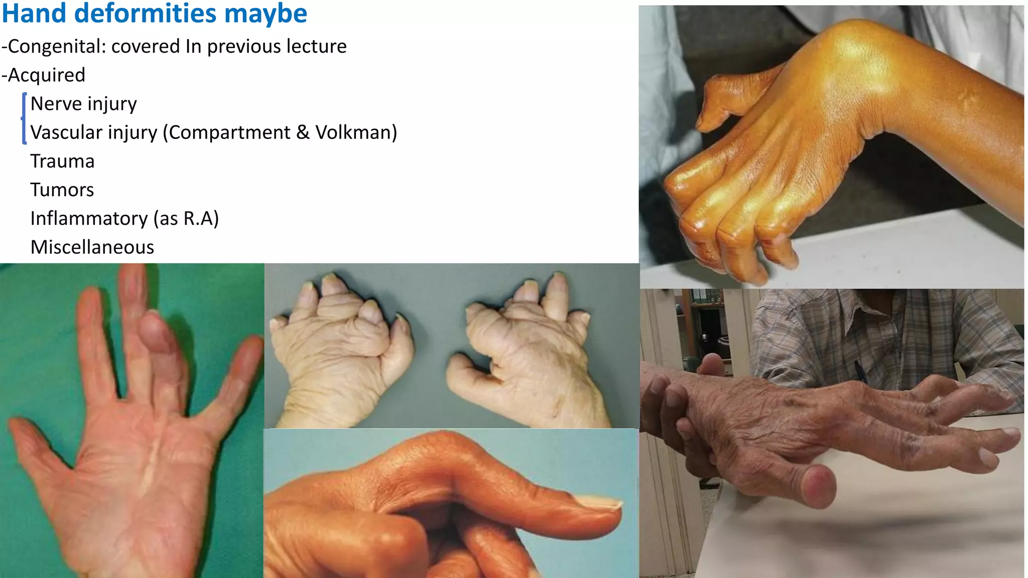
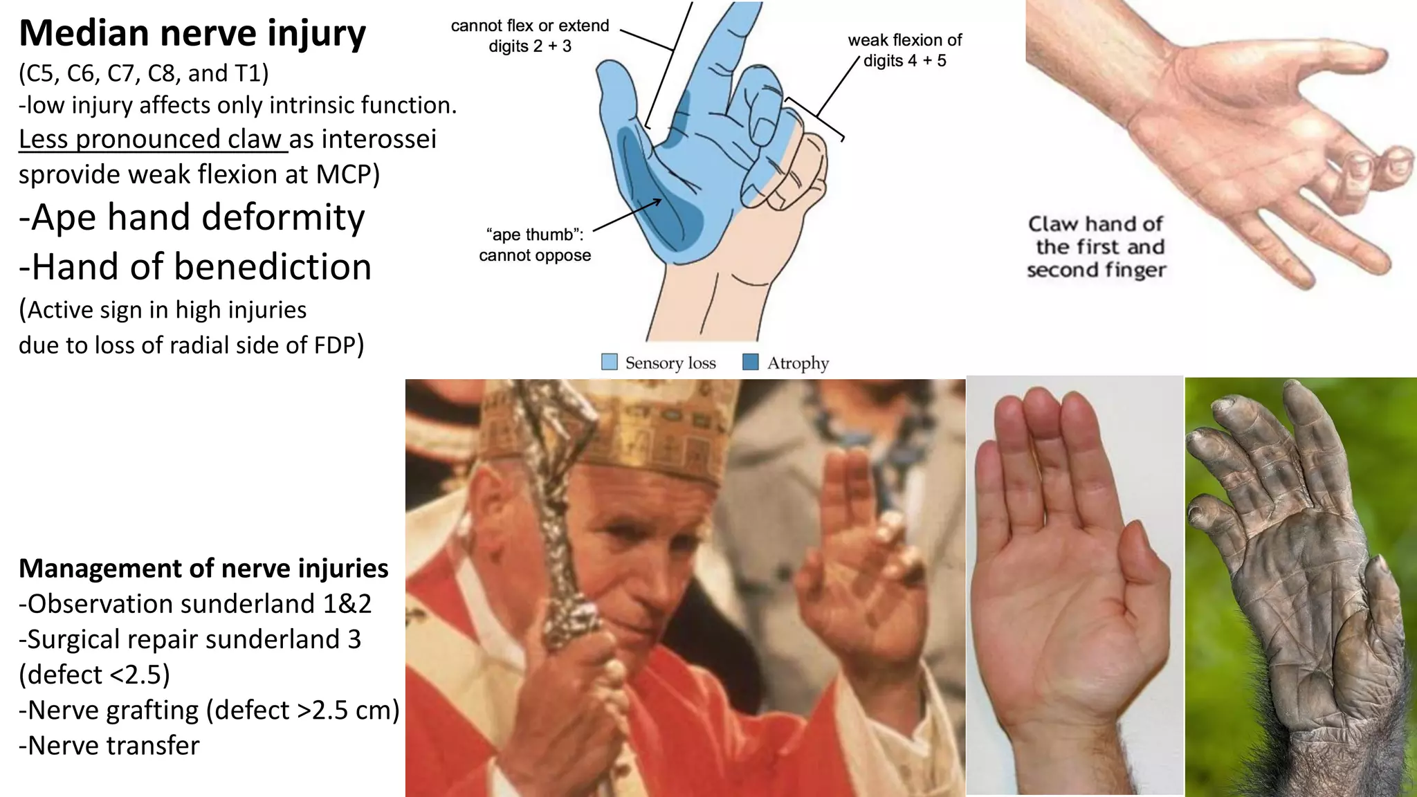
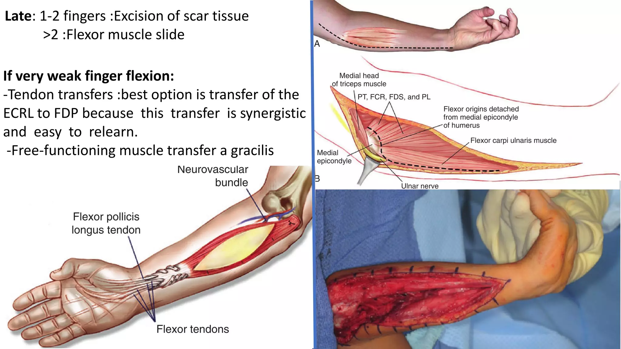
2. What is deformity!
• a physical distortion or disfigurement (Marriam webster)
• A permanent structural deviation from the normal shape, size, or
alignment, resulting in disfigurement; may be congenital or acquired.
(Farlex Partner Medical Dictionary)
4. The LUMBRICALS & INTEROSSEI are the
primary agents in Simultaneous flexion
at the metacarpophalangeal joints and
extension at the interphalangeal joint
VIA their attachment to the dorsal
digital expansion
Intrinsics
Extrinsics
Extension at MCP EDC
Flexion at IP FDS-FDP
6. (-)Loss of intrinsics → loss
of MCP flexion and IP Extension
(+) Strong extrinsic EDC →
unopposed extension of MCP
EDC is not a strong extensor of
the PIP (most IP Extension is
through Interossei
(+) Strong FDP and FDS→
unopposed flexion at the PIP
and DIP
MCP extension, PIP & DIP Flexion
7. Ulnar nerve injury (C8 and T1)
(loss of interossei and medial lumbricals)
Can’t flex MCP
Can’t Extend Interphalangeal
Paradox with functioning
extrinsic in distal injuries
8. Management of nerve injuries
-Observation sunderland 1&2
-Surgical repair sunderland 3
(defect <2.5)
-Nerve grafting (defect >2.5 cm)
-Nerve transfer
Median nerve injury
(C5, C6, C7, C8, and T1)
-low injury affects only intrinsic function.
Less pronounced claw as interossei
sprovide weak flexion at MCP)
-Ape hand deformity
-Hand of benediction
(Active sign in high injuries
due to loss of radial side of FDP)
9. Klumpke Palsy C8-T1
Paralysis in ulnar and median nerve
Loss of hand Intrinsics
Loss of flexors of the wrist and fingers (notably
flexor carpi ulnaris and ulnar half of the flexor
digitorum profundus)
muscular atrophy and tightening of Wrist
& finger flexors → claw hand and wrist
flexion
sensation along C8 and T1 dermatome.
12. Volkman ischemic contracture
It’s the end result of prolonged ischemia and
irreversible tissue necrosis in the forearm
Historically: main cause was SCH
(manipulation and casting in hyperflexion,
NOW→BBF
Ischemia→ fibrosis → contracture and
shortening of Extrinsics → intrinsic minus
Volkman Sign
Wrist extension→ Clawing→ allows finger
Wrist flextion→ allow finger extension
Volkman Angle: flex the wrist and extend the
fingers….gradually extend the wrist..the
angle at which extension ceases→Volkman
angle
13. The following three grades of
Volkmann contracture have been
described:
Mild (Deep flexor compartment
without neurologic deficit)
17. Late: 1-2 fingers :Excision of scar tissue
>2 :Flexor muscle slide
If very weak finger flexion:
-Tendon transfers :best option is transfer of the
ECRL to FDP because this transfer is synergistic
and easy to relearn.
-Free-functioning muscle transfer a gracilis
19. (+)spastic intrinsics
(interossei and lumbricals)
→ incresed of MCP flexion
and IP Extension
(-) weak extrinsics:
FDP, FDS Fail to provide
balancing flexion force at PIP, DIP
EDC Fail to provide balancing
extension force at MCP
MCP Flexion , PIP & DIP extension
29. Transverse
Retinacular
ligament
Oblique
Retinacular
ligament
Function: prevent excessive dorsal
translation of lateral bands.
Counteracts the Triangular ligament
Function: links motion
of DIP with PIP
Triangular ligament
Function preventing volar subluxation of lateral band
Counteract the transverse retinacular ligament
Sagittal Band
Function: keep extensor mechanism in the midline during
flexion of MP joint & prevents bowstringing
Triangular ligament and Transverse retinacular ligament balance the functioning of the lateral bands
31. Radial part of sagittal
band under more
stress due to ulnar
forces
32. Ulnar directed Stress + Tenosynovitis→ rupture of radial pa
of sagittal band
Extensor tendon subluxate ulnarly
Ulnar Intrinsic Contracture further worsening the
deformity→intrinsic Plus Position
-flexor tendons subluxate as well further augmenting the
deformity (due to ulnar deviation of the wrist, radial hand
deviation resulting in ulnar subluxation of fingers
33. Management
-night splints may slow the progression
-Surgery: Synovectomy, Extensor tendon centralization
(release of contracted ulnar sagittal band, radial sagittal tightened)
and intrinsic release
-Arthroplasty
35. Can also be caused by
Traumatic tendon injuries
→ damaged central slip
-Synovitis of PIP → central slip rupture and dorsal
capsule attenuation
-Increased PIP flexion → lateral band subluxate in
volar direction
-attenuation/tear of triangular ligament
-Oblique retinacular ligament contractures causing
extension contracture of DIP
38. -DIP synovitis →terminal tendon rupture and
dorsal capsule attenuation
-lateral band subluxate dorsal to PIP axis
-attenuation of the transverse retinacular ligament
-DIP flexion/PIP hyperextension
Can also be caused by
1ry→lax volar plate (ehler danlos)
Mallet finger
FDS laceration
40. Thumb deformities in RA
Type I Thumb Deformity (Boutonniere)
most common, Metacarpal phalangeal (MP) joint flexion
and interphalangeal (IP) joint hyperextension
Type II Same with adduction at the trapeziometacarpal
(TMC) joint
type III (Swan Neck)
•TMC adduction, MP hyperextension and IP flexion
Type IV (Gamekeeper's)
laxity of the ulnar collateral ligament at the thumb MP joint
related to synovitis, proximal phalanx of the thumb deviates
laterally and then the TMC joint goes into adduction
•Type V Thumb Deformity: isolated MCP hyper-extension
•Type VI Thumb Deformity Arthritis mutilans → bone loss
46. Cutaneous Ligaments
Facial components involved
in doputyren’s
-Pretindenious band
-Spiral band
-Lateral digital sheet
-Greyson lig.
-Natatory ligament
deep
47. • progressive thickening and shorting of the palmar fascia →
digital contractures, particularly MCP & PIP joints, usually 4th
and 5th digit
• cause is unknown, family history is often present. Males >
females in ratio and severity
• Other risk factors: include manual labor with vibration
exposure, prior trauma, alcoholism, smoking, DM,
hyperlipidemia, Peyronie disease, and CRPS
48. Grade 1 - Thickened nodule and
band in the palmar
aponeurosis; may have
associated skin abnormalities
Grade 2 - Development of
pretendinous and digital cords
with limitation of finger
extension
Grade 3 - Presence of flexion
contracture
Cardinal features are
the nodule & cord
Management:
-ROM Exercises, Collagenase injection
-Surgical resection (fasciectomy) +/- skin grafts
Problematic as spiral band spirals around NV bundle