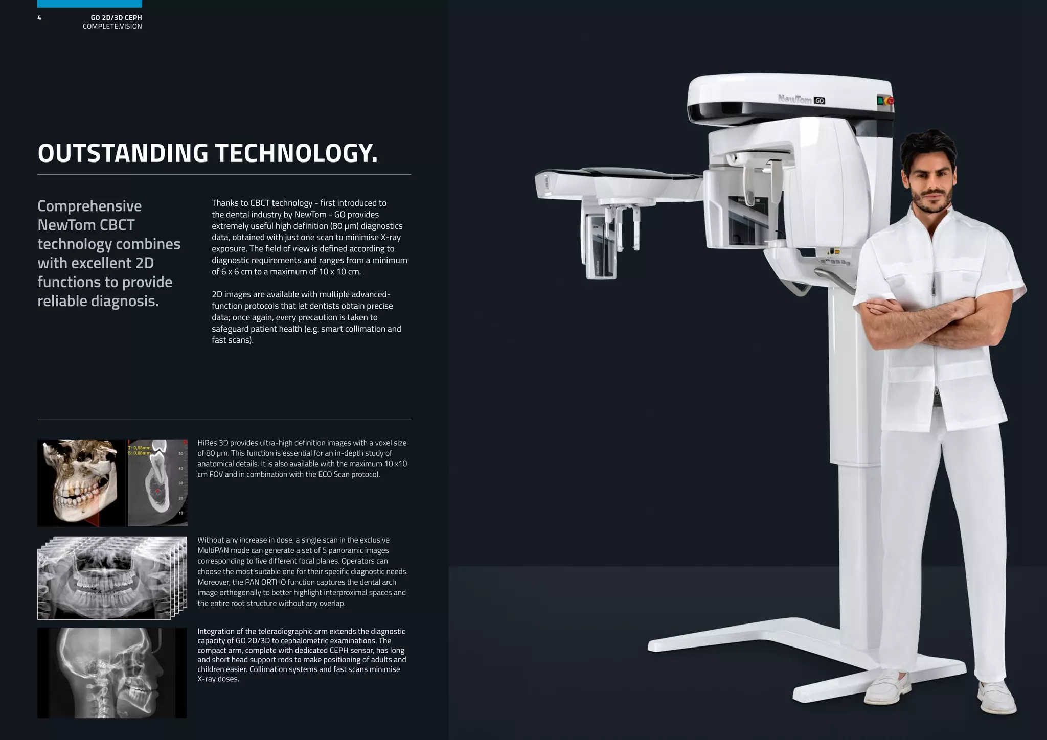
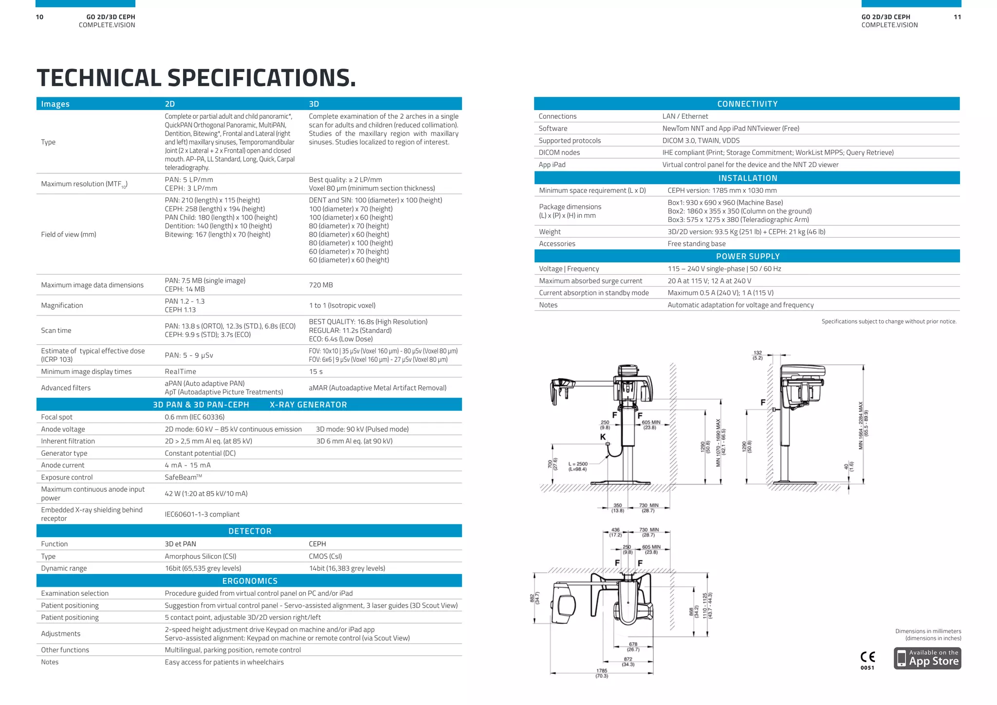
CEFLA s.c.
Via Selice Provinciale 23/a • 40026 Imola • Italy
t. +39 045 8202727 • 045 583500
info@newtom.it
newtom.it
NewTom GO
COMPLETE.VISION
2D/3D CEPH INTEGRATED IMAGING
07/2019NG3CGB191S00
Accordingtothestandardsinforce,inextra-EUareastheavailabilityandspecificationsofsomeproductsand/orcharacteristicsmayvary.Pleasecontactyourlocaldistributorforfurtherinformation.Picturesareforillustrationpurposeonly.
GO 2D/3D CEPH
COMPLETE.VISION
2
GO 2D/3D CEPH able to provide high resolution
images, the platform prioritises patient health thanks
to low exposure protocols and exclusive SafeBeam™
technology, which lets users adapt the dose to their
actual diagnostic needs and the size of the examined
anatomical area.
Excellent ergonomics and an adaptive alignment
system ensure correct positioning of the patient and
perfect focusing for clear, detailed images. A virtual
control panel guides the operator through each
stage of the examination. NNT is the technologically
advanced software platform to manage, process,
consult and share diagnostic images.
High quality images
that meet a wide
range of clinical
diagnostic needs,
all in one compact
device.
MINIMUM
X-RAY DOSE
ECO Dose functions and
SafeBeam™ technology,
which automatically
adapts to the dose
radiated to the patient,
put patient safety
above all else.
ACCESSIBLE
TECHNOLOGY
Guided procedures
and smart automatic
features have made
this sophisticated
technology available
to all.
BROAD DIAGNOSTIC
POTENTIAL
A combination of device
versatility and NewTom
solutions designed to
meet every diagnostic
need broaden the
opportunities available
to the surgery.
MAXIMUM
CONNECTIVITY
Acquired X-ray images
can easily be stored,
exported and shared
with specialist third
party software.
VERSATILE AND PRECISE.
GO 2D/3D CEPH
COMPLETE.VISION
4
Thanks to CBCT technology - first introduced to
the dental industry by NewTom - GO provides
extremely useful high definition (80 μm) diagnostics
data, obtained with just one scan to minimise X-ray
exposure. The field of view is defined according to
diagnostic requirements and ranges from a minimum
of 6 x 6 cm to a maximum of 10 x 10 cm.
2D images are available with multiple advanced-
function protocols that let dentists obtain precise
data; once again, every precaution is taken to
safeguard patient health (e.g. smart collimation and
fast scans).
Comprehensive
NewTom CBCT
technology combines
with excellent 2D
functions to provide
reliable diagnosis.
OUTSTANDING TECHNOLOGY.
Integration of the teleradiographic arm extends the diagnostic
capacity of GO 2D/3D to cephalometric examinations. The
compact arm, complete with dedicated CEPH sensor, has long
and short head support rods to make positioning of adults and
children easier. Collimation systems and fast scans minimise
X-ray doses.
Without any increase in dose, a single scan in the exclusive
MultiPAN mode can generate a set of 5 panoramic images
corresponding to five different focal planes. Operators can
choose the most suitable one for their specific diagnostic needs.
Moreover, the PAN ORTHO function captures the dental arch
image orthogonally to better highlight interproximal spaces and
the entire root structure without any overlap.
HiRes 3D provides ultra-high definition images with a voxel size
of 80 μm. This function is essential for an in-depth study of
anatomical details. It is also available with the maximum 10 x10
cm FOV and in combination with the ECO Scan protocol.
GO 2D/3D CEPH
COMPLETE.VISION
7GO 2D/3D CEPH
COMPLETE.VISION
6
To ensure accurate diagnoses in every situation it is
vital to observe procedures that ensure images are
always sharp and clear. GO 2D/3D has a single native
16-bit sensor that produces 2D and 3D images with
thousands of grey levels. Image quality is ensured by
advanced algorithms and protocols and by
high-tech image sequencing. The high frequency,
pulsed-emission generator adjusts exposure to obtain
the best scans with a minimum dose.
Moreover, the cephalometric exam collimation system
is based on automatic movement of the turret, which
rotates and lowers the sensor, creating an opening
for the X-rays directed at the 2D sensor on the
teleradiographic arm.
Solutions developed
to maximise
examination
quality, from
positioning systems
to automated
collimation.
With its five contact points, the 3D scan head
support helps staff position the patient correctly
and comfortably. Frontal and lateral contact points
can be adjusted to maximise both patient stability
during the scan and, consequently, the quality of the
obtained data.
A specific protocol allows for tomographic scans
of radiological templates, prostheses, models or
impressions after they have been positioned on a
special support.
AUTOMATIC AND ERGONOMIC.
GO 2D/3D CEPH
COMPLETE.VISION
9GO 2D/3D CEPH
COMPLETE.VISION
8
VIRTU AL
CONSO LE
SURGERY
SOFTWARE
PRINTERS
PROCESSING
DEV
ICES
REM
OTE
ASSISTANCE
3D/2DVIEWER1:1PRINT
RIS/PACS
M
ANAGEMENT
PLANNING
OTHER
AC
QUISITION
DISPLAY AND 3D SCANNER
3D
M
ILLING
MULTI-STATION
SPECIALIST
SOFTWARE
REPORTS
SURGERY
T
REATMENTSYSTEMS
2D/3D
IM
AGE MANAGEMENT
INFORMATIO
N
SYSTEMS
COMPLETE CONNECTIVITY.
Excellent connectivity and
integration with the modern
systems adopted by NewTom.
Workflow and clinical and
diagnostic activities become
much easier and highly
performing.
VIRTUAL CONSOLE
Settings required for acquisition can be easily
controlled from a remote virtual control panel
on the PC, laptop, Windows tablet or iPad.
RIS/PACS
IHE compliant system that allows
communication with RIS/PACS systems and
DICOM printers. Complete services available:
Print, Worklist, Storage Commitment, MPPS
and Query/Retrieve.
REMOTE ASSISTANCE
By appropriately configuring the device
to use the surgery’s Internet connection,
technical support can be provided from
remote, and device status can be monitored.
SURGERY MANAGEMENT SOFTWARE
An open system designed for fast, efficient
interfacing with the main dental surgery
management software solutions via various
standard VDDS, TWAIN and/or proprietary
NNTBridge modes.
3D/2D VIEWER
Examinations can be shared with colleagues
and patients by providing the Viewer directly
on CD, DVD or a USB storage device.
SPECIALIST PLANNING SOFTWARE
Exports in DICOM 3.0 format to specialist
planning software to process orthodontic
treatments, prostheses, implants,
orthognathic and maxillofacial surgery.
1:1 PRINT
Complete and flexible report for storing and
sharing colour reports on photographic paper
or grey scale reports on X-ray-equivalent
transparencies.
3D MILLING PRINTERS
Software modules are available to segment
the reconstructed volume and export to
STL format the surfaces required to create
3D models that can underpin planning and
treatment.
OTHER ACQUISITION DEVICES
Compatibility with TWAIN and DICOM
3.0 standards guarantees NNT software
management of images from other 2D/3D
image acquisition devices, such as video
cameras, sensors, PSP and CBCT scanners.
3D SCANNER
Prosthetically guided planning by integrating
(via the dedicated software module) data
in STL format from optical, intraoral or
laboratory scanners, with volumetric data.
MULTI-STATION DISPLAY AND
PROCESSING
Image storage on a shared database in a
local network that can be accessed from any
workstation and iPad (only 2D). Management
of multiple archives and access to password-
protected data.
GO 2D/3D CEPH
COMPLETE.VISION
11GO 2D/3D CEPH
COMPLETE.VISION
10
0051
TECHNICAL SPECIFICATIONS.
Images 2D 3D
Type
Complete or partial adult and child panoramic*,
QuickPAN Orthogonal Panoramic, MultiPAN,
Dentition, Bitewing*, Frontal and Lateral (right
and left) maxillary sinuses, Temporomandibular
Joint (2 x Lateral + 2 x Frontal) open and closed
mouth. AP-PA, LL Standard, Long, Quick, Carpal
teleradiography.
Complete examination of the 2 arches in a single
scan for adults and children (reduced collimation).
Studies of the maxillary region with maxillary
sinuses. Studies localized to region of interest.
Maximum resolution (MTF10
)
PAN: 5 LP/mm
CEPH: 3 LP/mm
Best quality: ≥ 2 LP/mm
Voxel 80 µm (minimum section thickness)
Field of view (mm)
PAN: 210 (length) x 115 (height)
CEPH: 258 (length) x 194 (height)
PAN Child: 180 (length) x 100 (height)
Dentition: 140 (length) x 10 (height)
Bitewing: 167 (length) x 70 (height)
DENT and SIN: 100 (diameter) x 100 (height)
100 (diameter) x 70 (height)
100 (diameter) x 60 (height)
80 (diameter) x 70 (height)
80 (diameter) x 60 (height)
80 (diameter) x 100 (height)
60 (diameter) x 70 (height)
60 (diameter) x 60 (height)
Maximum image data dimensions
PAN: 7.5 MB (single image)
CEPH: 14 MB
720 MB
Magnification
PAN 1.2 - 1.3
CEPH 1.13
1 to 1 (Isotropic voxel)
Scan time
PAN: 13.8 s (ORTO), 12.3s (STD.), 6.8s (ECO)
CEPH: 9.9 s (STD); 3.7s (ECO)
BEST QUALITY: 16.8s (High Resolution)
REGULAR: 11.2s (Standard)
ECO: 6.4s (Low Dose)
Estimate of typical effective dose
(ICRP 103)
PAN: 5 - 9 µSv
FOV: 10x10 | 35 µSv (Voxel 160 µm) - 80 µSv (Voxel 80 µm)
FOV: 6x6 | 9 µSv (Voxel 160 µm) - 27 µSv (Voxel 80 µm)
Minimum image display times RealTime 15 s
Advanced filters
aPAN (Auto adaptive PAN)
ApT (Autoadaptive Picture Treatments)
aMAR (Autoadaptive Metal Artifact Removal)
3D PAN & 3D PAN-CEPH X-RAY GENERATOR
Focal spot 0.6 mm (IEC 60336)
Anode voltage 2D mode: 60 kV – 85 kV continuous emission 3D mode: 90 kV (Pulsed mode)
Inherent filtration 2D > 2,5 mm Al eq. (at 85 kV) 3D 6 mm Al eq. (at 90 kV)
Generator type Constant potential (DC)
Anode current 4 mA - 15 mA
Exposure control SafeBeamTM
Maximum continuous anode input
power
42 W (1:20 at 85 kV/10 mA)
Embedded X-ray shielding behind
receptor
IEC60601-1-3 compliant
DETECTOR
Function 3D et PAN CEPH
Type Amorphous Silicon (CSI) CMOS (CsI)
Dynamic range 16bit (65,535 grey levels) 14bit (16,383 grey levels)
ERGONOMICS
Examination selection Procedure guided from virtual control panel on PC and/or iPad
Patient positioning Suggestion from virtual control panel - Servo-assisted alignment, 3 laser guides (3D Scout View)
Patient positioning 5 contact point, adjustable 3D/2D version right/left
Adjustments
2-speed height adjustment drive Keypad on machine and/or iPad app
Servo-assisted alignment: Keypad on machine or remote control (via Scout View)
Other functions Multilingual, parking position, remote control
Notes Easy access for patients in wheelchairs
INSTALLATION
Minimum space requirement (L x D) CEPH version: 1785 mm x 1030 mm
Package dimensions
(L) x (P) x (H) in mm
Box1: 930 x 690 x 960 (Machine Base)
Box2: 1860 x 355 x 350 (Column on the ground)
Box3: 575 x 1275 x 380 (Teleradiographic Arm)
Weight 3D/2D version: 93.5 Kg (251 lb) + CEPH: 21 kg (46 lb)
Accessories Free standing base
POWER SUPPLY
Voltage | Frequency 115 – 240 V single-phase | 50 / 60 Hz
Maximum absorbed surge current 20 A at 115 V; 12 A at 240 V
Current absorption in standby mode Maximum 0.5 A (240 V); 1 A (115 V)
Notes Automatic adaptation for voltage and frequency
Specifications subject to change without prior notice.
Dimensions in millimeters
(dimensions in inches)
CONNECTIVITY
Connections LAN / Ethernet
Software NewTom NNT and App iPad NNTviewer (Free)
Supported protocols DICOM 3.0, TWAIN, VDDS
DICOM nodes IHE compliant (Print; Storage Commitment; WorkList MPPS; Query Retrieve)
App iPad Virtual control panel for the device and the NNT 2D viewer

Go 2 d3d ceph_cataloge

  • 1.
    CEFLA s.c. Via SeliceProvinciale 23/a • 40026 Imola • Italy t. +39 045 8202727 • 045 583500 info@newtom.it newtom.it NewTom GO COMPLETE.VISION 2D/3D CEPH INTEGRATED IMAGING 07/2019NG3CGB191S00 Accordingtothestandardsinforce,inextra-EUareastheavailabilityandspecificationsofsomeproductsand/orcharacteristicsmayvary.Pleasecontactyourlocaldistributorforfurtherinformation.Picturesareforillustrationpurposeonly.
  • 2.
    GO 2D/3D CEPH COMPLETE.VISION 2 GO2D/3D CEPH able to provide high resolution images, the platform prioritises patient health thanks to low exposure protocols and exclusive SafeBeam™ technology, which lets users adapt the dose to their actual diagnostic needs and the size of the examined anatomical area. Excellent ergonomics and an adaptive alignment system ensure correct positioning of the patient and perfect focusing for clear, detailed images. A virtual control panel guides the operator through each stage of the examination. NNT is the technologically advanced software platform to manage, process, consult and share diagnostic images. High quality images that meet a wide range of clinical diagnostic needs, all in one compact device. MINIMUM X-RAY DOSE ECO Dose functions and SafeBeam™ technology, which automatically adapts to the dose radiated to the patient, put patient safety above all else. ACCESSIBLE TECHNOLOGY Guided procedures and smart automatic features have made this sophisticated technology available to all. BROAD DIAGNOSTIC POTENTIAL A combination of device versatility and NewTom solutions designed to meet every diagnostic need broaden the opportunities available to the surgery. MAXIMUM CONNECTIVITY Acquired X-ray images can easily be stored, exported and shared with specialist third party software. VERSATILE AND PRECISE.
  • 3.
    GO 2D/3D CEPH COMPLETE.VISION 4 Thanksto CBCT technology - first introduced to the dental industry by NewTom - GO provides extremely useful high definition (80 μm) diagnostics data, obtained with just one scan to minimise X-ray exposure. The field of view is defined according to diagnostic requirements and ranges from a minimum of 6 x 6 cm to a maximum of 10 x 10 cm. 2D images are available with multiple advanced- function protocols that let dentists obtain precise data; once again, every precaution is taken to safeguard patient health (e.g. smart collimation and fast scans). Comprehensive NewTom CBCT technology combines with excellent 2D functions to provide reliable diagnosis. OUTSTANDING TECHNOLOGY. Integration of the teleradiographic arm extends the diagnostic capacity of GO 2D/3D to cephalometric examinations. The compact arm, complete with dedicated CEPH sensor, has long and short head support rods to make positioning of adults and children easier. Collimation systems and fast scans minimise X-ray doses. Without any increase in dose, a single scan in the exclusive MultiPAN mode can generate a set of 5 panoramic images corresponding to five different focal planes. Operators can choose the most suitable one for their specific diagnostic needs. Moreover, the PAN ORTHO function captures the dental arch image orthogonally to better highlight interproximal spaces and the entire root structure without any overlap. HiRes 3D provides ultra-high definition images with a voxel size of 80 μm. This function is essential for an in-depth study of anatomical details. It is also available with the maximum 10 x10 cm FOV and in combination with the ECO Scan protocol.
  • 4.
    GO 2D/3D CEPH COMPLETE.VISION 7GO2D/3D CEPH COMPLETE.VISION 6 To ensure accurate diagnoses in every situation it is vital to observe procedures that ensure images are always sharp and clear. GO 2D/3D has a single native 16-bit sensor that produces 2D and 3D images with thousands of grey levels. Image quality is ensured by advanced algorithms and protocols and by high-tech image sequencing. The high frequency, pulsed-emission generator adjusts exposure to obtain the best scans with a minimum dose. Moreover, the cephalometric exam collimation system is based on automatic movement of the turret, which rotates and lowers the sensor, creating an opening for the X-rays directed at the 2D sensor on the teleradiographic arm. Solutions developed to maximise examination quality, from positioning systems to automated collimation. With its five contact points, the 3D scan head support helps staff position the patient correctly and comfortably. Frontal and lateral contact points can be adjusted to maximise both patient stability during the scan and, consequently, the quality of the obtained data. A specific protocol allows for tomographic scans of radiological templates, prostheses, models or impressions after they have been positioned on a special support. AUTOMATIC AND ERGONOMIC.
  • 5.
    GO 2D/3D CEPH COMPLETE.VISION 9GO2D/3D CEPH COMPLETE.VISION 8 VIRTU AL CONSO LE SURGERY SOFTWARE PRINTERS PROCESSING DEV ICES REM OTE ASSISTANCE 3D/2DVIEWER1:1PRINT RIS/PACS M ANAGEMENT PLANNING OTHER AC QUISITION DISPLAY AND 3D SCANNER 3D M ILLING MULTI-STATION SPECIALIST SOFTWARE REPORTS SURGERY T REATMENTSYSTEMS 2D/3D IM AGE MANAGEMENT INFORMATIO N SYSTEMS COMPLETE CONNECTIVITY. Excellent connectivity and integration with the modern systems adopted by NewTom. Workflow and clinical and diagnostic activities become much easier and highly performing. VIRTUAL CONSOLE Settings required for acquisition can be easily controlled from a remote virtual control panel on the PC, laptop, Windows tablet or iPad. RIS/PACS IHE compliant system that allows communication with RIS/PACS systems and DICOM printers. Complete services available: Print, Worklist, Storage Commitment, MPPS and Query/Retrieve. REMOTE ASSISTANCE By appropriately configuring the device to use the surgery’s Internet connection, technical support can be provided from remote, and device status can be monitored. SURGERY MANAGEMENT SOFTWARE An open system designed for fast, efficient interfacing with the main dental surgery management software solutions via various standard VDDS, TWAIN and/or proprietary NNTBridge modes. 3D/2D VIEWER Examinations can be shared with colleagues and patients by providing the Viewer directly on CD, DVD or a USB storage device. SPECIALIST PLANNING SOFTWARE Exports in DICOM 3.0 format to specialist planning software to process orthodontic treatments, prostheses, implants, orthognathic and maxillofacial surgery. 1:1 PRINT Complete and flexible report for storing and sharing colour reports on photographic paper or grey scale reports on X-ray-equivalent transparencies. 3D MILLING PRINTERS Software modules are available to segment the reconstructed volume and export to STL format the surfaces required to create 3D models that can underpin planning and treatment. OTHER ACQUISITION DEVICES Compatibility with TWAIN and DICOM 3.0 standards guarantees NNT software management of images from other 2D/3D image acquisition devices, such as video cameras, sensors, PSP and CBCT scanners. 3D SCANNER Prosthetically guided planning by integrating (via the dedicated software module) data in STL format from optical, intraoral or laboratory scanners, with volumetric data. MULTI-STATION DISPLAY AND PROCESSING Image storage on a shared database in a local network that can be accessed from any workstation and iPad (only 2D). Management of multiple archives and access to password- protected data.
  • 6.
    GO 2D/3D CEPH COMPLETE.VISION 11GO2D/3D CEPH COMPLETE.VISION 10 0051 TECHNICAL SPECIFICATIONS. Images 2D 3D Type Complete or partial adult and child panoramic*, QuickPAN Orthogonal Panoramic, MultiPAN, Dentition, Bitewing*, Frontal and Lateral (right and left) maxillary sinuses, Temporomandibular Joint (2 x Lateral + 2 x Frontal) open and closed mouth. AP-PA, LL Standard, Long, Quick, Carpal teleradiography. Complete examination of the 2 arches in a single scan for adults and children (reduced collimation). Studies of the maxillary region with maxillary sinuses. Studies localized to region of interest. Maximum resolution (MTF10 ) PAN: 5 LP/mm CEPH: 3 LP/mm Best quality: ≥ 2 LP/mm Voxel 80 µm (minimum section thickness) Field of view (mm) PAN: 210 (length) x 115 (height) CEPH: 258 (length) x 194 (height) PAN Child: 180 (length) x 100 (height) Dentition: 140 (length) x 10 (height) Bitewing: 167 (length) x 70 (height) DENT and SIN: 100 (diameter) x 100 (height) 100 (diameter) x 70 (height) 100 (diameter) x 60 (height) 80 (diameter) x 70 (height) 80 (diameter) x 60 (height) 80 (diameter) x 100 (height) 60 (diameter) x 70 (height) 60 (diameter) x 60 (height) Maximum image data dimensions PAN: 7.5 MB (single image) CEPH: 14 MB 720 MB Magnification PAN 1.2 - 1.3 CEPH 1.13 1 to 1 (Isotropic voxel) Scan time PAN: 13.8 s (ORTO), 12.3s (STD.), 6.8s (ECO) CEPH: 9.9 s (STD); 3.7s (ECO) BEST QUALITY: 16.8s (High Resolution) REGULAR: 11.2s (Standard) ECO: 6.4s (Low Dose) Estimate of typical effective dose (ICRP 103) PAN: 5 - 9 µSv FOV: 10x10 | 35 µSv (Voxel 160 µm) - 80 µSv (Voxel 80 µm) FOV: 6x6 | 9 µSv (Voxel 160 µm) - 27 µSv (Voxel 80 µm) Minimum image display times RealTime 15 s Advanced filters aPAN (Auto adaptive PAN) ApT (Autoadaptive Picture Treatments) aMAR (Autoadaptive Metal Artifact Removal) 3D PAN & 3D PAN-CEPH X-RAY GENERATOR Focal spot 0.6 mm (IEC 60336) Anode voltage 2D mode: 60 kV – 85 kV continuous emission 3D mode: 90 kV (Pulsed mode) Inherent filtration 2D > 2,5 mm Al eq. (at 85 kV) 3D 6 mm Al eq. (at 90 kV) Generator type Constant potential (DC) Anode current 4 mA - 15 mA Exposure control SafeBeamTM Maximum continuous anode input power 42 W (1:20 at 85 kV/10 mA) Embedded X-ray shielding behind receptor IEC60601-1-3 compliant DETECTOR Function 3D et PAN CEPH Type Amorphous Silicon (CSI) CMOS (CsI) Dynamic range 16bit (65,535 grey levels) 14bit (16,383 grey levels) ERGONOMICS Examination selection Procedure guided from virtual control panel on PC and/or iPad Patient positioning Suggestion from virtual control panel - Servo-assisted alignment, 3 laser guides (3D Scout View) Patient positioning 5 contact point, adjustable 3D/2D version right/left Adjustments 2-speed height adjustment drive Keypad on machine and/or iPad app Servo-assisted alignment: Keypad on machine or remote control (via Scout View) Other functions Multilingual, parking position, remote control Notes Easy access for patients in wheelchairs INSTALLATION Minimum space requirement (L x D) CEPH version: 1785 mm x 1030 mm Package dimensions (L) x (P) x (H) in mm Box1: 930 x 690 x 960 (Machine Base) Box2: 1860 x 355 x 350 (Column on the ground) Box3: 575 x 1275 x 380 (Teleradiographic Arm) Weight 3D/2D version: 93.5 Kg (251 lb) + CEPH: 21 kg (46 lb) Accessories Free standing base POWER SUPPLY Voltage | Frequency 115 – 240 V single-phase | 50 / 60 Hz Maximum absorbed surge current 20 A at 115 V; 12 A at 240 V Current absorption in standby mode Maximum 0.5 A (240 V); 1 A (115 V) Notes Automatic adaptation for voltage and frequency Specifications subject to change without prior notice. Dimensions in millimeters (dimensions in inches) CONNECTIVITY Connections LAN / Ethernet Software NewTom NNT and App iPad NNTviewer (Free) Supported protocols DICOM 3.0, TWAIN, VDDS DICOM nodes IHE compliant (Print; Storage Commitment; WorkList MPPS; Query Retrieve) App iPad Virtual control panel for the device and the NNT 2D viewer