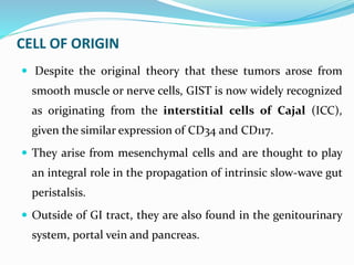
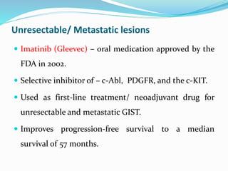
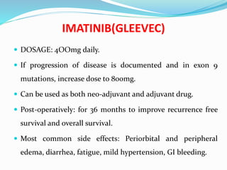
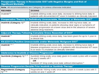
Dr. N.ANJAN KUMAR
MODERATOR : Dr. SAMBASHIVA RAO,
ASSOCIATE PROFESSOR,
DEPT. OF GENERAL SURGERY.
LAYOUT
 INTRODUCTION
 EPIDEMIOLOGY
 IMMUNO-HISTOCHEMISTRY
 RISK ASSESSMENT
 MOLECULAR MECHANISMS OF CARCINOGENESIS
 CLINICAL PRESENTATION
 IMAGING
 TREATMENT
 IMATINIB AND OTHER NEWER DRUGS
 OUTCOME
INTRODUCTION
• Golden and Stout in 1941: described the mesenchymal
tumors arising in bowel as tumors arising from smooth
muscle cells; leiomyoblastoma, leiomyoma and
leiomyosarcoma.
• Term GIST was 1st used by Mazur and Clark in 1983.
• In 1998 Japanese research workers (Hirota et al.) discovered
KIT mutations in GIST that possibly distinguish GIST from
other tumors.
 Most common mesenchymal tumours of the
Gastrointestinal tract.
 Classified separate from leiomyomas and
leiomyosarcomas in 1980s after demonstration by
immunohistochemistry and electron microscopy.
 Although they represent only 0.1–3% of all
gastrointestinal (GI) malignancies, they account for
80% of gastrointestinal mesenchymal neoplasms.
EPIDEMIOLOGY
 Annual incidence: 15 per million.
 Age: 50-80 years; median age: 6th decade.
 Gender: M=F.
 Mostly: sporadic.
 Associations:
- Carney’s triad (GIST, paragangliomas, pulmonary
chondromas)
- Neurofibromatosis
- Familial GIST syndrome
IMMUNO-HISTOCHEMISTRY
 Immuno-histochemical staining demonstrated decreased
staining for desmin and smooth muscle actin (markers
that are typically expressed in smooth muscle tumors).
 In the early 1990s, the extracellular membrane protein
CD34 was identified in GIST but only rarely in
leiomyomas, and the distinct classification became more
widely accepted.
 Further research identified immuno-histochemical
staining for the protein CD117, which is the tyrosine
kinase receptor known as KIT, with a sensitivity
greater than 95% and a high specificity.
 CD117 immuno-histochemistry staining is now widely
accepted as a criterion for a pathologic diagnosis of
GIST.
 Additional research has identified a new immuno-
histochemical stain called DOG1—literally Discovered
On GIST-1.
 Liegl et al. in 2009 examined the sensitivity and
specificity of a specific monoclonal antibody, DOG 1.1,
directed against DOG1 and suggested that this new
antibody may have improved sensitivity compared with
KIT for the diagnosis of GIST.
 95% of GISTs positive for KIT (CD117),
 60–70% positive for CD34,
 30–40% for SMA (SMA – smooth muscle actin), 5% for S100.
 1–2% positive for desmin or keratin.
 5% DOG1 antigen helps in identification of CD 117 negative.
 KIT positivity is usually diffuse and strong, with a cytoplasmic,
membranous or paranuclear ‘dotlike’ distribution.
CELL OF ORIGIN
 Despite the original theory that these tumors arose from
smooth muscle or nerve cells, GIST is now widely recognized
as originating from the interstitial cells of Cajal (ICC),
given the similar expression of CD34 and CD117.
 They arise from mesenchymal cells and are thought to play
an integral role in the propagation of intrinsic slow-wave gut
peristalsis.
 Outside of GI tract, they are also found in the genitourinary
system, portal vein and pancreas.
INCIDENCE
 Stomach: 50-60%
 Small intestine: 25%
 Duodenum – 10-20 %
 Jejunum – 27-37%
 Ileum : 27-53%
 Colorectal : 10%
 Oesophagus: 2%
 Other less common areas: Omentum, Mesentery,
pancreas, genitourinary tract, gall bladder, liver.
MALIGNANT POTENTIAL
 Initially GISTs were classified as benign and malignant.
 Later it has been recognised that all GISTs have some
potential to metastasise.
 Small lesions have a lower risk, can become invasive if left
untreated.
 A study by Fletcher et al. in 2002 characterized the
malignant potential of GISTs and is widely cited.
RISK ASSESSMENT FOR GIST
MOLECULAR MECHANISM OF CARCINOGENESIS
 Hirota and group (1998) described the role played by the
KIT receptor tyrosine kinase (RTK) in cell growth and
development.
 C-KIT(CD117) a type III receptor tyrokinase(RTK) that is
involved in the development and maintainance of RBC,
melanocytes,mast cells, germ cells and interstitial cells of
cajal(ICC).
 Basic pathophysiology: Gain of excess function mutation at
the tyrosine kinase receptor (KIT) on the cell membrane.
 Normal physiology consists of a monomeric tyrosine
kinase receptor that binds to an extracellular ligand,
stem cell factor (SCF), which then causes dimerization
of the receptor.
 This dimerization allows for subsequent
autophosphorylation, activating downstream
intracellular signaling pathways.
KIT protooncogene mutations lead to activation of the c-kit receptor
resulting in spontaneous receptor activation not requiring a ligand
EXONS INVOLVED IN KIT MUTATION IN GIST
 EXON 9 encoding the extracellular transmembrane
domain.
 EXON 11 encoding the intracellular juxtramembrane
domain.
 EXON 13 encoding the first portion of the split kinase
domain.
 EXON 17 encoding the kinase activation loop.
PDGFRA MUTATIONS
 Approximately 5% to 15% of GISTs have been found to be negative
for KIT mutations.This is due to weak exon 11 mutant staining.
 Platelet-derived growth factor receptor alpha (PDGFRA) was
identified as an alternative oncogene responsible for activation of
the intracellular phosphorylation cascade.
 WILD-TYPE TUMORS: wild-type mutations involve those GISTs
that are negative for both c-KIT and PDGFRA mutations but still
display gain-of-function activity and have similar histologic
features.
Gross Appearance of GIST
 Well circumscribed but unencapsulated
 Whorled fibroid-like or a softer more fleshy appearance on
the cut surface
 Occur in the submucosa , muscularis propria or serosa.
 Grow in an endophytic or exophytic way perpendicular
to the bowel lumen.
 Seeding of tumour deposits into the serosa or
omentum is a sign of malignancy.
CELLULAR MORPHOLOGY
Three relatively distinctive types
Spindle cell type – 70 percent
Epithelioid type – 20 percent, more commonly c-kit
negative and found in omentum and mesentery
Mixed type – 10 percent
Histologic type may be of prognostic significance, worse
with epithelioid.
ATYPICAL PRESENTATIONS OF GIST
PEDIATRIC GIST: Pediatric GISTs are exceptionally
very rare.
 Fewer than 200 cases of GISTs in patients under the
age of 18 years have been reported.
 The tumors are more often epithelioid instead of
spindle cell morphology .
 M<F.
 The most common presenting symptom is severe
anemia, and the most common site of tumor is the
stomach.
 GISTs tumors in pediatrics have a higher rate of
presenting with metastases.
 However, the tumors tend to follow a more indolent
course and have a more favorable long-term prognosis.
 Surgery remains the mainstay of treatment for
pediatric cases.
 One of the main biological differences is that pediatric
patients lack activating mutations in the oncogenes
that drive tumor formation in adults.
 Several more recent studies are identifying the role of
insulin like growth factor 1 receptor (IGF1R).
 Pediatric GIST is a relatively new and enigmatic entity
that requires further study.
FAMILIAL GIST
 Is an heriditary snydrome that increase a persons risk
of developing GIST.
 In familial gist tumors appear most ofen in small
intestine . The tumors are ofen diagnosed between the
age of 25 and 45 instead of after age 50 which is when
sporadic gist tends to develop.
 The two most common genes affected are KIT and
PDGFRE.
 Increasing research are finding mutations in other
genes such as SDH.
DIFFERENTIAL DIAGNOSES
 Leiomyoma,
 Leiomyosarcoma,
 Schwannoma,
 Neuroendocrine tumor,
 Malignant peripheral nerve sheath tumour,
 Gastrointestinal autonomic nerve tumor (GANT).
CLINICAL MANIFESTATIONS
- Often asymptomatic
- Discovered incidentally during endoscopic or barium studies.
Symptoms and signs depend on the site of tumor.
1. 1) Overt GI bleeding — 50 percent
2. 2)GI obstruction — 10-30 percent
3. 3)Abdominal pain — 20-50 percent
 4)Asymptomatic – 20 percent
 The vast majority of GIST metastases at presentation are
intra-abdominal, either with metastases to the liver,
omentum, or peritoneal cavity .
STAGING FOR GIST
IMAGING
CT scan:
 For initial evaluation and surveillance for recurrence.
 Most effective way to image primary lesions in the stomach.
 Also essential to stage the extent of disease completely and
accurately.
 Typically enhance with IV contrast.
 Primary GISTs are typically well−circumscribed masses
within the walls of hollow viscera.
MRI:
 Useful for the
assessment of liver
metastases.
 And also primary
perirectal disease .
Endoscopic Ultrasonography:
 Determines size and extent of the tumor .
 Useful technology because of their submucosal localization.
 Endoscopic ultrasound (EUS) is not necessary to evaluate a
confirmed GIST.
 Low risk – regular margins, tumor size 3 cm or smaller,
homogeneous echogenicity pattern.
 High risk- size larger than 4 cm, irregular extraluminal
borders, echogenic foci larger than 3 mm, cystic spaces larger
than 4 mm, ulceration, heterogeneity.
Endoscopy:GIST presenting as an asymptomatic submucosal
mass in the proximal along the lesser curvature.
Endoscopic ultrasound
ROLE OF EUS-FNA
 EUS− guided fine− needle aspiration (FNA) may be
attempted to establish diagnosis.
 Nevertheless, EUS−FNA is not consistently diagnostic.
 Additional cytologic morphology, immunohistochemistry,
and reverse−transcriptase polymerase chain reaction
analysis for KIT mutations may be required to confirm a
diagnosis.
PET-CT:
 Reveals small metastases and establish baseline metabolic
activity and assess therapy response.
 To aid assessment when radical surgery is required, particularly of
the rectum and oesophagus.
 Important role in monitoring response to therapy, emerging
resistance to medical treatment.
 Should be considered in all moderate to high risk patients who are
about to be started on imatinib.
 Recommended for all patients with metastatic disease, prior to
commencing imatinib.
PET-CT
 PET scan and CT scans in a patient with a GIST metastatic to the liver, before
(left) and after treatment with imatinib mesylate
ROLE OF BIOPSY:
 GIST lesions can be highly vascularized - present an
unacceptable risk for biopsy.
 Risk of tumor rupture, tumor cell seeding along the
biopsy tract, or spreading tumor cells via peritoneal or
mesenteric contamination.
 Biopsy not to be performed if resection is planned.
 Must be performed in cases of unresectable GIST to
make the diagnosis.
TREATMENT
SURVEILLANCE
SURGERY
BIOLOGICAL AGENTS
 For every small Gastric GIST <2cm-
1. EUS-FNA.
2. Abdominal/pelvic CT with contrast.
 Patients with no high risk EUS features can be considered
for endoscopic surveillance every 6 to 12 months interval.
 Patients with high risk EUS features should be considered
for complete surgical resection. They should be followed up
with contrast enhanced CT every 3 – 6 months for 3 to 5
years, then annually.
 Tumors >2 cms can be-
1. Localized or potentially resectable.
2. Definitively unresectable or with metastasis.
 For localized tumors, if preoperative imatinib is not
considered. Resection is advised followed by pathology
result and risk assessment.
 For high risk patients, imatinib should be considered.
 If pre-operative imatinib is considered, biopsy should
be taken to confirm the diagnosis.
 If biopsy confirms GIST, then-
1. It can be resectable without significant risk of
morbidity.
2. Or resectable with signicificant morbidity
/unresectable.These patients should be given
preoperative imatinib.
 After resection –
 Completely resected - consider imantinib in
intermediate and high risk patients otherwise observe
 R2 resection with or without preoperative imatinib-
continue imatinib
 Metastatic disease- continue imatinib.
 Debulking surgery in unresectable disease- in case of
intestinal obstruction or pressure effects due to large
tumour.
 High rate of recurrence: 40-50%
 Follow-up after surgical resection:
1. serial CT scans every 3 months for the first 2 years,
followed by,
2. annual CT scan upto 5 years.
MINIMALLY INVASIVE TARGETED APPROACHES
 Laparoscopic resection technique.
 2004 consensus guidelines recommend laparoscopic
procedures only for tumours less than 2cm in size.
 With advent of newer and safer techniques and better
expertise of surgeons, good results have been achieved for
tumours >5cm also.
 It is important to remove the specimen in a contained
sealed system to prevent tumour spillage.
Based on tumour location (Privette et
al.)
 Type I tumours- fundus or greater
curvature- Laparoscopic stapled
partial gastrectomy
 Type II tumours- antrum/
prepyloric region- Laparoscopic
distal gastrectomy
 Type III tumours- lesser curvature
and GI junction- Laparoscopic
transgastric resection
Unresectable/ Metastatic lesions
 Imatinib (Gleevec) – oral medication approved by the
FDA in 2002.
 Selective inhibitor of – c-Abl, PDGFR, and the c-KIT.
 Used as first-line treatment/ neoadjuvant drug for
unresectable and metastatic GIST.
 Improves progression-free survival to a median
survival of 57 months.
IMATINIB(GLEEVEC)
 DOSAGE: 4OOmg daily.
 If progression of disease is documented and in exon 9
mutations, increase dose to 800mg.
 Can be used as both neo-adjuvant and adjuvant drug.
 Post-operatively: for 36 months to improve recurrence free
survival and overall survival.
 Most common side effects: Periorbital and peripheral
edema, diarrhea, fatigue, mild hypertension, GI bleeding.
IMATINIB MESYLATE:MECHANISM OF ACTION
 Imitabin mesylate occupies
the ATP binding pocket of the
cKIT kinase domaine.
 This prevents substrate
phosphorylation and
signaling.
 A lack of signaling inhibits
proliferation and survival.
SIGNALING
P
PP P
ATP
Imatinib
mesylate
c KIT
Resistance to Imatinib
• Primary resistance : no achievement of stable disease or progressing
disease within 6 months of an initial clinical response (KIT exon 9
mutation or no detectable kinase mutation – wild-type tumors).
• Secondary resistance: disease progression after more than 6 months
clinical response (new acquired kinase mutation in KIT or PDGF-R
that interfere with Imatinib activity)
• Use of other kinase inhibitors (Sunitinib).
SUNITINIB
 Indicated in cases of tumours which are refractory to
Imatinib or in severe Imatinib toxicity
 37.5 mg daily dose or 50mg for 4 weeks with 2 week interval
 Inhibits CD117, PDGFRs, VEGFRs
 Prevents angiogenesis and tumorigenesis
 Adverse effects: HTN, palmar-plantar erythrodysesthesia
(hand-foot syndrome), oral cavity mucosal irritation
hypothyroidism
 Median time to tumour progression of 27 weeks.
Newer drugs:
 Regorafenib,
 Sorafenib,
 Nilotinib,
 Dasatinib,
 Pazopanib.
 In resistant tumours: embolisation of tumour
REFERENCES
FDA Algorithm for management of GIST
OUTCOME
 Surgery is the primary treatment modality for GIST .
 5 year survival is 50-65%.
 Recurrence if occurs is after a decade or more .
 If incomplete resection or metastatic at presentation
median survival would be <1 year, 5 year survival would
be <35%.
 Unresectable disease – median survival is 9-12 months.
 Commonest smooth muscle tumour .
 Better prognosis compared to Ca stomach.
 GISTs falling into pelvis can be mistaken for adnexal
tumours like ovarian tumours.
REFERENCES
1) Shackelford’s Surgery of the Alimentary Tract, 7th
edition.
2) Sabiston Textbook of Surgery, 19th edition.
3) NCCN guidelines version 1.2017. Gastrointestinal
stromal tumours (GIST)
4) Maingot’s abdominal operations 12th edition.
Thank you

Gist dr. anjan

  • 1.
    Dr. N.ANJAN KUMAR MODERATOR: Dr. SAMBASHIVA RAO, ASSOCIATE PROFESSOR, DEPT. OF GENERAL SURGERY.
  • 2.
    LAYOUT  INTRODUCTION  EPIDEMIOLOGY IMMUNO-HISTOCHEMISTRY  RISK ASSESSMENT  MOLECULAR MECHANISMS OF CARCINOGENESIS  CLINICAL PRESENTATION  IMAGING  TREATMENT  IMATINIB AND OTHER NEWER DRUGS  OUTCOME
  • 3.
    INTRODUCTION • Golden andStout in 1941: described the mesenchymal tumors arising in bowel as tumors arising from smooth muscle cells; leiomyoblastoma, leiomyoma and leiomyosarcoma. • Term GIST was 1st used by Mazur and Clark in 1983. • In 1998 Japanese research workers (Hirota et al.) discovered KIT mutations in GIST that possibly distinguish GIST from other tumors.
  • 4.
     Most commonmesenchymal tumours of the Gastrointestinal tract.  Classified separate from leiomyomas and leiomyosarcomas in 1980s after demonstration by immunohistochemistry and electron microscopy.  Although they represent only 0.1–3% of all gastrointestinal (GI) malignancies, they account for 80% of gastrointestinal mesenchymal neoplasms.
  • 5.
    EPIDEMIOLOGY  Annual incidence:15 per million.  Age: 50-80 years; median age: 6th decade.  Gender: M=F.  Mostly: sporadic.  Associations: - Carney’s triad (GIST, paragangliomas, pulmonary chondromas) - Neurofibromatosis - Familial GIST syndrome
  • 6.
    IMMUNO-HISTOCHEMISTRY  Immuno-histochemical stainingdemonstrated decreased staining for desmin and smooth muscle actin (markers that are typically expressed in smooth muscle tumors).  In the early 1990s, the extracellular membrane protein CD34 was identified in GIST but only rarely in leiomyomas, and the distinct classification became more widely accepted.
  • 7.
     Further researchidentified immuno-histochemical staining for the protein CD117, which is the tyrosine kinase receptor known as KIT, with a sensitivity greater than 95% and a high specificity.  CD117 immuno-histochemistry staining is now widely accepted as a criterion for a pathologic diagnosis of GIST.
  • 8.
     Additional researchhas identified a new immuno- histochemical stain called DOG1—literally Discovered On GIST-1.  Liegl et al. in 2009 examined the sensitivity and specificity of a specific monoclonal antibody, DOG 1.1, directed against DOG1 and suggested that this new antibody may have improved sensitivity compared with KIT for the diagnosis of GIST.
  • 9.
     95% ofGISTs positive for KIT (CD117),  60–70% positive for CD34,  30–40% for SMA (SMA – smooth muscle actin), 5% for S100.  1–2% positive for desmin or keratin.  5% DOG1 antigen helps in identification of CD 117 negative.  KIT positivity is usually diffuse and strong, with a cytoplasmic, membranous or paranuclear ‘dotlike’ distribution.
  • 10.
    CELL OF ORIGIN Despite the original theory that these tumors arose from smooth muscle or nerve cells, GIST is now widely recognized as originating from the interstitial cells of Cajal (ICC), given the similar expression of CD34 and CD117.  They arise from mesenchymal cells and are thought to play an integral role in the propagation of intrinsic slow-wave gut peristalsis.  Outside of GI tract, they are also found in the genitourinary system, portal vein and pancreas.
  • 11.
    INCIDENCE  Stomach: 50-60% Small intestine: 25%  Duodenum – 10-20 %  Jejunum – 27-37%  Ileum : 27-53%  Colorectal : 10%  Oesophagus: 2%  Other less common areas: Omentum, Mesentery, pancreas, genitourinary tract, gall bladder, liver.
  • 12.
    MALIGNANT POTENTIAL  InitiallyGISTs were classified as benign and malignant.  Later it has been recognised that all GISTs have some potential to metastasise.  Small lesions have a lower risk, can become invasive if left untreated.  A study by Fletcher et al. in 2002 characterized the malignant potential of GISTs and is widely cited.
  • 13.
  • 15.
    MOLECULAR MECHANISM OFCARCINOGENESIS  Hirota and group (1998) described the role played by the KIT receptor tyrosine kinase (RTK) in cell growth and development.  C-KIT(CD117) a type III receptor tyrokinase(RTK) that is involved in the development and maintainance of RBC, melanocytes,mast cells, germ cells and interstitial cells of cajal(ICC).  Basic pathophysiology: Gain of excess function mutation at the tyrosine kinase receptor (KIT) on the cell membrane.
  • 16.
     Normal physiologyconsists of a monomeric tyrosine kinase receptor that binds to an extracellular ligand, stem cell factor (SCF), which then causes dimerization of the receptor.  This dimerization allows for subsequent autophosphorylation, activating downstream intracellular signaling pathways.
  • 17.
    KIT protooncogene mutationslead to activation of the c-kit receptor resulting in spontaneous receptor activation not requiring a ligand
  • 18.
    EXONS INVOLVED INKIT MUTATION IN GIST  EXON 9 encoding the extracellular transmembrane domain.  EXON 11 encoding the intracellular juxtramembrane domain.  EXON 13 encoding the first portion of the split kinase domain.  EXON 17 encoding the kinase activation loop.
  • 19.
    PDGFRA MUTATIONS  Approximately5% to 15% of GISTs have been found to be negative for KIT mutations.This is due to weak exon 11 mutant staining.  Platelet-derived growth factor receptor alpha (PDGFRA) was identified as an alternative oncogene responsible for activation of the intracellular phosphorylation cascade.  WILD-TYPE TUMORS: wild-type mutations involve those GISTs that are negative for both c-KIT and PDGFRA mutations but still display gain-of-function activity and have similar histologic features.
  • 20.
    Gross Appearance ofGIST  Well circumscribed but unencapsulated  Whorled fibroid-like or a softer more fleshy appearance on the cut surface
  • 21.
     Occur inthe submucosa , muscularis propria or serosa.  Grow in an endophytic or exophytic way perpendicular to the bowel lumen.  Seeding of tumour deposits into the serosa or omentum is a sign of malignancy.
  • 23.
    CELLULAR MORPHOLOGY Three relativelydistinctive types Spindle cell type – 70 percent Epithelioid type – 20 percent, more commonly c-kit negative and found in omentum and mesentery Mixed type – 10 percent Histologic type may be of prognostic significance, worse with epithelioid.
  • 24.
    ATYPICAL PRESENTATIONS OFGIST PEDIATRIC GIST: Pediatric GISTs are exceptionally very rare.  Fewer than 200 cases of GISTs in patients under the age of 18 years have been reported.  The tumors are more often epithelioid instead of spindle cell morphology .  M<F.  The most common presenting symptom is severe anemia, and the most common site of tumor is the stomach.
  • 25.
     GISTs tumorsin pediatrics have a higher rate of presenting with metastases.  However, the tumors tend to follow a more indolent course and have a more favorable long-term prognosis.  Surgery remains the mainstay of treatment for pediatric cases.  One of the main biological differences is that pediatric patients lack activating mutations in the oncogenes that drive tumor formation in adults.
  • 26.
     Several morerecent studies are identifying the role of insulin like growth factor 1 receptor (IGF1R).  Pediatric GIST is a relatively new and enigmatic entity that requires further study.
  • 27.
    FAMILIAL GIST  Isan heriditary snydrome that increase a persons risk of developing GIST.  In familial gist tumors appear most ofen in small intestine . The tumors are ofen diagnosed between the age of 25 and 45 instead of after age 50 which is when sporadic gist tends to develop.  The two most common genes affected are KIT and PDGFRE.  Increasing research are finding mutations in other genes such as SDH.
  • 28.
    DIFFERENTIAL DIAGNOSES  Leiomyoma, Leiomyosarcoma,  Schwannoma,  Neuroendocrine tumor,  Malignant peripheral nerve sheath tumour,  Gastrointestinal autonomic nerve tumor (GANT).
  • 29.
    CLINICAL MANIFESTATIONS - Oftenasymptomatic - Discovered incidentally during endoscopic or barium studies. Symptoms and signs depend on the site of tumor. 1. 1) Overt GI bleeding — 50 percent 2. 2)GI obstruction — 10-30 percent 3. 3)Abdominal pain — 20-50 percent  4)Asymptomatic – 20 percent
  • 30.
     The vastmajority of GIST metastases at presentation are intra-abdominal, either with metastases to the liver, omentum, or peritoneal cavity .
  • 31.
  • 34.
    IMAGING CT scan:  Forinitial evaluation and surveillance for recurrence.  Most effective way to image primary lesions in the stomach.  Also essential to stage the extent of disease completely and accurately.  Typically enhance with IV contrast.  Primary GISTs are typically well−circumscribed masses within the walls of hollow viscera.
  • 36.
    MRI:  Useful forthe assessment of liver metastases.  And also primary perirectal disease .
  • 37.
    Endoscopic Ultrasonography:  Determinessize and extent of the tumor .  Useful technology because of their submucosal localization.  Endoscopic ultrasound (EUS) is not necessary to evaluate a confirmed GIST.  Low risk – regular margins, tumor size 3 cm or smaller, homogeneous echogenicity pattern.  High risk- size larger than 4 cm, irregular extraluminal borders, echogenic foci larger than 3 mm, cystic spaces larger than 4 mm, ulceration, heterogeneity.
  • 38.
    Endoscopy:GIST presenting asan asymptomatic submucosal mass in the proximal along the lesser curvature.
  • 39.
  • 40.
    ROLE OF EUS-FNA EUS− guided fine− needle aspiration (FNA) may be attempted to establish diagnosis.  Nevertheless, EUS−FNA is not consistently diagnostic.  Additional cytologic morphology, immunohistochemistry, and reverse−transcriptase polymerase chain reaction analysis for KIT mutations may be required to confirm a diagnosis.
  • 41.
    PET-CT:  Reveals smallmetastases and establish baseline metabolic activity and assess therapy response.  To aid assessment when radical surgery is required, particularly of the rectum and oesophagus.  Important role in monitoring response to therapy, emerging resistance to medical treatment.  Should be considered in all moderate to high risk patients who are about to be started on imatinib.  Recommended for all patients with metastatic disease, prior to commencing imatinib.
  • 42.
    PET-CT  PET scanand CT scans in a patient with a GIST metastatic to the liver, before (left) and after treatment with imatinib mesylate
  • 43.
    ROLE OF BIOPSY: GIST lesions can be highly vascularized - present an unacceptable risk for biopsy.  Risk of tumor rupture, tumor cell seeding along the biopsy tract, or spreading tumor cells via peritoneal or mesenteric contamination.  Biopsy not to be performed if resection is planned.  Must be performed in cases of unresectable GIST to make the diagnosis.
  • 44.
  • 45.
     For everysmall Gastric GIST <2cm- 1. EUS-FNA. 2. Abdominal/pelvic CT with contrast.  Patients with no high risk EUS features can be considered for endoscopic surveillance every 6 to 12 months interval.  Patients with high risk EUS features should be considered for complete surgical resection. They should be followed up with contrast enhanced CT every 3 – 6 months for 3 to 5 years, then annually.
  • 46.
     Tumors >2cms can be- 1. Localized or potentially resectable. 2. Definitively unresectable or with metastasis.  For localized tumors, if preoperative imatinib is not considered. Resection is advised followed by pathology result and risk assessment.  For high risk patients, imatinib should be considered.
  • 47.
     If pre-operativeimatinib is considered, biopsy should be taken to confirm the diagnosis.  If biopsy confirms GIST, then- 1. It can be resectable without significant risk of morbidity. 2. Or resectable with signicificant morbidity /unresectable.These patients should be given preoperative imatinib.
  • 48.
     After resection–  Completely resected - consider imantinib in intermediate and high risk patients otherwise observe  R2 resection with or without preoperative imatinib- continue imatinib  Metastatic disease- continue imatinib.
  • 49.
     Debulking surgeryin unresectable disease- in case of intestinal obstruction or pressure effects due to large tumour.  High rate of recurrence: 40-50%  Follow-up after surgical resection: 1. serial CT scans every 3 months for the first 2 years, followed by, 2. annual CT scan upto 5 years.
  • 50.
    MINIMALLY INVASIVE TARGETEDAPPROACHES  Laparoscopic resection technique.  2004 consensus guidelines recommend laparoscopic procedures only for tumours less than 2cm in size.  With advent of newer and safer techniques and better expertise of surgeons, good results have been achieved for tumours >5cm also.  It is important to remove the specimen in a contained sealed system to prevent tumour spillage.
  • 51.
    Based on tumourlocation (Privette et al.)  Type I tumours- fundus or greater curvature- Laparoscopic stapled partial gastrectomy  Type II tumours- antrum/ prepyloric region- Laparoscopic distal gastrectomy  Type III tumours- lesser curvature and GI junction- Laparoscopic transgastric resection
  • 52.
    Unresectable/ Metastatic lesions Imatinib (Gleevec) – oral medication approved by the FDA in 2002.  Selective inhibitor of – c-Abl, PDGFR, and the c-KIT.  Used as first-line treatment/ neoadjuvant drug for unresectable and metastatic GIST.  Improves progression-free survival to a median survival of 57 months.
  • 53.
    IMATINIB(GLEEVEC)  DOSAGE: 4OOmgdaily.  If progression of disease is documented and in exon 9 mutations, increase dose to 800mg.  Can be used as both neo-adjuvant and adjuvant drug.  Post-operatively: for 36 months to improve recurrence free survival and overall survival.  Most common side effects: Periorbital and peripheral edema, diarrhea, fatigue, mild hypertension, GI bleeding.
  • 54.
    IMATINIB MESYLATE:MECHANISM OFACTION  Imitabin mesylate occupies the ATP binding pocket of the cKIT kinase domaine.  This prevents substrate phosphorylation and signaling.  A lack of signaling inhibits proliferation and survival. SIGNALING P PP P ATP Imatinib mesylate c KIT
  • 55.
    Resistance to Imatinib •Primary resistance : no achievement of stable disease or progressing disease within 6 months of an initial clinical response (KIT exon 9 mutation or no detectable kinase mutation – wild-type tumors). • Secondary resistance: disease progression after more than 6 months clinical response (new acquired kinase mutation in KIT or PDGF-R that interfere with Imatinib activity) • Use of other kinase inhibitors (Sunitinib).
  • 56.
    SUNITINIB  Indicated incases of tumours which are refractory to Imatinib or in severe Imatinib toxicity  37.5 mg daily dose or 50mg for 4 weeks with 2 week interval  Inhibits CD117, PDGFRs, VEGFRs  Prevents angiogenesis and tumorigenesis  Adverse effects: HTN, palmar-plantar erythrodysesthesia (hand-foot syndrome), oral cavity mucosal irritation hypothyroidism  Median time to tumour progression of 27 weeks.
  • 57.
    Newer drugs:  Regorafenib, Sorafenib,  Nilotinib,  Dasatinib,  Pazopanib.  In resistant tumours: embolisation of tumour
  • 58.
  • 59.
    FDA Algorithm formanagement of GIST
  • 60.
    OUTCOME  Surgery isthe primary treatment modality for GIST .  5 year survival is 50-65%.  Recurrence if occurs is after a decade or more .  If incomplete resection or metastatic at presentation median survival would be <1 year, 5 year survival would be <35%.
  • 61.
     Unresectable disease– median survival is 9-12 months.  Commonest smooth muscle tumour .  Better prognosis compared to Ca stomach.  GISTs falling into pelvis can be mistaken for adnexal tumours like ovarian tumours.
  • 62.
    REFERENCES 1) Shackelford’s Surgeryof the Alimentary Tract, 7th edition. 2) Sabiston Textbook of Surgery, 19th edition. 3) NCCN guidelines version 1.2017. Gastrointestinal stromal tumours (GIST) 4) Maingot’s abdominal operations 12th edition.
  • 63.