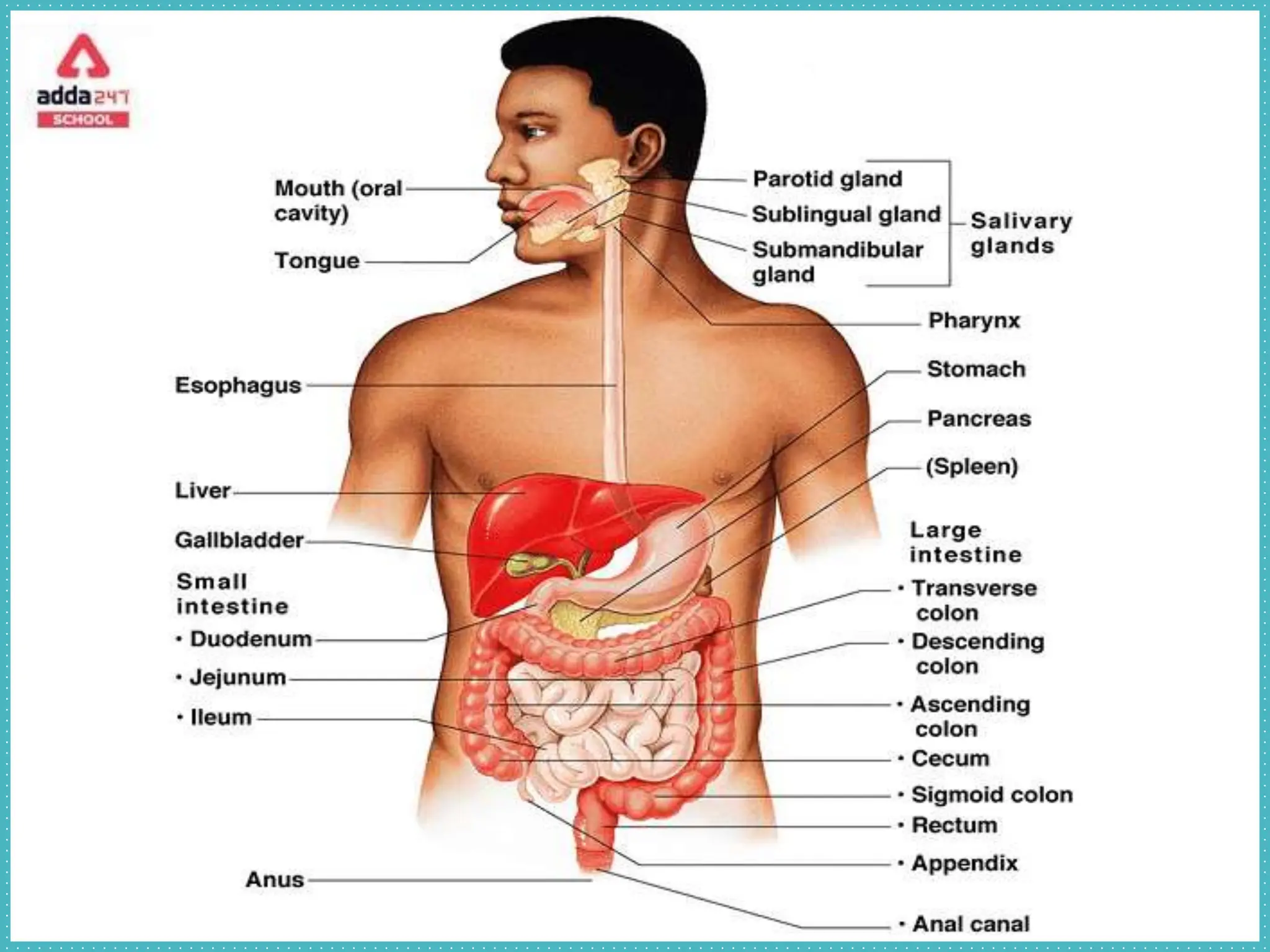
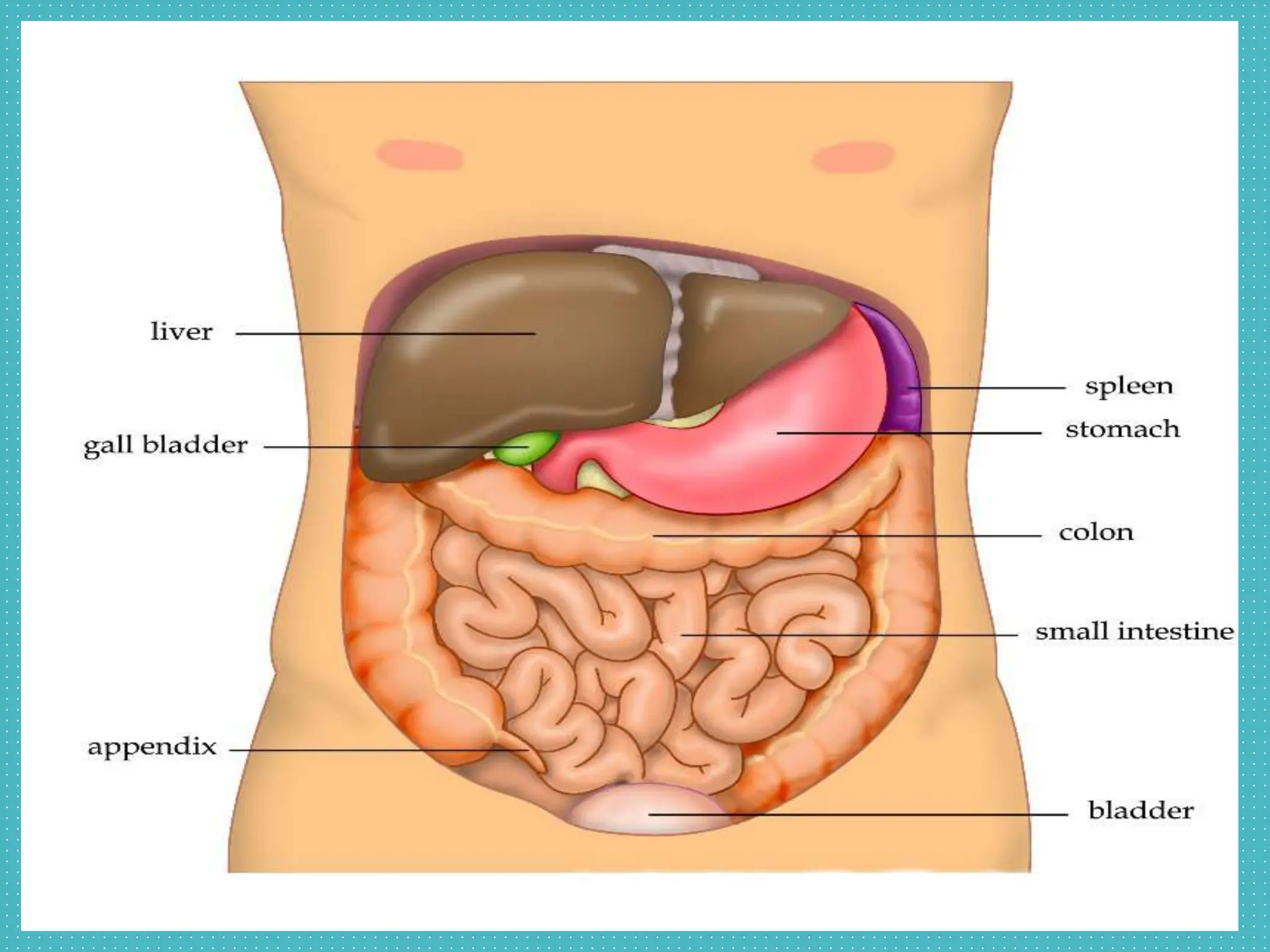
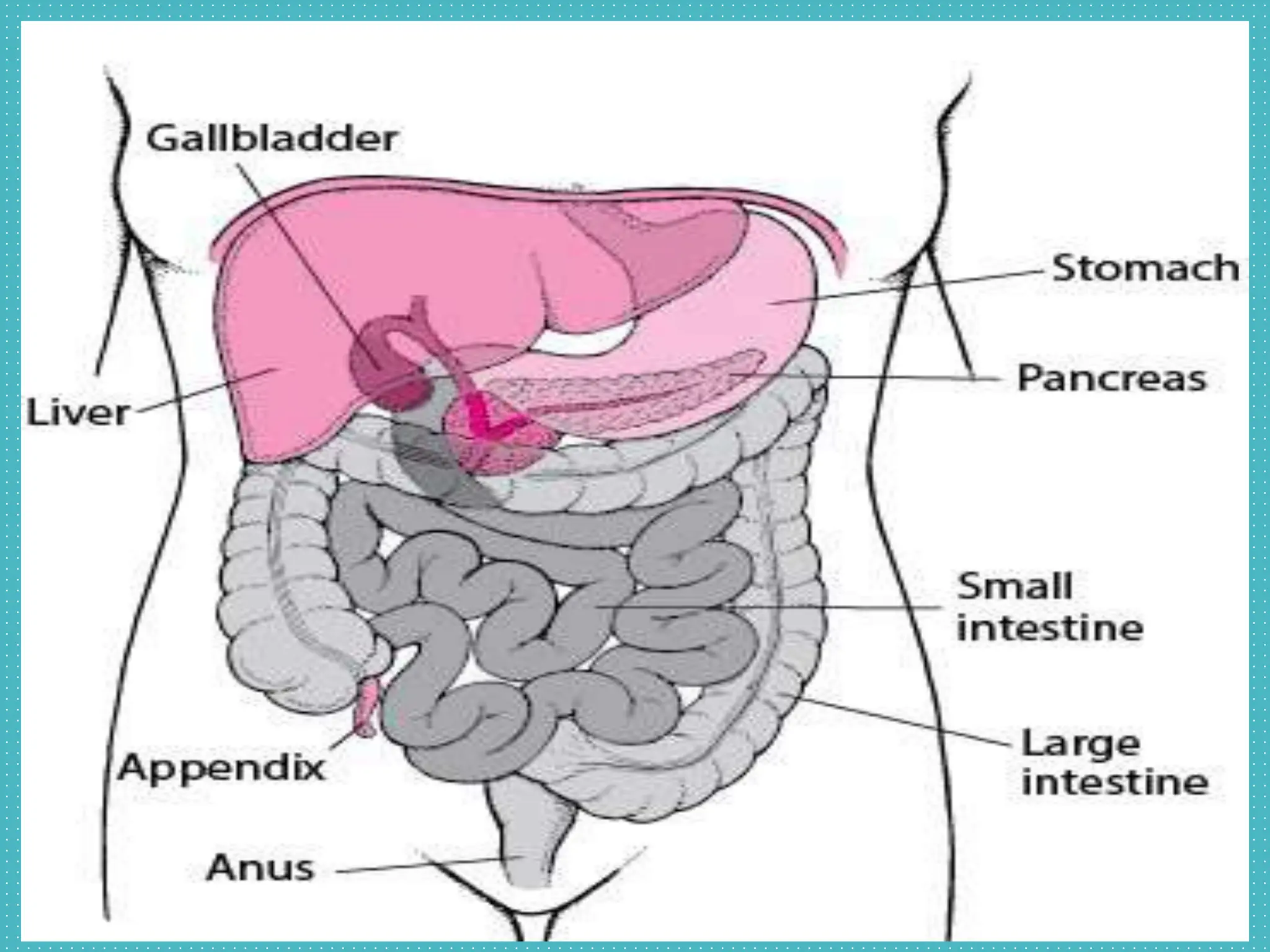
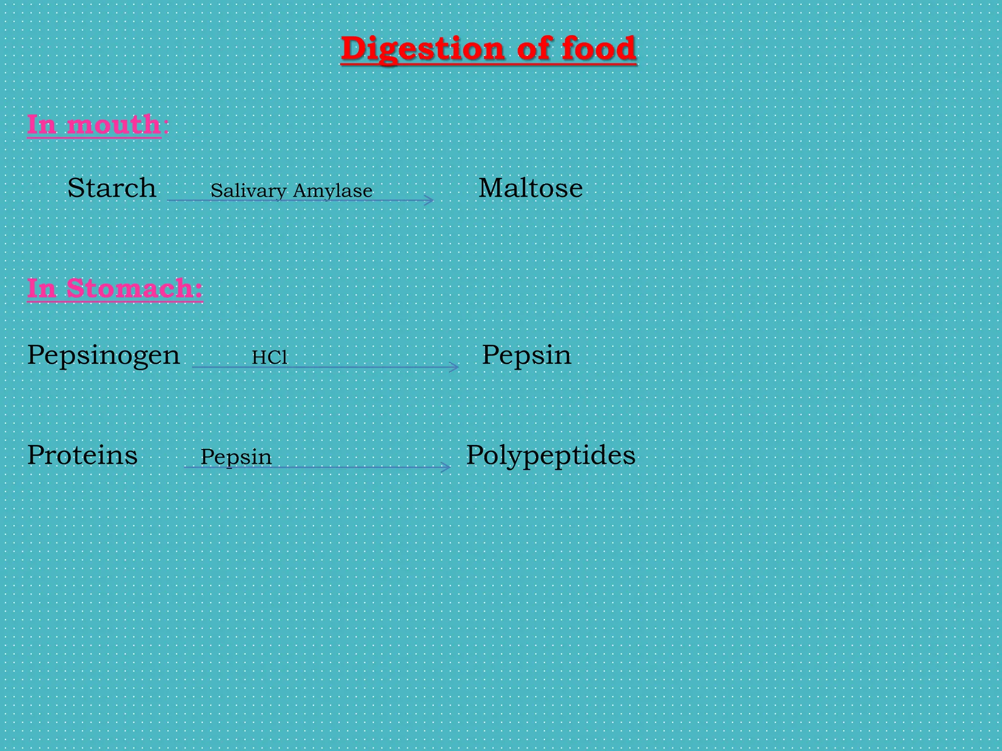
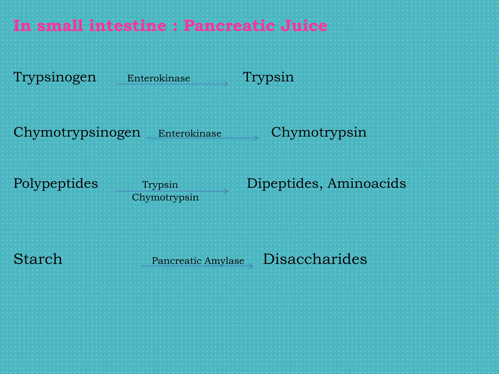
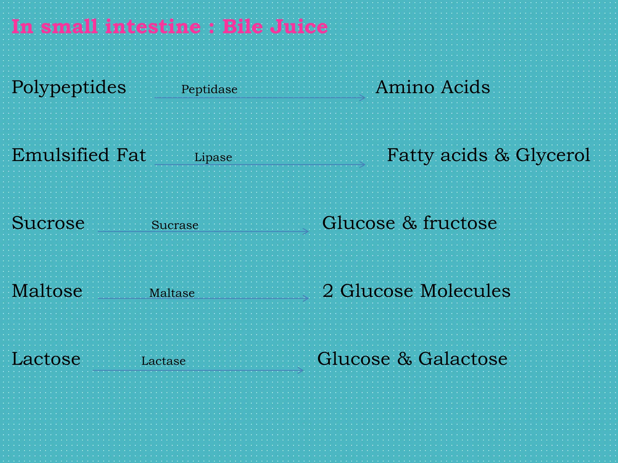
The digestive system breaks down food through a multi-step process. The alimentary canal begins at the mouth and runs through the throat, chest, abdomen and pelvis, ending at the anus. Accessory organs like the liver, pancreas and salivary glands help break down food through mechanical and chemical digestion. Food is broken into smaller molecules that can be absorbed and used by the body before waste is eliminated. The digestive system is made up of organs like the mouth, esophagus, stomach and intestines that work together through processes like ingestion, digestion, absorption and elimination.
Anatomy & Physiology of GIT: It covers Organs of the Digestive system, Structure of the Alimentary canal, Mouth, Salivary glands, Pharynx, Oesophagus, Stomach, Small intestine, Large intestine, Rectum & Anal canal, Pancreas, Liver, Biliary tract
The document summarizes the structure and function of the digestive system. It begins with an overview of digestion and absorption. It then describes each section of the gastrointestinal tract in detail, including the mouth, esophagus, stomach, and intestines. For each section, it discusses the layers, muscles, glands, and role in digestion.
The document describes the structure and function of the digestive system, including the organs that make up the gastrointestinal tract such as the mouth, esophagus, stomach, small intestine, and large intestine. It discusses the roles of these organs in ingestion, digestion, absorption, and elimination of food. In addition, it provides details on the layers of the gut wall, nerves and blood supply to the digestive organs, and glands that secrete enzymes to break down food.
The digestive system begins at the mouth and includes the esophagus, stomach, and small and large intestines. Food is ingested and broken down through both mechanical and chemical digestion. Mechanical digestion begins with chewing in the mouth. Chemical digestion involves enzymes that break down carbohydrates, proteins, and fats in specific areas of the digestive tract. Digested nutrients are then absorbed through the walls of the tract and transported throughout the body.
Anatomy and physiology of GI system and Diagnostic techniquesharshraman1989
The document describes the human digestive system. It begins with an introduction and overview of the digestive tract. It then details each part of the tract, including the mouth, esophagus, stomach, and small and large intestines. For each organ, it discusses structure, function, blood supply, and associated glands and tissues. The document provides a comprehensive overview of the digestive system and the processes of ingestion, digestion, absorption, and elimination.
The digestive system extends from the mouth to the anus. It contains organs that ingest, digest, and absorb food and eliminate waste. The gastrointestinal tract lining contains three layers - epithelium, connective tissue lamina propria, and smooth muscle muscularis mucosae. Saliva produced by salivary glands in the mouth begins digesting starch and buffers acidic foods. The tongue maneuvers food and contains taste buds. Teeth crush and grind food before it enters the esophagus.
digestive system nursing [Autosaved].pptxNeha630537
The digestive system converts food into nutrients that can be absorbed and used by the body. Food passes through the alimentary canal, which includes the mouth, esophagus, stomach, and intestines. In the mouth, food is ingested and mixed with saliva from the salivary glands. The stomach aids in digestion as food moves into the small intestine, where nutrients are absorbed into the bloodstream. Undigested waste is then eliminated from the large intestine. Accessory organs like the liver, pancreas, and gallbladder also secrete substances that help break down food.
The document summarizes the structure and organization of the gastrointestinal tract (GIT). It describes the main organs of the GIT as well as accessory organs. It then discusses the general structure of the GIT walls, which are composed of four layers - the outer adventitia/serous layer, muscle layer, submucosal layer, and inner mucosal layer. Each layer is described in detail. The document also provides a detailed overview of the structure and features of the mouth as the starting point of the GIT.
Gastrointestinal Tract (GIT)//DIGESTIVE SYSTEM Wasim Ak
The digestive tract or gastrointestinal tract ( GIT) is composed of mouth , pharynx, oesophagus, stomach , small intestine and large intestine .
This GIT will helps in digestion of food and absorption of needed nutrients into our body .
The document provides an overview of the digestive system, including its main functions and components. The digestive system consists of the alimentary canal and digestive glands. The alimentary canal runs from the mouth to the anus and includes the mouth, pharynx, esophagus, stomach, small intestine, and large intestine. Each part has specific structures and functions related to ingestion, digestion, absorption, and elimination of food.
This my original work on Anatomy of digestive system, therefore it is strongly forbidden to copy, share and foreword without the permission of the authors to the third person or anybody else.
The document provides information about the digestive system, including the organs and processes involved. It describes the main parts of the digestive tract from mouth to anus. It details the layers of the digestive tract walls and explains the roles of the salivary glands, teeth, tongue, and liver and pancreas in digestion. Accessory organs help break down food while the stomach, small intestine, and large intestine further digest and absorb nutrients before waste is eliminated.
The digestive system breaks down food into molecules small enough for absorption. It consists of the gastrointestinal tract (mouth, esophagus, stomach, small and large intestines) and accessory organs (teeth, tongue, salivary glands, liver, gallbladder, pancreas). Food is ingested, digested through both mechanical and chemical breakdown, absorbed, and waste is eliminated. The GI tract contains four tissue layers - mucosa, submucosa, muscularis, and serosa. Digestion involves ingestion, secretion, propulsion, digestion, absorption, and defecation.
The document provides an overview of the anatomy and functions of the digestive system. It begins with definitions of digestion and the organs involved. The digestive system breaks down food through both mechanical and chemical digestion in the alimentary canal and accessory organs. The alimentary canal walls consist of four layers - adventitia, muscle, submucosa, and mucosa. Accessory organs include salivary glands, liver, and pancreas. Digestion involves ingestion, propulsion, digestion, absorption, and elimination.
Digestive system Mouth Buccal cavity Tongue Teeth Salivary glands Pharynx Oesophagus Stomach Small intestine Large intestine Rectum Anus Liver Gall bladder Pancreas Absorption Digestion
The document summarizes the key components and functions of the human digestive system. It describes the organs that make up the gastrointestinal tract, including the mouth, esophagus, stomach, and intestines. It explains the four main activities of digestion: ingestion, propulsion, digestion, and absorption. Each organ is then described in more detail, outlining its structure, blood supply, nerve innervation, and role in digestion. The accessory organs that contribute to digestion, such as the salivary glands and liver, are also briefly discussed.
Digestive System Review anatomy for alliedDr Musadiq
The document summarizes key aspects of the digestive system, including:
1) It describes the main stages of digestion as ingestion, digestion, absorption, and defecation.
2) It explains the two types of digestion - mechanical and chemical digestion - and how they break down food.
3) It provides an overview of the mouth, esophagus, small intestine, and large intestine, outlining their functions and features.
The document discusses the structure and function of the digestive system. It begins by describing the digestive system as a long tube running from the mouth to the anus. It then explains the five main processes of digestion: ingestion, propulsion, digestion, absorption, and elimination. The document goes on to describe the main organs that make up the digestive system, including the mouth, esophagus, stomach, and intestines. It provides details on the roles and contents of saliva, gastric juice, and the layers of the digestive tract walls.
The digestive system consists of the gastrointestinal tract and accessory organs. The gastrointestinal tract includes the mouth, esophagus, stomach, and intestines. Accessory organs include the teeth, tongue, salivary glands, liver, gallbladder and pancreas. The digestive system breaks down food into smaller molecules that can be absorbed and used by the body. Digestion involves both mechanical and chemical breakdown of food.
The document provides an overview of the histology of the gastrointestinal tract. It describes the five main phases of GIT function and the major organs that make up the digestive system, including the oral cavity, esophagus, stomach, small and large intestines, rectum, and anus. For each organ, it discusses the epithelial lining, layers (mucosa, submucosa, muscularis externa), and cell types (e.g. gastric glands in stomach). It also describes the tongue papillae, esophagus layers, stomach regions and glands, and small intestine function. In summary, it provides a detailed histological analysis of the gastrointestinal tract.
The document summarizes the key components and functions of the digestive system. It describes the organs that make up the alimentary canal including the mouth, esophagus, stomach, small intestine, and large intestine. It also discusses the accessory organs that contribute to digestion like the teeth, tongue, salivary glands, liver, gallbladder and pancreas. The document outlines the processes of digestion including ingestion, secretion, mixing, propulsion, digestion, absorption and defecation. It provides details on the layers of the digestive tract and peristalsis.
The digestive system contains both the alimentary canal (gastrointestinal tract) and accessory organs. The alimentary canal includes the mouth, esophagus, stomach, and small and large intestines. Accessory organs that aid digestion include the teeth, tongue, liver, gallbladder and pancreas. The digestive tract walls are composed of four main layers - mucosa, submucosa, muscularis externa, and serosa. Digestion involves both mechanical and chemical breakdown of food, with enzymes from the mouth, stomach, pancreas and small intestine facilitating the process.
The digestive system consists of the alimentary canal and accessory organs. The alimentary canal extends from the mouth to the anus and includes the mouth, esophagus, stomach, small intestine, and large intestine. Accessory organs that aid in digestion include the teeth, tongue, salivary glands, liver, gallbladder, and pancreas. Digestion involves the mechanical and chemical breakdown of food so that nutrients can be absorbed in the small intestine and remaining waste is eliminated as feces.
The document discusses the process of digestion in humans. It describes the major organs that make up the digestive system, including the mouth, esophagus, stomach, small intestine, large intestine, and accessory digestive glands like the liver, pancreas, and salivary glands. It explains the functions of these organs in breaking down food and absorbing nutrients.
The document summarizes the structure and functions of the digestive system. It describes the primary digestive organs including the mouth, pharynx, esophagus, stomach, small intestine and large intestine. It also discusses the accessory digestive organs such as teeth, tongue, salivary glands, liver and pancreas. It provides details on the parts, functions and process of digestion in each organ of the gastrointestinal tract.
The document summarizes the histological features of the gastrointestinal tract. It begins with the oral cavity, describing the epithelium and structures found in the lips, tongue, and oropharynx. It then discusses the esophagus and stomach, noting the transitions in epithelial lining and layers of muscle. The small and large intestines are also summarized, focusing on the absorptive roles played by the villi in the small intestines and the colon in the large intestines.
This document provides an overview of the key parts and functions of the digestive system, including the mouth, pharynx, esophagus, and stomach. It describes the structures and roles of the mouth including teeth, tongue, and salivary glands. It then discusses the pharynx and esophagus, and their roles in swallowing and moving food to the stomach. Finally, it details the anatomy of the stomach including its layers, glands that secrete digestive juices, and temporary storage and chemical breakdown of food before it passes to the small intestine.
The digestive system breaks down ingested food into nutrients that can be absorbed and used by the body. It consists of the alimentary canal and accessory organs. The alimentary canal includes the mouth, esophagus, stomach, and small and large intestines. In the mouth, teeth and tongue break down food and saliva contains enzymes that begin digestion. The stomach stores, churns, and breaks down food further with acid and enzymes. The small intestine completes digestion and absorbs nutrients into the bloodstream through fingerlike villi and microvilli. The large intestine absorbs water before waste is excreted. Accessory organs include the liver, pancreas and gallbladder which produce bile and enzymes to aid digestion
Shock is a life-threatening condition where tissues do not receive adequate oxygen due to low blood flow. There are three stages of shock: compensatory, progressive, and refractory. Shock can be classified as hypovolemic, cardiogenic, neurogenic, anaphylactic, or septic depending on the cause. Treatment focuses on restoring adequate circulation through fluid resuscitation, vasopressors, or inotropes while also treating the underlying cause of shock.
Pain is defined as an unpleasant sensory and emotional experience associated with actual or potential tissue damage. It is subjective and influenced by developmental, physiological, social, spiritual, psychological, and cultural factors. There are several types of pain including acute pain which has a sudden onset, chronic pain which persists over 6 months, and pain classified by origin such as cutaneous, deep somatic, and visceral pain. Pain is assessed using tools like pain scales and can be managed through pharmacological interventions like opioids, non-pharmacological methods like massage, heat/cold therapy, acupressure, hypnosis, relaxation, exercise, and distraction.
The document discusses several oral disorders including cheilitis, stomatitis, gingivitis, glossitis, parotitis, and dental caries. Cheilitis is an inflammation of the lips with various causes such as overexposure to sunlight, nutritional deficiencies, infections, and drugs. Stomatitis is the inflammation of the mouth lining that can be caused by malnutrition, infections, medications, and other factors. Gingivitis is the non-destructive inflammation of gums typically caused by plaque buildup leading to bleeding and swelling.
Liver abscess is a pus-filled mass in the liver that usually develops due to a bacterial or parasitic infection spreading from the intestines or bile ducts. Common causes include infections from E. coli, Klebsiella, and Entamoeba histolytica. Abscesses form when infecting organisms destroy liver cells, causing necrosis and an inflammatory response where leukocytes fill the cavity with pus and dead cells. Symptoms include fever, abdominal pain, nausea, jaundice, and weight loss. Diagnosis involves blood tests, imaging like ultrasound or CT, and aspiration of pus. Treatment consists of IV antibiotics targeting the suspected bacteria plus drainage of abscesses percutaneously or surgically.
This document summarizes gastrointestinal bleeding, including its causes, risk factors, clinical manifestations, diagnostic studies, prevention, and treatment. It defines GI bleeding as bleeding anywhere in the GI tract. The major causes discussed are esophageal varices, gastric/duodenal ulcers, hemorrhagic gastritis, polyps, and medications like NSAIDs. Diagnostic studies include stool tests, endoscopy, and blood tests. Treatment depends on the source but may include endoscopic therapy, surgery, or drug therapy with antacids, H2 blockers, PPIs, vasopressin, or octreotide to stop bleeding and promote healing.
Malabsorption is a condition that prevents the absorption of nutrients through the small intestine, resulting in impaired absorption of micro and macro nutrients. It can occur due to deficiencies in enzymes, bacterial overgrowth, damage to the intestinal mucosa from diseases like celiac and Crohn's, or reduced surface area from surgery. Clinical manifestations include steatorrhea, weight loss, diarrhea, and deficiencies leading to anemia, bone disease, and neurological symptoms. Diagnosis involves tests of stool, breath, and biopsy of the small intestine. Treatment depends on the underlying cause but may include specialized nutrient-rich diets and antibiotics.
Anal fissure is a longitudinal tear or crack in the lining of the anal canal distal to the dentate line. It is usually caused by local trauma from hard bowel movements but can also be caused by infections or other medical conditions. Symptoms include painful defecation, burning sensation, and bleeding. Diagnosis is made through history and physical exam. Conservative treatment focuses on warm sitz baths, increased fiber and fluid intake, stool softeners, and topical nitroglycerin to help the fissure heal in 2-4 weeks. Surgery may be needed if conservative measures fail and include procedures like coagulation therapy, sphincterotomy, or fissure excision.
The document provides an overview of the excretory/urinary system. It describes the key organs including the kidneys, ureters, urinary bladder, and urethra. The kidneys filter waste from the blood to form urine, which passes through the ureters to the bladder for storage, then exits through the urethra. The kidneys contain nephrons, which are the functional filtering units. Filtration occurs through the glomerulus, then substances are selectively reabsorbed or secreted to regulate water and electrolyte levels as urine is formed.
Peritonitis is an inflammatory process of the peritoneum, the membrane lining the abdominal cavity. It can be primary, secondary, or tertiary based on the cause. Secondary peritonitis occurs when the contents of abdominal organs leak into the cavity, as from a ruptured appendix or perforated ulcer. This leads to bacterial proliferation, edema, and exudation of fluid in the cavity. Symptoms include abdominal pain, tenderness, distension, fever, and altered bowel movements. Treatment focuses on identifying and eliminating the cause, combating infection, and preventing complications through fluid replacement, antibiotics, and sometimes surgery.
This document provides information about colorectal cancer including its definition, causes, symptoms, diagnostic tests, staging, complications, prevention, and management. Colorectal cancer develops in the inner lining of the colon or rectum and is the third leading cause of cancer death for males over 50. Risk factors include age, family history, inflammatory bowel disease, diet, obesity, and smoking. Symptoms often do not appear until advanced stages and can include changes in bowel habits, blood in stool, and weight loss. Diagnostic tests include colonoscopy, biopsy, and blood tests. Treatment depends on staging and may involve surgery to remove the tumor, chemotherapy, or radiation.
This document discusses Gastro-Esophageal Reflux Disease (GERD). It defines GERD as a condition where stomach acid leaks back up into the esophagus, causing symptoms or damage. Approximately 5-7% of the world experiences GERD. Risk factors include obesity, pregnancy, and connective tissue disorders. Treatment involves lifestyle modifications like avoiding aggravating foods, medications like antacids and proton pump inhibitors, and sometimes surgery. Nursing management includes educating patients on treatments and lifestyle changes.
Hemorrhoids, also known as piles, are swollen veins in the anus and lower rectum that can cause symptoms like itching, pain and bleeding. They are classified as either internal or external hemorrhoids depending on their location. Internal hemorrhoids are further classified by their degree of prolapse. Risk factors include prolonged sitting or standing, obesity, pregnancy and liver diseases. Symptoms are usually treated conservatively with fiber, fluids, creams and sitz baths while surgical procedures like rubber band ligation or hemorrhoidectomy may be used for more severe cases. Nursing care focuses on preventing constipation and infection.
Ulcerative colitis is a long-term condition characterized by inflammation and ulcers of the colon and rectum. The causes are not fully known but may include genetic and environmental factors. Symptoms include bloody diarrhea, abdominal pain, and weight loss. Diagnosis involves medical imaging, endoscopy, and biopsy of the colon. Treatment focuses on reducing inflammation, managing symptoms, and correcting nutritional deficiencies through medications, dietary changes, and sometimes surgery to remove all or part of the colon. Nursing care involves managing symptoms, preventing complications, providing education on lifestyle changes and treatment plans, and supporting patients through the challenges of living with a chronic condition.
This document provides information about hiatal hernia, including its definition as a portion of the stomach herniating through the diaphragm, types (sliding and paraesophageal), causes such as weakening muscles or increased abdominal pressure, symptoms like heartburn or dysphagia, potential complications, diagnostic tests, and management through surgery or lifestyle changes. The most common type is sliding hiatal hernia, which occurs when the stomach and junction slide into the chest and back into the abdomen. Surgical repair is recommended for symptomatic patients and involves wrapping the stomach around the esophagus.
This document discusses cardiac tamponade, which is the compression of the heart caused by fluid accumulation in the pericardial sac. It defines cardiac tamponade and describes its causes, signs, symptoms, diagnostic tests, and treatments. Key causes include infection, cancer, surgery, and medication side effects. Diagnosis involves echocardiography and other imaging tests. Treatment involves draining excess fluid via pericardiocentesis, a procedure where a needle is used to drain the fluid for diagnostic and therapeutic purposes. Nursing care focuses on monitoring vital signs, administering oxygen, IV fluids, and medications to support heart function.
Cholecystitis is an inflammation of the gallbladder that is usually caused by gallstones blocking the cystic duct. There are three main types: acute calculous cholecystitis caused by gallstones; acalculous cholecystitis seen in critically ill patients without stones; and chronic cholecystitis from repeated acute episodes. Symptoms include pain in the right upper abdomen, fever, jaundice, and Murphy's sign on palpation. Diagnosis involves blood tests, imaging like ultrasound and CT. Treatment focuses on pain relief, antibiotics, and sometimes surgery to remove the gallbladder for severe or recurrent cases.
This document provides an overview of gastritis, including its introduction, types, etiology, pathophysiology, clinical manifestations, diagnostic studies, and management. Gastritis is inflammation of the stomach lining that can be acute or chronic. Acute gastritis lasts hours to days while chronic gastritis results from repeated irritation. Causes include medications, diet, infections like H. pylori, and medical conditions. Symptoms vary depending on the type but may include nausea, vomiting, abdominal pain, and bleeding. Diagnosis involves endoscopy, blood tests, and stool tests. Treatment focuses on identifying and eliminating causes as well as using antacids, H2 blockers, PPIs, and antibiotics to
Cor pulmonale is defined as enlargement of the right ventricle due to pulmonary hypertension caused by lung diseases. It accounts for 6-7% of adult heart diseases, with 50% of cases caused by chronic obstructive pulmonary disease. The main causes are lung diseases like emphysema and bronchitis that damage the lungs and lead to high blood pressure in the pulmonary artery and right ventricle over time. This puts strain on the right ventricle and causes it to enlarge and fail. Symptoms include dyspnea, cough, wheezing, leg swelling, and fatigue. Diagnosis involves physical exam, imaging tests of the heart and lungs, and assessing pulmonary artery pressures. Treatment focuses on managing the
Anemia is a blood disorder where the blood has a reduced ability to carry oxygen due to lower red blood cell count or hemoglobin levels. Common causes include iron deficiency, blood loss, and impaired red blood cell production. Symptoms range from fatigue to shortness of breath and depend on severity. Diagnosis involves blood tests showing low hemoglobin and identifying the underlying cause. Treatment focuses on treating the underlying condition and may involve oral or intravenous iron supplementation, vitamin supplements, blood transfusions, or medications.
Cardiogenic shock occurs when the heart is unable to pump enough blood to meet the body's needs due to systolic or diastolic dysfunction. It can be caused by myocardial infarction, cardiomyopathy, arrhythmias, or structural problems of the heart. Signs include tachycardia, hypotension, pulmonary congestion, and impaired tissue perfusion. Treatment focuses on restoring oxygen supply and demand balance through fluids, inotropes, vasodilators, and mechanical circulatory support if needed.
Muktapishti is a traditional Ayurvedic preparation made from Shoditha Mukta (Purified Pearl), is believed to help regulate thyroid function and reduce symptoms of hyperthyroidism due to its cooling and balancing properties. Clinical evidence on its efficacy remains limited, necessitating further research to validate its therapeutic benefits.
share - Lions, tigers, AI and health misinformation, oh my!.pptxTina Purnat
• Pitfalls and pivots needed to use AI effectively in public health
• Evidence-based strategies to address health misinformation effectively
• Building trust with communities online and offline
• Equipping health professionals to address questions, concerns and health misinformation
• Assessing risk and mitigating harm from adverse health narratives in communities, health workforce and health system
Promoting Wellbeing - Applied Social Psychology - Psychology SuperNotesPsychoTech Services
A proprietary approach developed by bringing together the best of learning theories from Psychology, design principles from the world of visualization, and pedagogical methods from over a decade of training experience, that enables you to: Learn better, faster!
Here is the updated list of Top Best Ayurvedic medicine for Gas and Indigestion and those are Gas-O-Go Syp for Dyspepsia | Lavizyme Syrup for Acidity | Yumzyme Hepatoprotective Capsules etc
Local Advanced Lung Cancer: Artificial Intelligence, Synergetics, Complex Sys...Oleg Kshivets
Overall life span (LS) was 1671.7±1721.6 days and cumulative 5YS reached 62.4%, 10 years – 50.4%, 20 years – 44.6%. 94 LCP lived more than 5 years without cancer (LS=2958.6±1723.6 days), 22 – more than 10 years (LS=5571±1841.8 days). 67 LCP died because of LC (LS=471.9±344 days). AT significantly improved 5YS (68% vs. 53.7%) (P=0.028 by log-rank test). Cox modeling displayed that 5YS of LCP significantly depended on: N0-N12, T3-4, blood cell circuit, cell ratio factors (ratio between cancer cells-CC and blood cells subpopulations), LC cell dynamics, recalcification time, heparin tolerance, prothrombin index, protein, AT, procedure type (P=0.000-0.031). Neural networks, genetic algorithm selection and bootstrap simulation revealed relationships between 5YS and N0-12 (rank=1), thrombocytes/CC (rank=2), segmented neutrophils/CC (3), eosinophils/CC (4), erythrocytes/CC (5), healthy cells/CC (6), lymphocytes/CC (7), stick neutrophils/CC (8), leucocytes/CC (9), monocytes/CC (10). Correct prediction of 5YS was 100% by neural networks computing (error=0.000; area under ROC curve=1.0).
Osteoporosis - Definition , Evaluation and Management .pdfJim Jacob Roy
Osteoporosis is an increasing cause of morbidity among the elderly.
In this document , a brief outline of osteoporosis is given , including the risk factors of osteoporosis fractures , the indications for testing bone mineral density and the management of osteoporosis
- Video recording of this lecture in English language: https://youtu.be/kqbnxVAZs-0
- Video recording of this lecture in Arabic language: https://youtu.be/SINlygW1Mpc
- Link to download the book free: https://nephrotube.blogspot.com/p/nephrotube-nephrology-books.html
- Link to NephroTube website: www.NephroTube.com
- Link to NephroTube social media accounts: https://nephrotube.blogspot.com/p/join-nephrotube-on-social-media.html
Rasamanikya is a excellent preparation in the field of Rasashastra, it is used in various Kushtha Roga, Shwasa, Vicharchika, Bhagandara, Vatarakta, and Phiranga Roga. In this article Preparation& Comparative analytical profile for both Formulationon i.e Rasamanikya prepared by Kushmanda swarasa & Churnodhaka Shodita Haratala. The study aims to provide insights into the comparative efficacy and analytical aspects of these formulations for enhanced therapeutic outcomes.
TEST BANK For Community Health Nursing A Canadian Perspective, 5th Edition by...Donc Test
TEST BANK For Community Health Nursing A Canadian Perspective, 5th Edition by Stamler, Verified Chapters 1 - 33, Complete Newest Version Community Health Nursing A Canadian Perspective, 5th Edition by Stamler, Verified Chapters 1 - 33, Complete Newest Version Community Health Nursing A Canadian Perspective, 5th Edition by Stamler Community Health Nursing A Canadian Perspective, 5th Edition TEST BANK by Stamler Test Bank For Community Health Nursing A Canadian Perspective, 5th Edition Pdf Chapters Download Test Bank For Community Health Nursing A Canadian Perspective, 5th Edition Pdf Download Stuvia Test Bank For Community Health Nursing A Canadian Perspective, 5th Edition Study Guide Test Bank For Community Health Nursing A Canadian Perspective, 5th Edition Ebook Download Stuvia Test Bank For Community Health Nursing A Canadian Perspective, 5th Edition Questions and Answers Quizlet Test Bank For Community Health Nursing A Canadian Perspective, 5th Edition Studocu Test Bank For Community Health Nursing A Canadian Perspective, 5th Edition Quizlet Test Bank For Community Health Nursing A Canadian Perspective, 5th Edition Stuvia Community Health Nursing A Canadian Perspective, 5th Edition Pdf Chapters Download Community Health Nursing A Canadian Perspective, 5th Edition Pdf Download Course Hero Community Health Nursing A Canadian Perspective, 5th Edition Answers Quizlet Community Health Nursing A Canadian Perspective, 5th Edition Ebook Download Course hero Community Health Nursing A Canadian Perspective, 5th Edition Questions and Answers Community Health Nursing A Canadian Perspective, 5th Edition Studocu Community Health Nursing A Canadian Perspective, 5th Edition Quizlet Community Health Nursing A Canadian Perspective, 5th Edition Stuvia Community Health Nursing A Canadian Perspective, 5th Edition Test Bank Pdf Chapters Download Community Health Nursing A Canadian Perspective, 5th Edition Test Bank Pdf Download Stuvia Community Health Nursing A Canadian Perspective, 5th Edition Test Bank Study Guide Questions and Answers Community Health Nursing A Canadian Perspective, 5th Edition Test Bank Ebook Download Stuvia Community Health Nursing A Canadian Perspective, 5th Edition Test Bank Questions Quizlet Community Health Nursing A Canadian Perspective, 5th Edition Test Bank Studocu Community Health Nursing A Canadian Perspective, 5th Edition Test Bank Quizlet Community Health Nursing A Canadian Perspective, 5th Edition Test Bank Stuvia
2. Introduction :
The Digestive system describes alimentary canal, its
accessory organs & variety of digestive process that prepare
food eaten in the diet for absorption.
The Alimentary canal begins at the mouth, passes through
thorax, abdomen, pelvis and ends at Anus.
It has all basic structures that is modified at different levels.
The digestive process gradually break down the foods eaten
until they are in a form suitable for absorption.
3. Functions / Activities of Digestive system:
- Ingestion : this is taking of food into alimentary canal (Eating
& Drinking).
- Propulsion : this mixes the contents & moves them along
alimentary tract.
- Digestion :
o Mechanical breakdown of food by mastication
(Chewing)
o Chemical digestion of food into small molecules by the
action of enzymes produced by glands & accessory
organs.
4. - Absorption : it is the process by which products of digestion
pass through the walls of some organs of alimentary canal ,
into the blood and lymph capillaries for circulation & use by
body cells.
- Elimination : food that has been eaten, but cannot be
digested & absorbed is excreted in the form of Faeces by the
process of Defaecation.
5. Alimentary canal / GI Tract
It is a long tube through which food passes.
It commences at mouth and terminates at Anus.
In Adults, it is around 5 meters in Length.
The Parts are :
- Mouth
- Pharynx
- Oesophagus
- Stomach
- Small intestine
- Large intestine
- Rectum &
- Anal canal
6.
7. Accessory Organs:
Various secretions are released into the GI Tract, some by
glands in the membrane lining
(Gastric Juice by glands in the stomach lining) and some by
glands situated outside the tract.
They consists of
- Three pairs of Salivary glands
- The pancreas
- The liver & Biliary tract.
These organs & glands are linked physiologically as well as
anatomically in the digestion & absorption.
8. Structure of Alimentary / GI Tract:
The layers of the walls of alimentary canal follow consistent
pattern from the Oesophagus onwards.
The walls of GI tract are formed by four layers of tissue.
- Serosa or Adventitia : outer covering.
- Muscle Layer (Muscularis Mucosa)
- Submucosa
- Mucosa : Mucosal Lining.
9. Serosa / Adventitia :
o This is the outermost layer.
o In thorax, it consists of loose fibrous tissue & in abdomen,
the organs are covered by serous membrane called
“Peritoneum”.
Peritoneum:
It is the largest serous membrane of the body.
It is a single membrane, forming a close sac & containing a
small amount of serous fluid.
It provides a physical barrier to local spread of infection &
prevent involving of other abdominal structures.
10. Peritoneum :
It has two layers.
- The Parietal peritoneum lines abdominal wall.
- The visceral peritoneum covers the organs within the
abdominal & pelvic cavities.
The Two layers are in close contact, friction between them is
prevented by the presence of peritoneal fluid (a serous fluid
secreted by peritoneal cells).
11.
12. o The abdominal organs are covered to varying degrees by the
loops and folds of the visceral peritoneum, which attaches
firmly to the abdominal wall.
o If the organs are completely covered by the visceral layer,
then it is said to be intraperitoneal (stomach, intestine,
liver) and they are double fold.
o If the organs are covered on the anterior / superior side only,
then it is said to be retroperitoneal (Pelvic organs,
Pancreas, spleen, kidneys adrenal glands).
13. Muscle Layer / Muscularis Mucosa
This consists of Two layers of smooth muscle.
The outer layer consists of muscle fibres arranged in
Longitudinally, where as inner layer consists of circular
fibres.
Between these Two layers there are blood vessels, lymph
vessels and plexus of sympathetic & parasympathetic
nerves.
14. Submucosa :
This layer consists of loose areolar connective tissue
containing collagen and elastic fibres which binds the
muscle layer to the mucosa.
It includes blood vessels, nerves, lymph vessels & varying
amount of lymphoid tissue.
15. Mucosa :
It is the lining of GI Tract. Its most superficial layer is mucous
membrane.
It has mainly three functions.
- Protection
- Secretion
- Absorption.
16. o Inner most layer consists of stratified squamous epithelium
with mucous secreting glands below the surface.
o Mucous lubricates the walls of the tract & provides a
physical barrier that protects from damaging effects of
digestive enzymes.
o Under the epithelial lining there are varying amount of
lymphoid tissue that protect against ingested microbes.
17. Nerve Supply :
- The alimentary canal & accessory organs are supplied by
nerves from both Sympathetic & Parasympathetic divisions.
- Their actions are generally antagonistic to each other & one
has a greater influence than the other according to the body
needs.
- Increased parasympathetic activity to digestive organs
promotes digestive processes & increased sympathetic activity
inhibits them.
18. Mouth :
Mouth or Oral Cavity is formed by:
- Anteriorly by Lips
- Posteriorly it is continuous with Oropharynx.
- Laterally by the muscles of Cheek.
- Superiorly by bony hard palate & muscular soft palate.
- Inferiorly by the muscular tongue.
19. Oral cavity is lined with mucous membrane consisting of
stratified squamous epithelium containing small mucus –
secreting glands.
The palate forms the roof of mouth & is divided into the
Anterior hard palate and Posterior soft palate.
The Hard palate is formed by the Maxilla & Palatine bones.
The Soft palate is muscular in nature and blends with walls
of Pharynx at the sides.
Uvula is a curved fold of muscle covered with mucous
membrane, hanging from the middle of free border of soft
palate.
20.
21. Tongue :
The tongue is composed of voluntary muscle.
It is attached by its base to the hyoid bone and by a fold of
its mucous membrane covering called Frenulum to the floor
of mouth.
The superior surface consists of stratified squamous
epithelium with numerous papillae (little Projections).
Many of these contain sensory receptors for the sense of
taste in taste buds.
22. Blood Supply:
Arterial supply : Lingual Branch of external carotid artery
Venous drainage : Lingual vein which joins the internal jugular
vein.
Nerve Supply:
The Hypoglossal Nerve – voluntary muscle of tongue.
Lingual branch of Mandibular Nerve – somatic sensation (Pain,
Temperature, Touch)
Facial & Glossopharyngeal Nerves – for taste.
Functions :
It plays an important role in :
Chewing (Mastication), swallowing (Deglutition), speech and
Taste.
23. Teeth:
o They are embedded in the sockets of alveolar ridges of the
Mandible and Maxilla.
o There are two sets of teeth :
- Temporary or Deciduous teeth
- Permanent teeth
o There are 20 Temporary Teeth, 10 in each Jaw. They begin to
erupt at about 6 months and should be present by 24
months.
o The permanent teeth begin to replace Temporary Teeth
between 6 and 13 years.
o The dentition consists of 32 teeth and usually complete by
the age of 20 years.
24. Structure :
Shape of teeth may vary, but structure is same. It consists of
The Crown : part that protrudes from Gum.
The Root : part embedded in the bone.
The Neck : narrowed region where the crown merges with root.
In the centre of tooth, there is a pulp cavity containing blood
vessels, lymph vessels & Nerves.
It is surrounded by hard Ivory like substance called Dentine.
Dentine of the crown is covered by a thin layer of very hard
substance called Enamel.
Root of the tooth are covered with substance resembling bone
called Cementum, which secures tooth in its socket.
25.
26.
27. Blood Supply :
Arterial Supply : Maxillary arteries.
Venous Drainage : small branches of internal Jugular veins.
Nerve Supply:
Upper Teeth : Branches of Maxillary Nerves.
Lower Teeth : branches of Mandibular Nerves.
Functions:
Teeth have different shape depending on their functions.
Incisors & Canines are cutting teeth where as premolar & molar
teeth with broad, flat surfaces used for grinding / chewing of
Food.
28. Salivary Glands:
There are three main pairs of salivary glands.
- Parotid glands
- Submandibular glands
- Sublingual glands.
There are numerous smaller salivary glands scattered around
the mouth.
Parotid Glands:
These are situated one on each side of the face, just below
the external acoustic meatus.
Each gland has parotid duct opening into mouth at the level
of second upper molar teeth.
29. Submandibular Glands:
These lie one on each side of the face under the angle of Jaw.
The submandibular ducts open on the floor of mouth, one on
each side of Frenulum of Tongue.
Sublingual Glands:
These lie under the mucous membrane of the floor of mouth in
front of submandibular glands.
They have numerous small ducts that open into floor of mouth.
30. Composition of Saliva:
Saliva is the combined secretion from salivary glands & the
small mucous secreting glands of the oral mucosa.
About 1.5 litres of saliva is produced daily & it consists of :
- Water
- Mineral salts
- Salivary Amylase : a digestive enzyme.
- Mucus
- Antimicrobial substances : Antibodies & enzyme Lysozyme.
31. Secretion of Saliva:
It is controlled by Autonomic nervous system.
Parasympathetic stimulation causes profuse secretion of
watery saliva with low content of enzymes & organic
substances.
Sympathetic stimulation results in secretion of small amount
rich in organic material.
Reflex secretion occurs when there is food in mouth.
Secretion also occurs during sight, smell & even thought of
smell.
32. Functions of Saliva:
- Chemical digestion of polysaccharides (enzyme amylase
begins breakdown of complex sugars into disaccharide
maltose).
- Lubrication of Food.
- Cleaning & Lubrication of Mouth.
- Non specific defence.
- Taste (dry food stimulate sense of taste only after mixing with
Saliva)
33. Pharynx:
It is divided into three parts, namely Nasopharynx,
Oropharynx & Laryngopharynx.
Oro and Laryngopharynx are passages common to both
Respiratory & Digestive system.
Food passes into the pharynx & then to the oesophagus
below, with which it is continuous.
It consists of three layers; inner, middle & outer layer.
34. Inner mucosa is made up of stratified squamous epithelium. This
provides thick sturdy, lining.
Middle layer consists of connective tissue which becomes thinner
towards lower end, contains blood & lymph vessels with nerves.
Outer layer consists of involuntary muscles that are involved in
swallowing.
Blood supply:
Artery : branches of Facial arteries.
Venous drainage : Facial veins & Jugular veins.
Nerve Supply:
Parasympathetic : Glossopharyngeal & Vagus Nerves
Sympathetic : Cervical Ganglia
35. Oesophagus :
It is a narrow muscular tube extending from pharynx to
stomach, descends in front of vertebral column goes through
superior and posterior mediastinum.
It begins with lower part of the neck at the inferior border of
cricoid cartilage (C-6), extending to the Cardiac orifice of
stomach (T-11).
It lies in the median plane in thorax behind Trachea and
Heart.
36.
37. Dimensions :
Length : 25 cm or 10 Inches.
Width : 2 cm
Lumen : Its flattened Antero-posteriorly.
- Normally its kept closed (collapsed) and opens (dilates)
only during the passage of Food.
38. - It is continuous with Pharynx & just below the diaphragm it
joins stomach.
- The upper and lower ends of Oesophagus are closed by
sphincters.
A) the upper oesophageal sphincter prevents passage of
air into the oesophagus during inspiration &
aspiration of oesophageal contents.
B) the lower oesophageal sphincter (cardiac) prevents the
reflux of gastric contents into the oesophagus.
- There is no thickening of muscle in this area.
- When intra abdominal pressure raised (inspiration &
defaecation), the tone of lower oesophageal sphincter
increases.
39. Structure:
There are Four layers of tissue.
the outer covering (The Adventitia) consists of elastic fibrous
tissue that attaches oesophagus to surrounding structures.
The proximal third is lined by stratified squamous epithelium
for protection during swallowing.
The Distal third is covered by columnar epithelium.
The middle third is covered by mixture of two.
40. Blood Supply :
Artery :
Thoracic region : paired oesophageal arteries.
Abdominal region : branches from inferior phrenic arteries &
left gastric branch of coeliac artery.
Venous Drainage :
Thoracic region : Azygos & hemiazygos veins.
Abdominal region : Left gastric vein.
41. Functions of Mouth, Pharynx & Oesophagus:
1) Formation of Bolus.
When food is taken into mouth, it is chewed (masticated) by
teeth and moved around the mouth by tongue and muscles
of cheeks.
It is mixed with saliva & formed into soft mass or Bolus
ready for swallowing.
The length of time that food remains in the mouth largely
depends on the consistency of food.
42. 2) Swallowing :
It occurs in three stages after chewing is complete & the bolus
has been formed.
oral stage :
With the mouth closed, voluntary muscles of tongue & cheeks
push the bolus backwards into the pharynx.
Pharyngeal stage :
The muscles of pharynx are stimulated by reflex action
initiated in the walls oropharynx & coordinated by the
swallowing centre in the Medulla.
Involuntary contraction of these muscles propels the Bolus
down into the oesophagus.
43. Oesophageal Stage :
o The presence of Bolus in pharynx stimulates a wave of
peristalsis that propels the bolus through the oesophagus to
stomach.
o Peristaltic waves pass only after the swallowing begins, until
that the walls are relaxed.
o After the initiation of peristalsis, the lower oesophageal
sphincter guarding the entrance to the stomach relaxes to
allow descending bolus to pass into stomach.
44.
45. STOMACH :
The stomach is a J – Shaped, muscular, hollow organ & dilated
portion of the alimentary tract situated in the epigastric, umbilical &
left hypochondriac regions of the abdominal cavity.
Organs associated with Stomach:
Anterior : left lobe of Liver.
Posterior : Abdominal Aorta, Pancreas, Spleen, Left kidney & adrenal
gland.
Superior : Diaphragm, oesophagus & left lobe of Liver.
Inferior : Transverse colon & small Intestine.
To the Left : diaphragm & spleen
To the right : Liver & Duodenum.
46.
47. Structure :
It is continuous with oesophagus at lower oesophageal
sphincter & with duodenum at pyloric sphincter.
It has two curvatures.
- Lesser curvature is short & is continuation of the posterior
wall of Oesophagus.
- Just before the pyloric sphincter, it curves upwards to
complete J shape and forms greater curvature.
Stomach is divided into Three regions:
o The Fundus
o The body
o The pylorus
48. Cardia is not an anatomically distinct region of stomach.
The fundus is formed in the upper curved part.
Body is the main, central region of stomach.
Pylorus is the lower section of stomach that empties
contents into duodenum.
At the distal end of pylorus, is the pyloric sphincter guarding
opening between stomach & duodenum. When stomach is
inactive / empty the pyloric sphincter is relaxed and open,
when stomach contains food sphincter is closed.
49.
50. Walls of stomach:
It has Four layers of tissue that comprise basic structure of
Alimentary canal with some modifications.
Muscularis :
it consists of three layers of smooth muscle fibres.
- An outer layer of Longitudinal fibres
- A Middle layer of circular fibres
- An Inner layer of Oblique fibres.
This arrangement allows churning action characteristic of
gastric activity.
51. Mucosa :
When stomach is empty, the numerous membrane lining
thrown into longitudinal folds or Rugae.
When stomach is full, rugae are ironed out giving surface a
smooth, velvety appearance.
Numerous gastric glands are situated below surface in
mucous membrane. They contain specialised cells including
chief cells & parietal cells which secrete gastric juice
constituents.
52.
53. Gastric Juice:
About 2 litres of gastric juice are secreted daily by specialised
secretory glands in mucosa.
It consists of :
Water – liquifies food.
Mineral salts
Mucous – prevents mechanical injury to stomach.
Hydrochloric acid – acidifies food, kills ingested microbes,
provide acid environment for pepsin.
Intrinsic Factor – absorption of Vit B12
Inactive enzyme precursors – Pepsinogen.
54. Functions of Stomach :
Temporary storage : stomach size varies with volume of food it
contains (1.5 litres in adults). After food consumption, it
accumulates in stomach in layers.
Chemical digestion : pepsin breaks proteins into polypeptides.
Mechanical breakdown: three muscle layers enable the
stomach to act as a churn, gastric juice is added and
contents are liquefied into Chyme.
Limited absorption: water, alcohol, lipid soluble drugs.
Non specific defense against microbes.
Preparation of Iron for absorption.
(to be continued….)
55. Production & Secretion of Intrinsic Factor.
Regulation of passage of gastric contents into the duodenum.
Secretion of hormone Gastrin – it circulates in the blood and
stimulates gastric glands to produce more gastric Juice.
Blood Supply :
Arterial Supply: it is by Left Gastric artery a branch of Coeliac
artery, Right Gastric artery and Gastroepiploic artery.
Venous drainage : branches of Portal vein.
56. Pancreas:
It is a creamy pink gland weighing about 60 gm.
It is about 12 – 15 cm long & situated in the Epigastric and
left hypogastric regions of the abdominal cavity.
It consists of broad Head, a Body & a Narrow Tail.
Head lies in the curve of Duodenum, body behind the
stomach & tail in front of the left Kidney.
Abdominal Aorta & Inferior vena cava lie behind the gland.
It is both an exocrine and an endocrine gland.
57.
58. Exocrine Pancreas :
• This consists of large number of Lobules, made up of small
Acini (it composed of secretory cells).
• Each Lobules is drained by tiny ducts, which unite to form
pancreatic duct and they open into Duodenum.
• Just before they enter Duodenum, pancreatic duct joins
common bile duct to form Hepato-pancreatic Ampulla.
• The duodenal opening of ampulla is controlled by Sphincter
of Oddi.
• Function of exocrine pancreas is to produce pancreatic juice
containing enzymes which digest Carbohydrates, Proteins &
Fat.
59. Endocrine Pancreas :
o They consists of a group of specialised cells called
Pancreatic Islets (Islets of Langerhans).
o These cells have no ducts, hence Hormones directly
diffuse into the blood circulation.
o They secrete hormones like Insulin, glucagon which are
concerned with regulation of blood sugar level.
60. Blood Supply :
Artery : splenic & mesenteric arteries
Venous drainage : pancreatic vein that join other veins to
form Portal vein.
Nerve Supply :
Parasympatheic N.S.: its stimulation increase pancreatic
secretion.
Sympathetic N.S.: Its stimulation depress pancreatic
secretion.
61.
62. Liver :
It is the Largest gland in the body.
It is reddish brown in colour and weighs between 1 to 2.3 kg.
It is situated in the upper part of the abdominal cavity, it
occupies greater part of Right hypochondriac region, part of
epigastric region & extends into left hypochondriac region.
Its upper & anterior surfaces are smooth and curved to fit
under the diaphragm.
66. The liver is enclosed by thin inelastic Capsule and a layer of
peritoneum.
Liver is held in position partly by ligaments and partly by
pressure of the organs in Abdominal cavity.
Liver has Four lobes.
The Two most obvious are – large right lobe and smaller,
wedge shaped left lobe.
The other Two : the Caudate & the Quadrate lobes are found
on posterior surface.
67. Portal Fissure :
It is located on posterior surface of the liver where various
structures enter & leave the gland.
- Portal veins enters carrying blood from stomach, spleen,
pancreas, small and large intestines.
- Hepatic artery enters carrying oxygenated blood.
- Nerve fibres.
- Right & left hepatic ducts carrying bile from liver to gall
bladder.
- Lymph vessels.
68. Blood Supply :
Artery : Hepatic artery and portal veins carry pure blood
Venous Drainage: it is by variable number of hepatic veins
which join Inferior vena cava.
69. Structure :
The lobes of the Liver are made up of tiny functional units
called Lobules, which are visible to the naked eye.
Lobules are Hexagonal in outline & are formed by cuboidal
cells called “Hepatocytes”.
Hepatocytes are arranged in pairs of columns of cells.
Between two pairs of columns of cells, we have sinusoids,
blood vessels with incomplete walls.
Among the lining cells, there are Hepatic Macrophages
(Kupffer cells), whose function is to ingest & destroy worn out
blood vessels & any foreign particles present in the blood
flowing through the Liver.
70. Blood drains from sinusoids into central or centrilobular
veins.
These then merge with veins from other lobules forming
progressively large veins until they become Hepatic veins.
Each column of hepatocytes has blood sinusoid on one side
& bile canaliculus on the other.
The canaliculi joins up to form large bile canals until they
form right & left hepatic ducts, which drain bile from the
Liver.
71. Functions :
The Liver is extremely active metabolically with multiple inter
related functions.
- Carbohydrate metabolism.
- Fat metabolism.
- Protein metabolism.
- Breakdown of erythrocytes
- Detoxification of drugs & toxic substances.
- Inactivation of hormones
- Production of heat.
- Secretion of Bile.
72. Storage : it stores substances like
o Glycogen
o Fat soluble vitamins (A,D,E,K)
o Iron , Copper
o Water soluble vitamins like B12.
73. Bile Juice :
Bile is secreted by liver everyday. Around 500 to 1000 ml of
juice is secreted daily.
It consists of :
Water
Mineral salts
Mucus
Bile pigments (Bilirubin)
Bile salts
Cholesterol.
Functions : - Digestion of Fat.
- Excretion of Bilirubin.
74. Gall Bladder :
It is a pear shaped sac attached to the posterior surface of
Liver by connective tissue.
It has Fundus, Body or main part and Neck, which is
continuous with cystic duct.
Wall of gall bladder has same layers as that of Alimentary
tract.
Blood supply : artery – cystic artery, A branch of Hepatic
artery.
Vein : cystic vein which joins Portal vein.
75.
76. Functions :
- Storage of Bile.
- Concentration of the bile by upto 10 or 15 fold by the
absorption of water through walls of gall bladder.
- Release of stored bile.
77.
78. Bile Ducts:
The right and Left hepatic ducts join to form the common
Hepatic duct outside the portal Fissure.
The hepatic duct passes downwards for about 3cm & then it
is joined by the Cystic duct from Gall bladder.
The cystic & hepatic ducts merge to form common bile duct,
which passes downwards behind pancreas.
Common bile duct is joined by main pancreatic duct at
Hepatopancreatic ampulla & opens into duodenum.
The common bile duct is about 7.5cm long & has a diameter
of about 6mm.
79. Small Intestine :
It is continuous with the stomach at the pyloric sphincter.
It is about 2.5 cm in diameter and around 05 metres long.
It leads into the large intestine at the Ileocaecal valve.
It lies in the abdominal cavity surrounded by large intestine.
Small intestine comprised of Three continuous parts.
- Duodenum
- Jejunum
- Ileum
80.
81. Duodenum :
It is about 25cm long and curves around the head of the
pancreas.
Secretions from gall bladder & Pancreas merge in common
structure – hepatopancreatic ampulla that enter duodenum.
Jejunum :
This is the middle section of small intestine & its about 2
metres long.
Ileum :
This terminal section is about 3 metres long & ends at the
ileocaecal valve.
82. Structure of Small Intestine :
The walls of intestine are composed of the four layers with some
modifications.
Peritoneum: The Mesentry, a double layer of peritoneum
attaches Jejunum and Ileum to the posterior abdominal wall. It
is fan shaped. The large blood vessels & Nerves lie on posterior
abdominal wall.
Mucosa: the surface area of mucosa is greatly increased by
permanent circular folds, Villi & Microvilli. They promote mixing
of Chyme as they pass along.
(..continued)
83. The Villi are tiny Finger like projections of the mucosal layer
into the intestinal lumen, about 0.5 – 1mm long.
Their covering consists of columnar epithelial cells or
enterocytes, with tiny Microvilli (I micro meter long) on their
free border.
Goblet cells which secrete mucus are interspersed between
the enterocytes.
The villi contain network of blood capillaries & central lymph
capillaries.
These lymph capillaries are called lacteals, because
absorbed fat gives the lymph a milky appearance.
84. The intestinal glands are simple tubular glands situated
below the surface between the villi which are made up of
epithelial cells.
These epithelial cells produce digestive enzymes that lodge in
the microvilli & together with intestinal juice complete
digestion of Carbohydrates, Fats & proteins.
Blood Supply :
Artery : Superior Mesenteric artery
Venous drainage : superior mesenteric vein which joins
portal vein.
85.
86. Intestinal Juice :
o About 1500 ml of intestinal juice is secreted daily by glands
of small intestine.
o It is slightly basic and consists of water, lubricating mucus &
bicarbonate to neutralise gastric acid.
o Mechanical stimulation of intestinal glands is the main
stimulus to the secretion of intestinal juice.
87. Functions :
- Onward movement of its contents by peristalsis.
- Secretion of intestinal juice.
- Completion of chemical digestion of CHO, Fats & Proteins.
- Protection against infection by microbes.
- Secretion of Hormones CCK & secretin.
- Absorption of nutrients.
88. Pancreatic Juice:
It is secreted by exocrine pancreas. It enters the duodenum
at duodenal papilla.
It consists of :
- Water
- Mineral salts
- Enzymes : Amylase, Lipase, Nuclease
- Inactive enzyme precursors : Trypsinogen,
Chymotrypsinogen.
Pancreatic juice is alkaline in nature, because it contains
significant amount of bicarbonate ions which are basic.
89. Functions :
Digestion of proteins (Trypsin & Chymotrypsin)
Digestion of Carbohydrates (Pancreatic Amylase)
Digestion of Fats (Lipase)
Control of Secretion:
The secretion of Pancreatic juice is stimulated by Secretin &
cholecystokinin, which are produced by endocrine cells in the
walls of Duodenum.
90. Large Intestine :
• It is about 1.5 metres long. It begins at caecum in the right
iliac fossa & terminating at the rectum and anal canal.
• Its lumen is about 6.5 cm in diameter, larger than that of
small intestine.
• It forms an arch around round the coiled – up small
intestine.
• It is divided into The caecum, colon, Rectum & anal canal.
91. Caecum :
It is the first part of large intestine.
It is a dilated region that has blind end inferiorly & is
continuous with ascending colon superiorly.
The vermiform appendix (meaning worm like) is a fine tube,
closed at one end, which leads from caecum.
It is about 8 – 9 cm long and has the same structure of large
intestine, but contains more lymphoid tissue.
The appendix has no digestive functions.
It can cause significant problems when it becomes inflamed.
92. Colon :
The colon has 4 parts, which have same structure & functions.
The Ascending colon passes upwards from the caecum to the
level of liver, where it curves acutely to left at hepatic flexure.
The transverse colon extends across abdominal cavity in
front of the duodenum & stomach to the area of spleen,
where it forms splenic flexure and curves acutely downwards.
The descending colon passes down the left side of abdominal
cavity, then curves towards midline.
The sigmoid colon is ‘S’ shaped curve in the pelvic cavity that
continues downwards to become Rectum.
93. Rectum :
• It is slightly dilated section of large intestine.
• It is about 13 cm long.
• It starts from sigmoid colon & terminates at Anal canal.
Anal Canal :
• This is a short passage about 3.8 cm long in adults.
• It leads from rectum to the exterior.
• Two muscular sphincters control the Anus :
• Internal sphincter consisting of smooth muscle, is under the
control of Autonomic Nervous system.
• External sphincter formed by skeletal muscle, is under
voluntary control.
94. Structure :
- All the four layers which are described in the basic structure
of GI Tract are present in caecum, colon, rectum & anus.
- The arrangement of longitudinal fibres are modified in
caecum & colon.
- In rectum, the anal sphincters are formed by thickening of
circular muscle layer.
- In submucosal layer, there is more lymphoid tissue(for more
protection against organisms).
- In mucosal lining of colon & rectum, there are large number
of mucus secreting goblet cells within tubular glands. They
produce mucus, which lubricates passage of solid faeces.
95. Blood supply :
Arterial :
Superior Mesenteric artery – caecum, Ascending colon,
transverse colon.
Inferior Mesenteric artery – descending colon, sigmoid colon
Middle & inferior Rectal arteries – distal part of Rectum &
Anus.
Venous drainage :
Superior & Inferior mesenteric veins
Veins draining the distal part of Rectum & Anus join internal
Iliac veins.
96. Functions :
Absorption : absorption of water (by osmosis), mineral salts,
vitamins and some drugs.
Microbial activity : large intestine normally contain certain
types of bacteria like, E.Coli, Enterobacter aerogens,
streptococcus faecalis & clostridium perfringens. They
synthesize vitamin K & Folic acid. Few gases are produced by
bacterial fermentation of unabsorbed nutrients (Hydrogen,
carbon dioxide, Methane).
Mass movement : large intestine does not exhibit peristaltic
movement. Only at long interval (4 – 6 times a day) a wave of
strong peristalsis sweeps along with transverse colon forcing
its content to descending & sigmoid colon.
97. Defaecation :
o Rectum is usually empty. But when mass movement forces
the contents of sigmoid colon into rectum, the nerve endings
in the walls are stimulated.
o The external anal sphincter is under conscious control
through Pudendal nerve.
o Defaecation involves involuntary contraction of muscles of
rectum & relaxation of internal anal sphincter.
98. Constituents of Faeces :
It is a semisolid brown mass. The brown colour is due to
presence of Stercobilin.
60 – 70 % of the weight is due to presence of water.
Fibre (indigestible cellular plant & animal material)
Dead & live microbes
Epithelial cells shed from the walls of the tract.
Fatty acids
Mucus secreted by epithelial lining of Large intestine.
99. Metabolism :
It constitutes all the chemical reactions that occur in the body
to provide chemical energy essential for all cellular activities.
Metabolism involves two types of processes.
- Catabolism
- Anabolism
Catabolism : it breaks down large molecules into smaller ones,
releasing chemical energy, which is stored as Adenosine
Triphosphate (ATP) & heat. The heat generated maintains core
body temperature at the optimum level for chemical activity.
100. Anabolism : this is building up or synthesis of large molecules
from smaller ones require source of energy (ATP).
Energy :
All body cells require energy to carry out their metabolic
process.
It includes multiplication for replacement of worn – out cells,
muscle contraction & synthesis of glandular secretions.
The energy produced in the body measured & expressed in
either joules or kilocalories.
1 gm of Carbohydrate provides 4 kcal
1 gm of Protein provides 4 kcal
1 gm of Fat provides 9 kcal of energy.
101. Metabolic rate :
It is the rate at which energy is released from the fuel
molecules inside the cells.
It can be estimated by measuring oxygen uptake or carbon
dioxide excretion.
Basal Metabolic Rate (BMR) is the rate of the metabolism
when the individual is at rest in a warm environment & is in
the post absorptive state (i.e. has not had a meal for at least
12 hours).
In this state, energy released is sufficient to meet only
essential needs of vital organs (Heart, Lungs, Brain, Kidneys).
102. Carbohydrate Metabolism:
o Digested carbohydrate (Glucose) is absorbed into blood
capillaries of the villi of small intestine. It is transported by
portal circulation to Liver.
o Glucose is oxidised to provide chemical energy in the form of
ATP that is necessary for metabolic activity.
o If the glucose is in excess of requirements, then will be
converted to insoluble polysaccharide Glycogen by hormone
insulin in Liver & Skeletal Muscles.
o Glucagon, Adrenaline & Thyroxine are associated with
breakdown of glycogen into glucose.
o One molecule of Glucose can generate 38 ATP after complete
break down in the presence of Oxygen.
103. Protein Metabolism :
Dietary protein consists of number of Amino acids. About 20
of them are identified, out of which 9 are Essential amino
acids i.e. they cannot be synthesized in the body & need to
be supplemented through food.
Others are Non – Essential amino acids, because they can be
synthesized by many tissues.
Digestion breaks down protein into amino acids, then
absorbed into blood by villi in small intestine.
Amino acids are transported in the portal circulation to the
Liver & then into general circulation.
104. It makes them available to all body cells & tissues. Different
cells choose particular amino acids required for building or
repairing their specific type of tissues and for synthesizing
their secretions.
Ex : Antibodies, Enzymes, hormones.
In adults, 80 – 100 gm of protein are broken down & replaced
every day. The entire intestinal mucosa is replaced about
every 5 days.
105. Fat Metabolism :
Fats are digested & absorbed as fatty acids and glycerol into
Lacteals are transported via cisterna chyli to the blood
stream and then to Liver.
Fatty acids and glycerol circulating in the blood are used by
cells to provide energy & synthesize some secretions.
In Liver, some fatty acids & glycerol are used to provide
energy & heat. Some are recombined to form Triglycerides,
the form, in which Fat is stored.
106. Signs / symptoms of GI Disorders :
1. Anorexia – loss of appetite that reduces eating.
2. Constipation – passage of faeces less frequently than
normal.
3. Diarrhoea – unusual frequent passage of loose watery
faeces / stools.
4. Dysphagia – difficulty in swallowing.
5. Haematemesis – vomiting of blood either fresh or partly
digested.
6. Melaena – passing blood in the faeces which are black &
tarry.
7. Vomiting – an involuntary reflex in which there is a forceful
ejection of stomach contents through mouth.
107. GI System Diseases:
Gingivitis :
It is an inflammation of Gums, which may be acute or chronic.
It occurs in response to accumulation of bacterial plaque
around the teeth. It cause bleeding gums.
Dental Caries:
Tooth decays with discoloration & then formation of cavities. It
occurs when bacteria present on the plaque on teeth act on
sugar, forming acid which destroy the hard parts of teeth.
108. Mumps :
It is an acute inflammatory condition of the parotid salivary
glands. It is caused by mumps virus, which is one of the
parainfluenza group.
GERD (Gastro – Esophageal Reflux Disease):
It is the most common cause for Heartburn or indigestion. It is
caused by persistent regurgitation of acidic gastric contents
acidic gastric juice into the oesophagus causing irritation,
inflammation & painful ulceration.
109. Gastritis :
It is an inflammation of stomach, which can be acute or
chronic.
It is due to consumption of irritant drugs (aspirin) or alcohol,
Helicobacter Pylori infection, physiological stress, and others.
Peptic ulcer :
A sore that develops on the lining of the Oesophagus, stomach
or small intestine.
Ulcer occurs when stomach acid damages the lining of the
digestive tract. Helicobacter Pylori infection is very common
affecting 50 – 60 % of the victims.
110. Appendicitis :
A condition in which the appendix becomes inflammed, filled
with pus and causes pain. Appendix is a finger shapes pouch
that projects from your colon on the lower right side of
abdomen. Microbial infection, hard fecal matter imposition,
kinking, inflammatory exudate with fibrin and phagocytes are
few common causes.
Hernia:
Protrusion of the organ or part of organ through a weak point
or aperture in the surrounding structures. Here, a piece of
bowel protrudes through a weak point on anterior abdominal
wall or an existing opening.
111. Pancreatitis :
It is an inflammation of pancreas cuased due to acting of
activated enzymes, when they are still in pancreas. Common
causes are pacreatic cancer, viral infections, hypercalcemia,
severe hypothermia, drugs…
Hepatitis :
It is a serious liver infection and inflammation of liver due to
death of hepatocytes. It is caused due to viral infections, toxic
substances, circulatory disturbances,…
112. Cholecystitis :
It is an inflammation of gall bladder associated with presence of
gall stones.
Cholangitis :
It is an inflammation of bile ducts caused by bacterial infection
and is accompanied by abdominal pain, fever & jaundice.
Jaundice :
It is not a disease, but yellowing of the skin & mucous
membrane is a sign of abnormal bilirubin metabolism and
excretion.
113. Digestion of food
In mouth:
Starch Salivary Amylase Maltose
In Stomach:
Pepsinogen HCl Pepsin
Proteins Pepsin Polypeptides
114. In small intestine : Pancreatic Juice
Trypsinogen Enterokinase Trypsin
Chymotrypsinogen Enterokinase Chymotrypsin
Polypeptides Trypsin Dipeptides, Aminoacids
Chymotrypsin
Starch Pancreatic Amylase Disaccharides
115. In small intestine : Bile Juice
Polypeptides Peptidase Amino Acids
Emulsified Fat Lipase Fatty acids & Glycerol
Sucrose Sucrase Glucose & fructose
Maltose Maltase 2 Glucose Molecules
Lactose Lactase Glucose & Galactose