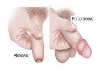
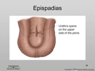
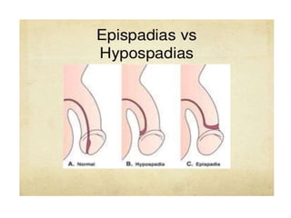
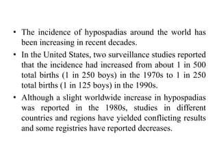
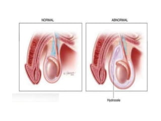
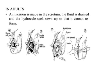
SlideShare a Scribd company logo
1 of 80
GENETO URINARY
DISSODERS
MANOJ M.G
LECTURER (MEDICAL SURGICAL NURSING)
DEPARTMENT OF NURSING AND MIDWIFERY
WOLLEGA UNIVERSITY
Urinary Incontinence
• Urinary incontinence is involuntary loss of urine from the
bladder), with most of them experiencing overactive bladder
syndrome.
• It is mainly due to
 Increased bladder pressure
 Lowered urethral resistance
 Pelvic muscle relaxation
 Impaired neural control
 Bladder problems
Types of Incontinence
Stress incontinence:
• is the involuntary loss of urine through an intact
urethra as a result of sneezing, coughing, or
changing position.
• It predominantly affects women who have had
vaginal deliveries and is thought to be the result
of decreasing ligament and pelvic floor support of
the urethra and decreasing or absent estrogen
levels within the urethral walls and bladder base.
• In men, stress incontinence is often experienced
after a radical prostatectomy for prostate cancer
because of the loss of urethral compression that
the prostate had supplied before the surgery, and
possibly bladder wall irritability.
Urge incontinence:
• is the involuntary loss of urine associated with
a strong urge to void that cannot be
suppressed.
• The patient is aware of the need to void but is
unable to reach a toilet in time.
• This can occur in a patient with neurologic
dysfunction that impairs inhibition of bladder
contraction or in a patient without overt
neurologic dysfunction.
Reflex incontinence:
• Is the involuntary loss of urine due to hyper
reflexia in the absence of normal sensations
usually associated with voiding.
• This commonly occurs in patients with spinal
cord injury because they have neither
neurologically mediated motor control of the
detrusor nor sensory awareness of the need to
void.
Overflow incontinence:
• is the involuntary loss of urine associated with
over distention of the bladder.
• Such over distention results from the bladder's
inability to empty normally, despite frequent
urine loss.
• Both neurologic abnormalities (eg, spinal cord
lesions) and factors that obstruct the outflow of
urine (eg, tumors, strictures, and prostatic
hyperplasia) can cause overflow incontinence
(Muller, 2005).
Functional incontinence:
• Refers to those instances in which lower
urinary tract function is intact but other factors,
such as severe cognitive impairment (eg,
Alzheimer's dementia), make it difficult for the
patient to identify the need to void or physical
impairments make it difficult or impossible
• Iatrogenic incontinence:
• refers to the involuntary loss of urine due to
extrinsic medical factors, predominantly
medications.
• One such example is the use of alpha-adrenergic
agents to decrease blood pressure.
• In some people with an intact urinary system,
these agents adversely affect the alpha receptors
responsible for bladder neck closing pressure; the
bladder neck relaxes to the point of incontinence
with a minimal increase in intra-abdominal
pressure, thus mimicking stress incontinence
• Some patients have several types of urinary
incontinence. This mixed incontinence is usually a
combination of stress and urge incontinence.
• Only with appropriate recognition of the problem,
assessment, and referral for diagnostic evaluation and
treatment can the outcome of incontinence be
determined.
• All people with incontinence should be considered
for evaluation and treatment.
Assessment and Diagnostic Findings
• Once incontinence is recognized, a thorough history is
necessary.
• This includes a detailed description of the problem and
a history of medication use (alpha-adrenergic agents).
• The patient's voiding history, a diary of fluid intake and
output, and bedside tests may be used to help determine
the type of urinary incontinence involved.
• Extensive urodynamic tests may be performed to assess
the flow of urine in the urinary tract .
• Urinalysis and urine culture are performed to identify
infection.
• Urinary incontinence may be transient or reversible if
the underlying cause is successfully treated and the
voiding pattern reverts to normal.
Medical Management
• Management depends on the type of urinary
incontinence and its causes.
• Management of urinary incontinence may be
behavioral, pharmacologic, or surgical in nature.
Surgical Management
• Surgical correction may be indicated in patients who
have not achieved continence using behavioral and
pharmacologic therapy.
• Surgical options vary according to the underlying
anatomy and the physiologic problem.
• Most procedures involve lifting and stabilizing the
bladder or urethra to lengthen the urethra.
• Women with stress incontinence may undergo an
anterior vaginal repair to reposition the urethra.
• Procedures to compress the urethra and increase
resistance to urine flow include placement of
periurethral bulking agents such as artificial collagen.
PHIMOSIS AND PARAPHIMOSIS
Phimosis
• Inability to retract a foreskin. Normally foreskin is
non-retractile at birth.
• Foreskin adhesions spontaneous break down and
form smegma pearls( sebaceous secretions) over next
2-10yrs (95% by age 16).
• Pathological phimosis usually from recurrent
infection or adhesions from forcible early foreskin
retraction may cause urinary obstruction, haematuria
or local pain.
Epidemiology
• Unusual in circumcised males
• In older DM patients often results from chronic
balanoposthitis (inflammation of foreskin and glans
penis)
• ↑Risk of penile Ca (very rare)
Presentation
• There may be swelling redness and tenderness of the
prepuce with purulent discharge.
Investigations
• Urethral swab. Urine MC&S. (microscopy, culture
and sensitivity)
Management
Physiological phimosis:
• No intervention is necessary.
• Advise against forcible retraction
• Personal hygiene is very important.
• Advise gentle cleaning under the foreskin
Pathological phimosis:
• Topical steroid application to the preputial
ring to treat ‘phimosis’ has reported success
rates between 33% – 95%.
• It may be necessary to slit the dorsal or ventral
foreskin or "preputial plasty". (Is a plastic
surgery on foreskin of the penis,to widen a
narrow non retractile foreskin which cannot
comfortably be drawn back off head of the
penis because of constriction.)
• Most paediatric urologists circumcise the
foreskin for pathological phimosis.
Circumcision
• Indications:
Absolute: Penile malignancy, traumatic foreskin injury
where it cannot be salvaged
Medical: Balanitis Xerotica Obliterans (is
dermatological condition of the penis and occurs
mainly on the foreskin and glans penis,atropic white
patches appears in the affected area), sev recurrent
balanoposthitis, recurrent febrile UTI’s with an
abnormal urinary tract
• Complications: Bleeding, sepsis, pain, stenosis,
urethral injury
Paraphimosis
• Unable to replace retracted foreskin, usually glans has
swelled. Urological emergency.
• Risk factors: Phimosis, penile piercing, urinary
catheterisation & not replacing foreskin
Presentation
• There is oedema around the constricting band that is
usually the prepuce.
• There may be pain on erection.
Management
• Analgesia+, LA lubrication, Ice pack for 5-
30min., gentle compression of glans with saline
gauze and manual reduction by sustained traction
of the prepuce over the glans.
• If fails, dorsal slit (preputial plasty ,best if
urologist does it).
• Decompression alternatives includes multiple
punctures in the oedematous foreskin
Complications
• Failure to remove the paraphimosis will result in
necrosis of the glans.
EPISPADIAS AND HYPOSPADIAS
EPISPADIAS
• An epispadias is a rare type of malformation of
the penis in which the urethra ends in an opening on
the upper aspect (the dorsum) of the penis.
• It can also develop in females when the urethra
develops too far anteriorly.
• It occurs in around 1 in 120,000 male and 1 in
500,000 female births.
• Classification of epispadias is based on the location
of the urethral meatus.
• It can be positioned on the glans (glanular),
• along the shaft of the penis (penile) or near the pubic
bone (penopubic).
• The position of the meatus is very important,
predicting the capacity of the bladder to store urine.
Causes
• Epispadias is an uncommon and partial form of a spectrum of
failures of abdominal and pelvic fusion in the first months of
embryogenesis known as the exstrophy - epispadias
complex.
• It occurs as a result of defective migration of the genital
tubercle primordii(body tissue it forms the ventral reproductive
region of mamalian embroys of both sex) to the cloacal
membrane(it is the membrane that covers embryonic cloaca
during the devevelopment of he urinary and reproductive
organs. It is formed by ectoderm and endoderm coming into
contact with each other), and so malformation of the genital
tubercle, at about the 5th week of gestation
Presentation
• Most cases involve a small and bifid penis which
requires surgical closure soon after birth, often
including a reconstruction of the urethra.
• Where it is part of a larger exstrophy, not only the
urethra but also the bladder (bladder exstrophy) or the
entire perineum (cloacal exstrophy) are open and
exposed on birth, requiring closure
• Women can also have this type of congenital
malformation.
• Epispadias of the female may occur when the
urethra develops too far anteriorly, exiting in the
clitoris or even more forward.
• For females, this may not cause difficulty in
urination but may cause problems with sexual
satisfaction.
• Frequently, the clitoris is bifurcated at the site of
urethral exit, and therefore clitoral sensation is
less intense during sexual intercourse due to
frequent stimulation during urination
Treatment
• The main treatment for isolated epispadias is a
comprehensive surgical repair of the genito-urinary
area usually during the first 7 years of life, including
reconstruction of the urethra, closure of the penile
shaft and mobilisation of the corpora.
• There are two popular surgical techniques that
achieve these objectives.
• The first is the modified Cantwell technique, which
involves partial disassembly of the penis and
placement of the urethra in a more normal position.
• The second technique and most recent evolution of
the modern epispadias repair is the complete
disassembly of the penis into its separate components
- two corpora cavernosa (either of two masses of
erectile tissue forming bulk of the penis or clitoris)
and a single corpus spongiosum ( mass of erectile
tissues).
• Following disassembly, the three components are
reassembled such that the urethra is in the most
functional and normal position and dorsal chordee (is
a condition in which the head of the penis curves
downword or upword) is corrected.
• Both techniques provide a straight urethra positioned
on the underside of the penis, correction of chordee
and an acceptable cosmetic result.
• The disassebmbly technique has a lower complication
rate and facilitates bladder and bladder neck repair.
• Sometimes, boys with the exstrophy-epispadias
complex are born with a very small or severely
underdeveloped penis. In these situations, surgical
reconstruction of the penis is more difficult, and
sometimes reserved for total phalloplasty
(construction or reconstruction of penis).
HYPOSPADIAS
• Hypospadias is a male birth defect in which the opening
of the tube that carries urine from the body (urethra)
develops abnormally, usually on the underside of the
penis, anywhere from below the end of the penis to the
scrotum.
• It is a rare congenital defect, more common in males than
in females
• Instead of opening at the tip of the glans of the penis, a
hypospadic urethra opens anywhere along a line
(the urethral groove) running from the tip along the
underside (ventral aspect) of the shaft to the junction of
the penis and scrotum or perineum.
• In females, the urethra opens into the vagina (rare).
• The incidence of hypospadias around the world has
been increasing in recent decades.
• In the United States, two surveillance studies reported
that the incidence had increased from about 1 in 500
total births (1 in 250 boys) in the 1970s to 1 in 250
total births (1 in 125 boys) in the 1990s.
• Although a slight worldwide increase in hypospadias
was reported in the 1980s, studies in different
countries and regions have yielded conflicting results
and some registries have reported decreases.
Etiology
• In most cases, the cause of this birth defect is not fully
understood.
• Treatment with hormones such as progesterone during
pregnancy may increase the risk of hypospadias.
• Certain hormonal fluctuations, such as failure of the fetal
testes to produce enough testosterone or the failure of the
body to respond to testosterone, increase the risk of
hypospadias and other genetic problems.
• Sometimes hypospadias is inherited.
• There also may be an increased risk of hypospadias in male
infants from late pregnancy or those conceived with in vitro
fertilization (IVF). Correlation with IVF may be due to the
mother's exposure to progesterone, a natural hormone, or to
progestin, a synthetic form of progesterone, administered
during the IVF process.
Clinical Manifestations
• Inability to void with penis in normal elevated position.
• In females, urine dribbling from vagina.
Diagnostic Evaluation
• Usually not difficult to diagnose because of visual
anomaly.
• Assess glans penis for possible hypospadias before
circumcision.
• Severe cases require genotypic/phenotypic sex
determination, chromosomal, and hormonal studies.
• Renal ultrasound,voiding cystourethrography to
determine associated defects.
Treatment
• Distal forms (glanular, subglanular, midshaft) are
repaired between 12 and 18 months of age.
• Success rate is approximately 90 to 95%.
• Surgical repair of distal hypospadias is almost always
successful after a single procedure, usually performed
by a pediatric urologist or a plastic surgeon until the
child is one year old.
• When the hypospadias is severe degree, or there are
associated birth defects such as chordee the best
management can be more complicated.
• A endocrine evaluation should be performed to
detect intersex conditions or hormone deficiencies.
• If the penis is small, testosterone or human chorionic
gonadotropin (hCG) injections may be given to
enlarge it prior to surgery.
• Surgical repair of severe hypospadias may require
multiple procedures and mucosal grafting.
• Preputial skin is often used for grafting and
circumcision should be avoided prior to repair.
HYDROCELE
HYDROCELE
• A hydrocele is a collection of fluid in a sac in the scrotum next
to the testicle.
• The normal testis is surrounded by a smooth protective tissue
sac.
• It makes a small amount of “lubricating fluid” to allow the
testes to move freely.
• Excess of fluid normally drains away into the veins of the
scrotum.
• If the balance is altered between the amount of fluid made,
and the amount that is drained, some fluid accumulates as
hydrocele.
• This will often cause the scrotum to look big or swollen.
• A hydrocele can be on either one side or on both sides of the
scrotum.
WHAT CAUSES A HYDROCELE?
In Children
• During pregnancy, the testicles in boy babies actually
grow inside the abdominal cavity, not in the scrotum.
• Four months before birth, a tunnel formed by the
smooth lining of the intestinal cavity, pushes down
into the scrotum.
• Between 1 and 2 months before birth, the testicle
moves down through this tunnel to be anchored in the
scrotum.
• The tunnel should close after the testicles move
through it.
• Sometimes when it seals off, some fluid is trapped
around the testicles of the scrotum. This trapped fluid
is called a non-communicating hydrocele.
• Sometimes the tunnel closes down but does not
completely. As a result, at times the fluid can drip
down and accumulate in the scrotum to cause it to
look bigger while at other times it can drain back into
the abdominal cavity and thus make the scrotum look
normal in size. This is called a communicating
hydrocele because there is still a tunnels
communicating between the belly and the scrotum.
In Adults
• Sometimes, and more commonly in older men,
inflammation, trauma of the testicle and
epididymis or fluid or blood can cause obstruction
within the spermatic cord.
• Occasionally a hydrocele may be associated with
an inguinal hernia.
• Many occur for no obvious reason.
• A hydrocele results in a painless, swollen
scrotum, one or both sides, that feels like a water
filled balloon.
• Epididymitis; inflamation of the tube at the back of
the testicle that stores and carries sperm)
• Hernia
• Injury
• Orchitis : inflamation of one or both testicles
• Surgery in the genital area
• Testicular torsion: spermatic cord twist
• Varicocele : enlargement of veins in the scotum
• Testicular cancer
• Diagnosis is confirmed
by examination,
ultrasound or by
transillumination
• (using a shining
flashlight through the
swollen part of the
scrotum. If the scrotum is
full of clear fluid, the
scrotum will light up).
HOW IS A HYDROCELE TREATED?
• Hydroceles are usually not dangerous.
• They are usually only treated when they cause
infection, discomfort or embarrassment.
• The best treatment for the hydrocele is surgery.
• Surgical repair or removal of the fluid filled sac is
performed
• through an incision in the scrotum under a general
anaesthetic to prevent further accumulation of fluid.
Benefits of surgery:
• -Cosmetic
• -Reduction of scrotal size
• -Pain relief
Drainage
• The fluid can be drained easily
with a needle and syringe.
• However, following this
procedure, it is common for the
sac of the hydrocele to refill
with fluid within a few months.
• Draining now and then may be
suitable though, if you are not
fit for surgery or if you do not
want an operation.
• The risks associated to this procedure are: recurrence of
the hydrocele in 100% of the cases (the fluid builds up
again in a month or two), infection, haemorrhage and
increase of adhesions in the area that make more difficult
and with more chances of complications the surgical
repair.
THE HYDROCELE REPAIR OPERATION
• Patient will be anaesthetized and pain free.
• Either a full general anaesthetic (where you will be asleep
throughout the procedure) or spinal anaesthetic (where
you are awake but unable to feel anything form the waist
down) will be used.
• All methods minimise pain; anaesthetist will explain the
pros and cons of each type of anaesthetic to patient.
IN CHILDREN
• The repair involves making and incision just below
the belt line and dissecting down to the hole in the
muscle layer.
• The hydrocele lying is removed from the scrotum and
any communication with the belly cavity is closes
with stiches.
• The deeper tissues and skin are the sewn together
with dissolvable sutures that are hidden under the
skin so that there are no stiches to be removed.
• An incision is made just above the groin.
• The open communication between the abdominal
cavity and scrotum will be repaired.
• The muscles are tightened to prevent a hernia.
• The fluid is removed from the scrotum, and the
incision is closed.
• The stiches dissolve, no stiches have to be removed
after surgery.
IN ADULTS
• An incision is made in the scrotum, the fluid is drained
and the hydrocele sack sewn up so that it cannot re-
form.
ARE THERE ANY SIDE EFFECTS
• As with any medical treatment there are some
potential complications with hydrocelectomy.
• Patients should be reassured that, although all these
complications are well recognized, the majority of
patients do not suffer any problems after a urological
procedure.
COMMON ( GREATER THAN 1 IN 10)
• Swelling of the scrotum lasting for several days
Seepage of yellowish fluid from the wound several
days after surgery
OCCASIONAL (BETWEEN 1 IN 10 AND 1 IN 50)
• Blood collection around testes which resolves slowly
or requires surgical removal.
• Possible infection of the incision or the testis
requiring further treatment with antibioitics or
surgical drainage.
RARE ( LESS THAN 1 IN 50)
• Recurrence of fluid collection
• Chronic pain in the testicle or scrotum
HOSPITAL-ACQUIRED INFECTION
• Clostridium difficile bowel infection (0.01% - 1 in
10,000) , bloodstream infection (0.02%- 1 in 5000)
PAIN CONTROL
• Expect some discomfort.
• provide strong pain killing and/or local anaesthetic drug in
theatre, which will reduce pain for the first few hours.
• pain will be closely monitored to ensure that is kept to a
minimum.
• The doctor can give you some antibiotics to take for 5 days as
directed.
TORSION TESTICLE
TORSION TESTICLE
•Testicular Torsion: Ischemia of the testicles and
surrounding structures within the scrotum secondary to
twisting of the spermatic cord.
Causes/Risk Factors
Bell Clapper Deformity: A condition where the testis and
epididymis(it carries sperm from testis) fail to anchor to the
tunica vaginalis (pouch of serous membrane covers the
testis). The deformity predisposes the testis to swing and
rotate within the scrotum, increasing risk of torsion.
Other Risk Factors or Possible Causes:
•Most common ages 12-16
•Cold Temperatures
•Physical Activities
•Trauma to Scrotum
• Incidence- 1:4000 males
Clinical Manifestation
Sudden and severe pain/tenderness in the scrotum.
Usually of duration less than six hours.
Swelling within the Scrotum
There is often an absent or decreased cremasteric reflex
(the cremasteric reflex is elicited by lightly stroking the
inner part of the thigh just above the the scrotum. If the
reflex is intact the cremaster muscle(the muscle of
spermatic cord,by which the testical can be patially
raised) pulls the testis on the side stroked up).
Abdominal Pain
Nausea and Vomiting
CLINICAL DIAGNOSIS
• History collection
• Physical examination
• Blood Flow:
– Ultrasound
– color doppler
MANAGEMENT
•Recognize the severity of testicular torsion and if there is high
suspicion (a correlating clinical exam and/or risk factors)
advocate and prepare (obtain IV access and preoperative serum
samples) for immediate surgical exploration.
•When there is lower suspicion for torsion a Doppler ultrasound
scan of the scrotum should be preformed and a urine sample for
urinalysis should be gathered to rule out a urinary tract infection
or epididymitis (other less emergent genitourinary disorders).
REPRESENTS SURGICAL EMERGENCY
• Requires immediate orchidopexy(fix the testicle in scortum)
• Contralateral side should be repaired at the same time
CYSTOCELE
CYSTOCELE
• Cystocele is a downward displacement (protrusion) of
the bladder into the vagina
Pathophysiology and Etiology
• Associated with obstetric trauma to fascia, muscle,
and ligaments during childbirth (results in poor
support).
• Typically becomes apparent years later, when genital
atrophy associated with aging occurs.
• May also be caused by congenital defect or may
appear after hysterectomy.
Clinical Manifestations
• May be asymptomatic in early stages.
• Pelvic pressure or heaviness, backache, nervousness,
fatigue.
• Urinary symptoms urgency, frequency, incontinence,
incomplete emptying.
• Aggravated by coughing, sneezing, standing for long
periods, and obesity, which increase intra-abdominal
pressure.
• Relieved by resting or by lying down.
Diagnostic Evaluation
• Pelvic examination identifies condition.
• Urinalysis and culture are done to rule out
infection.
Management
• Vaginal pessary plastic device inserted into vagina
as temporary treatment to support pelvic organs.
– Prolonged use may lead to necrosis and ulceration.
– Should be removed and cleaned every 1 to 2 months.
• Estrogen therapy after menopause to decrease
genital atrophy.
• Surgery if cystocele is large and interferes with
bladder functioning.
– May do anterior vaginal colporrhaphy (repair of
anterior vaginal wall).
– Complications of surgery include urinary retention,
bleeding (requires vaginal packing).
Complications
• Urinary incontinence and infection
NURSING PROCESES
Nursing Assessment
• Assess for growth and development during
reconstruction.
• Obtain history from prenatal and birth record, family,
and child.
• Assess feeding and crying patterns, indicating
potential obstruction or abdominal pain.
• Assess urinary elimination pattern to determine
degree of disorder.
• Assess for associated congenital defects
• Assess the family for coping ability.
Nursing Diagnoses
• Impaired Urinary Elimination related to anatomical
defect
• Deficient Knowledge related to surgery
• Disturbed Body Image related to appearance of
genitalia
• Risk for Infection related to surgical incision and
drainage tubes
• Risk for Injury related to complications of surgery
Nursing Interventions
Facilitating Urine Output Preoperatively
• Protect the bladder area from trauma and infection.
• Observe the infant closely for signs of infection.
• Involve other members of the health care team for
parental support because of the psychosocial
implications of a child who has special needs.
• Assist the parents in dealing with their emotional
reactions regarding the child's defect.
• Prepare child and parents for the proposed surgery.
Preventing Infection
• Administer antibiotics and I.V. fluids as ordered.
• Maintain patency of catheters. Provide catheter care
as directed.
• Administer wound care using aseptic technique.
Inspect incision for drainage or signs of infection
Maintaining Fluid Volume
• Administer fluids as ordered.
• Monitor vital signs for hypotension or tachycardia.
• Assess patient's skin turgor and mucous membranes
for signs of dehydration.
• Measure and record accurate intake and output.
Providing Postoperative Care to Prevent Complications
• Provide care for the ureteral and urethral catheters. Observe and
record the amount of urinary drainage, catheter positions, and
bladder spasms.
• Care for the child, who is placed in a body cast for several weeks or
in a traction system.
• Observe for complications.
– Urinary or incisional infections
– Fistulae in the suprapubic or penile incisions
• Recommend long-term support for children and families to help
them deal with such fears as appearance of genitalia, potential
inability to reproduce, rejection by peers, and sexual function.
Ongoing discussion groups for parents and children may be helpful.
• Teach the parents how to care for the child at home and make
appropriate referrals.
Family Education and Health Maintenance
• Provide community nursing referral as needed to
support family and make sure that family understands
the importance of follow up with the various
specialists.
• Act as a liaison as necessary to coordinate care.
• Evaluation: Expected Outcomes
• No skin breakdown or infection
• Incision healing well without fistula

More Related Content

Similar to Genitourinary disorders By Dr. Gurmessa.pptx

Urinary incontinence
Urinary incontinenceUrinary incontinence
Urinary incontinenceDoha Rasheedy
 
gynaecology,Urinary incontenince.(dr.hana)
gynaecology,Urinary incontenince.(dr.hana)gynaecology,Urinary incontenince.(dr.hana)
gynaecology,Urinary incontenince.(dr.hana)student
 
Endometriosis chandni
Endometriosis chandniEndometriosis chandni
Endometriosis chandniChandniThampi
 
Rectal prolapse.pptx
Rectal prolapse.pptxRectal prolapse.pptx
Rectal prolapse.pptxPradeep Pande
 
Uterine prolapse management
Uterine  prolapse managementUterine  prolapse management
Uterine prolapse managementVishnu Ambareesh
 
Disorders of Gu system by Abhi
Disorders of Gu system by AbhiDisorders of Gu system by Abhi
Disorders of Gu system by AbhiAbhishek Joshi
 
Evaluation of the Urologic Patient.pptx
Evaluation of the Urologic Patient.pptxEvaluation of the Urologic Patient.pptx
Evaluation of the Urologic Patient.pptxMoosenMooseni
 
Urinary incontinence new
Urinary incontinence  newUrinary incontinence  new
Urinary incontinence newDoha Rasheedy
 
Evidence Based Management of Endometrioma
Evidence Based Management of EndometriomaEvidence Based Management of Endometrioma
Evidence Based Management of EndometriomaSalah Roshdy AHMED
 
Endometriosis presented by Saima Q.pptx
Endometriosis presented by Saima Q.pptxEndometriosis presented by Saima Q.pptx
Endometriosis presented by Saima Q.pptxAvi9801
 
ECTOPIC PREGNANCY-1.ppt
ECTOPIC PREGNANCY-1.pptECTOPIC PREGNANCY-1.ppt
ECTOPIC PREGNANCY-1.pptMishiSoza
 
Mullarian agenesis
Mullarian agenesisMullarian agenesis
Mullarian agenesisBasem Hamed
 
Urinary incontinence
Urinary incontinence Urinary incontinence
Urinary incontinence racheetha
 

Similar to Genitourinary disorders By Dr. Gurmessa.pptx (20)

Urinary incontinence
Urinary incontinenceUrinary incontinence
Urinary incontinence
 
gynaecology,Urinary incontenince.(dr.hana)
gynaecology,Urinary incontenince.(dr.hana)gynaecology,Urinary incontenince.(dr.hana)
gynaecology,Urinary incontenince.(dr.hana)
 
Endometriosis chandni
Endometriosis chandniEndometriosis chandni
Endometriosis chandni
 
Rectal prolapse.pptx
Rectal prolapse.pptxRectal prolapse.pptx
Rectal prolapse.pptx
 
Uterine prolapse management
Uterine  prolapse managementUterine  prolapse management
Uterine prolapse management
 
Disorders of Gu system by Abhi
Disorders of Gu system by AbhiDisorders of Gu system by Abhi
Disorders of Gu system by Abhi
 
Evaluation of the Urologic Patient.pptx
Evaluation of the Urologic Patient.pptxEvaluation of the Urologic Patient.pptx
Evaluation of the Urologic Patient.pptx
 
Congenital anomalies ppt
Congenital anomalies pptCongenital anomalies ppt
Congenital anomalies ppt
 
Urinary incontinence new
Urinary incontinence  newUrinary incontinence  new
Urinary incontinence new
 
Evidence Based Management of Endometrioma
Evidence Based Management of EndometriomaEvidence Based Management of Endometrioma
Evidence Based Management of Endometrioma
 
RECTAL prolapse.pptx
RECTAL prolapse.pptxRECTAL prolapse.pptx
RECTAL prolapse.pptx
 
Ureterocele,
Ureterocele,Ureterocele,
Ureterocele,
 
Endometriosis presented by Saima Q.pptx
Endometriosis presented by Saima Q.pptxEndometriosis presented by Saima Q.pptx
Endometriosis presented by Saima Q.pptx
 
colacal and ARM.pptx
colacal and ARM.pptxcolacal and ARM.pptx
colacal and ARM.pptx
 
Hydronephrosis.pptx
Hydronephrosis.pptxHydronephrosis.pptx
Hydronephrosis.pptx
 
ECTOPIC PREGNANCY-1.ppt
ECTOPIC PREGNANCY-1.pptECTOPIC PREGNANCY-1.ppt
ECTOPIC PREGNANCY-1.ppt
 
Mullarian agenesis
Mullarian agenesisMullarian agenesis
Mullarian agenesis
 
Gynecology 5th year, 4th lecture (Dr. Sallama Kamil)
Gynecology 5th year, 4th lecture (Dr. Sallama Kamil)Gynecology 5th year, 4th lecture (Dr. Sallama Kamil)
Gynecology 5th year, 4th lecture (Dr. Sallama Kamil)
 
Urinary incontinence
Urinary incontinence Urinary incontinence
Urinary incontinence
 
Epispadias
EpispadiasEpispadias
Epispadias
 

More from RebiraWorkineh

Roper_logan_tierney presentation by Rebira.pptx
Roper_logan_tierney presentation by Rebira.pptxRoper_logan_tierney presentation by Rebira.pptx
Roper_logan_tierney presentation by Rebira.pptxRebiraWorkineh
 
Nursing Theory Seminar presentation by Rebira
Nursing Theory Seminar presentation by RebiraNursing Theory Seminar presentation by Rebira
Nursing Theory Seminar presentation by RebiraRebiraWorkineh
 
Drugs for Schiezophrenia by Rebira Workineh.pptx
Drugs for Schiezophrenia by Rebira Workineh.pptxDrugs for Schiezophrenia by Rebira Workineh.pptx
Drugs for Schiezophrenia by Rebira Workineh.pptxRebiraWorkineh
 
Urinary Catheterization presentation by Rebira.pptx
Urinary Catheterization presentation by Rebira.pptxUrinary Catheterization presentation by Rebira.pptx
Urinary Catheterization presentation by Rebira.pptxRebiraWorkineh
 
7. constipation seminar presentation by Master Diriba pptx
7. constipation seminar presentation by Master Diriba pptx7. constipation seminar presentation by Master Diriba pptx
7. constipation seminar presentation by Master Diriba pptxRebiraWorkineh
 
Rebira Workineh Article Critical Appraisal.pptx
Rebira Workineh Article Critical Appraisal.pptxRebira Workineh Article Critical Appraisal.pptx
Rebira Workineh Article Critical Appraisal.pptxRebiraWorkineh
 
Hypertensive heart disease presentation.pptx
Hypertensive heart disease presentation.pptxHypertensive heart disease presentation.pptx
Hypertensive heart disease presentation.pptxRebiraWorkineh
 
Glaucoma Seminar Presentation by Rebira.pptx
Glaucoma  Seminar Presentation by Rebira.pptxGlaucoma  Seminar Presentation by Rebira.pptx
Glaucoma Seminar Presentation by Rebira.pptxRebiraWorkineh
 
Hernia Presentation presentation by Rebira pptx
Hernia Presentation presentation by Rebira pptxHernia Presentation presentation by Rebira pptx
Hernia Presentation presentation by Rebira pptxRebiraWorkineh
 
Wound Management Presentation by Rebira.pptx
Wound Management Presentation by Rebira.pptxWound Management Presentation by Rebira.pptx
Wound Management Presentation by Rebira.pptxRebiraWorkineh
 
Chronic Glomerulo Nephritis Presentation.pptx
Chronic Glomerulo Nephritis Presentation.pptxChronic Glomerulo Nephritis Presentation.pptx
Chronic Glomerulo Nephritis Presentation.pptxRebiraWorkineh
 
Hernia Presentation by Master Rebira W.pptx
Hernia Presentation by Master Rebira W.pptxHernia Presentation by Master Rebira W.pptx
Hernia Presentation by Master Rebira W.pptxRebiraWorkineh
 
Fluid and electrolyte by Dr. Dereje.pptx
Fluid and electrolyte by Dr. Dereje.pptxFluid and electrolyte by Dr. Dereje.pptx
Fluid and electrolyte by Dr. Dereje.pptxRebiraWorkineh
 
Rx of Thyrodism by Dr. Wako.2014.0028.pdf
Rx of Thyrodism by Dr. Wako.2014.0028.pdfRx of Thyrodism by Dr. Wako.2014.0028.pdf
Rx of Thyrodism by Dr. Wako.2014.0028.pdfRebiraWorkineh
 
COPD, asthma presentation by Mr. Getupdf
COPD, asthma presentation by Mr. GetupdfCOPD, asthma presentation by Mr. Getupdf
COPD, asthma presentation by Mr. GetupdfRebiraWorkineh
 
Capter-5 Medical surgical nursing II by Dr. Letapdf
Capter-5 Medical surgical nursing II by Dr. LetapdfCapter-5 Medical surgical nursing II by Dr. Letapdf
Capter-5 Medical surgical nursing II by Dr. LetapdfRebiraWorkineh
 
Stroke presentation by Dr. Alemayew 4.ppt
Stroke presentation by Dr. Alemayew 4.pptStroke presentation by Dr. Alemayew 4.ppt
Stroke presentation by Dr. Alemayew 4.pptRebiraWorkineh
 
2. Congestive heart failure by Dawit.pptx
2. Congestive heart failure by Dawit.pptx2. Congestive heart failure by Dawit.pptx
2. Congestive heart failure by Dawit.pptxRebiraWorkineh
 
Acute SDH Seminar Presentation by Rebira W..pptx
Acute SDH  Seminar Presentation by Rebira W..pptxAcute SDH  Seminar Presentation by Rebira W..pptx
Acute SDH Seminar Presentation by Rebira W..pptxRebiraWorkineh
 
Seminar Presentation On Appendicitis (1).pptx
Seminar Presentation On Appendicitis (1).pptxSeminar Presentation On Appendicitis (1).pptx
Seminar Presentation On Appendicitis (1).pptxRebiraWorkineh
 

More from RebiraWorkineh (20)

Roper_logan_tierney presentation by Rebira.pptx
Roper_logan_tierney presentation by Rebira.pptxRoper_logan_tierney presentation by Rebira.pptx
Roper_logan_tierney presentation by Rebira.pptx
 
Nursing Theory Seminar presentation by Rebira
Nursing Theory Seminar presentation by RebiraNursing Theory Seminar presentation by Rebira
Nursing Theory Seminar presentation by Rebira
 
Drugs for Schiezophrenia by Rebira Workineh.pptx
Drugs for Schiezophrenia by Rebira Workineh.pptxDrugs for Schiezophrenia by Rebira Workineh.pptx
Drugs for Schiezophrenia by Rebira Workineh.pptx
 
Urinary Catheterization presentation by Rebira.pptx
Urinary Catheterization presentation by Rebira.pptxUrinary Catheterization presentation by Rebira.pptx
Urinary Catheterization presentation by Rebira.pptx
 
7. constipation seminar presentation by Master Diriba pptx
7. constipation seminar presentation by Master Diriba pptx7. constipation seminar presentation by Master Diriba pptx
7. constipation seminar presentation by Master Diriba pptx
 
Rebira Workineh Article Critical Appraisal.pptx
Rebira Workineh Article Critical Appraisal.pptxRebira Workineh Article Critical Appraisal.pptx
Rebira Workineh Article Critical Appraisal.pptx
 
Hypertensive heart disease presentation.pptx
Hypertensive heart disease presentation.pptxHypertensive heart disease presentation.pptx
Hypertensive heart disease presentation.pptx
 
Glaucoma Seminar Presentation by Rebira.pptx
Glaucoma  Seminar Presentation by Rebira.pptxGlaucoma  Seminar Presentation by Rebira.pptx
Glaucoma Seminar Presentation by Rebira.pptx
 
Hernia Presentation presentation by Rebira pptx
Hernia Presentation presentation by Rebira pptxHernia Presentation presentation by Rebira pptx
Hernia Presentation presentation by Rebira pptx
 
Wound Management Presentation by Rebira.pptx
Wound Management Presentation by Rebira.pptxWound Management Presentation by Rebira.pptx
Wound Management Presentation by Rebira.pptx
 
Chronic Glomerulo Nephritis Presentation.pptx
Chronic Glomerulo Nephritis Presentation.pptxChronic Glomerulo Nephritis Presentation.pptx
Chronic Glomerulo Nephritis Presentation.pptx
 
Hernia Presentation by Master Rebira W.pptx
Hernia Presentation by Master Rebira W.pptxHernia Presentation by Master Rebira W.pptx
Hernia Presentation by Master Rebira W.pptx
 
Fluid and electrolyte by Dr. Dereje.pptx
Fluid and electrolyte by Dr. Dereje.pptxFluid and electrolyte by Dr. Dereje.pptx
Fluid and electrolyte by Dr. Dereje.pptx
 
Rx of Thyrodism by Dr. Wako.2014.0028.pdf
Rx of Thyrodism by Dr. Wako.2014.0028.pdfRx of Thyrodism by Dr. Wako.2014.0028.pdf
Rx of Thyrodism by Dr. Wako.2014.0028.pdf
 
COPD, asthma presentation by Mr. Getupdf
COPD, asthma presentation by Mr. GetupdfCOPD, asthma presentation by Mr. Getupdf
COPD, asthma presentation by Mr. Getupdf
 
Capter-5 Medical surgical nursing II by Dr. Letapdf
Capter-5 Medical surgical nursing II by Dr. LetapdfCapter-5 Medical surgical nursing II by Dr. Letapdf
Capter-5 Medical surgical nursing II by Dr. Letapdf
 
Stroke presentation by Dr. Alemayew 4.ppt
Stroke presentation by Dr. Alemayew 4.pptStroke presentation by Dr. Alemayew 4.ppt
Stroke presentation by Dr. Alemayew 4.ppt
 
2. Congestive heart failure by Dawit.pptx
2. Congestive heart failure by Dawit.pptx2. Congestive heart failure by Dawit.pptx
2. Congestive heart failure by Dawit.pptx
 
Acute SDH Seminar Presentation by Rebira W..pptx
Acute SDH  Seminar Presentation by Rebira W..pptxAcute SDH  Seminar Presentation by Rebira W..pptx
Acute SDH Seminar Presentation by Rebira W..pptx
 
Seminar Presentation On Appendicitis (1).pptx
Seminar Presentation On Appendicitis (1).pptxSeminar Presentation On Appendicitis (1).pptx
Seminar Presentation On Appendicitis (1).pptx
 

Recently uploaded

EPANDING THE CONTENT OF AN OUTLINE using notes.pptx
EPANDING THE CONTENT OF AN OUTLINE using notes.pptxEPANDING THE CONTENT OF AN OUTLINE using notes.pptx
EPANDING THE CONTENT OF AN OUTLINE using notes.pptxRaymartEstabillo3
 
Hierarchy of management that covers different levels of management
Hierarchy of management that covers different levels of managementHierarchy of management that covers different levels of management
Hierarchy of management that covers different levels of managementmkooblal
 
Pharmacognosy Flower 3. Compositae 2023.pdf
Pharmacognosy Flower 3. Compositae 2023.pdfPharmacognosy Flower 3. Compositae 2023.pdf
Pharmacognosy Flower 3. Compositae 2023.pdfMahmoud M. Sallam
 
Solving Puzzles Benefits Everyone (English).pptx
Solving Puzzles Benefits Everyone (English).pptxSolving Puzzles Benefits Everyone (English).pptx
Solving Puzzles Benefits Everyone (English).pptxOH TEIK BIN
 
ECONOMIC CONTEXT - PAPER 1 Q3: NEWSPAPERS.pptx
ECONOMIC CONTEXT - PAPER 1 Q3: NEWSPAPERS.pptxECONOMIC CONTEXT - PAPER 1 Q3: NEWSPAPERS.pptx
ECONOMIC CONTEXT - PAPER 1 Q3: NEWSPAPERS.pptxiammrhaywood
 
CARE OF CHILD IN INCUBATOR..........pptx
CARE OF CHILD IN INCUBATOR..........pptxCARE OF CHILD IN INCUBATOR..........pptx
CARE OF CHILD IN INCUBATOR..........pptxGaneshChakor2
 
Types of Journalistic Writing Grade 8.pptx
Types of Journalistic Writing Grade 8.pptxTypes of Journalistic Writing Grade 8.pptx
Types of Journalistic Writing Grade 8.pptxEyham Joco
 
Meghan Sutherland In Media Res Media Component
Meghan Sutherland In Media Res Media ComponentMeghan Sutherland In Media Res Media Component
Meghan Sutherland In Media Res Media ComponentInMediaRes1
 
KSHARA STURA .pptx---KSHARA KARMA THERAPY (CAUSTIC THERAPY)————IMP.OF KSHARA ...
KSHARA STURA .pptx---KSHARA KARMA THERAPY (CAUSTIC THERAPY)————IMP.OF KSHARA ...KSHARA STURA .pptx---KSHARA KARMA THERAPY (CAUSTIC THERAPY)————IMP.OF KSHARA ...
KSHARA STURA .pptx---KSHARA KARMA THERAPY (CAUSTIC THERAPY)————IMP.OF KSHARA ...M56BOOKSTORE PRODUCT/SERVICE
 
Final demo Grade 9 for demo Plan dessert.pptx
Final demo Grade 9 for demo Plan dessert.pptxFinal demo Grade 9 for demo Plan dessert.pptx
Final demo Grade 9 for demo Plan dessert.pptxAvyJaneVismanos
 
Alper Gobel In Media Res Media Component
Alper Gobel In Media Res Media ComponentAlper Gobel In Media Res Media Component
Alper Gobel In Media Res Media ComponentInMediaRes1
 
MARGINALIZATION (Different learners in Marginalized Group
MARGINALIZATION (Different learners in Marginalized GroupMARGINALIZATION (Different learners in Marginalized Group
MARGINALIZATION (Different learners in Marginalized GroupJonathanParaisoCruz
 
भारत-रोम व्यापार.pptx, Indo-Roman Trade,
भारत-रोम व्यापार.pptx, Indo-Roman Trade,भारत-रोम व्यापार.pptx, Indo-Roman Trade,
भारत-रोम व्यापार.pptx, Indo-Roman Trade,Virag Sontakke
 
POINT- BIOCHEMISTRY SEM 2 ENZYMES UNIT 5.pptx
POINT- BIOCHEMISTRY SEM 2 ENZYMES UNIT 5.pptxPOINT- BIOCHEMISTRY SEM 2 ENZYMES UNIT 5.pptx
POINT- BIOCHEMISTRY SEM 2 ENZYMES UNIT 5.pptxSayali Powar
 
Roles & Responsibilities in Pharmacovigilance
Roles & Responsibilities in PharmacovigilanceRoles & Responsibilities in Pharmacovigilance
Roles & Responsibilities in PharmacovigilanceSamikshaHamane
 
Painted Grey Ware.pptx, PGW Culture of India
Painted Grey Ware.pptx, PGW Culture of IndiaPainted Grey Ware.pptx, PGW Culture of India
Painted Grey Ware.pptx, PGW Culture of IndiaVirag Sontakke
 
Employee wellbeing at the workplace.pptx
Employee wellbeing at the workplace.pptxEmployee wellbeing at the workplace.pptx
Employee wellbeing at the workplace.pptxNirmalaLoungPoorunde1
 

Recently uploaded (20)

EPANDING THE CONTENT OF AN OUTLINE using notes.pptx
EPANDING THE CONTENT OF AN OUTLINE using notes.pptxEPANDING THE CONTENT OF AN OUTLINE using notes.pptx
EPANDING THE CONTENT OF AN OUTLINE using notes.pptx
 
Hierarchy of management that covers different levels of management
Hierarchy of management that covers different levels of managementHierarchy of management that covers different levels of management
Hierarchy of management that covers different levels of management
 
Pharmacognosy Flower 3. Compositae 2023.pdf
Pharmacognosy Flower 3. Compositae 2023.pdfPharmacognosy Flower 3. Compositae 2023.pdf
Pharmacognosy Flower 3. Compositae 2023.pdf
 
TataKelola dan KamSiber Kecerdasan Buatan v022.pdf
TataKelola dan KamSiber Kecerdasan Buatan v022.pdfTataKelola dan KamSiber Kecerdasan Buatan v022.pdf
TataKelola dan KamSiber Kecerdasan Buatan v022.pdf
 
Solving Puzzles Benefits Everyone (English).pptx
Solving Puzzles Benefits Everyone (English).pptxSolving Puzzles Benefits Everyone (English).pptx
Solving Puzzles Benefits Everyone (English).pptx
 
ECONOMIC CONTEXT - PAPER 1 Q3: NEWSPAPERS.pptx
ECONOMIC CONTEXT - PAPER 1 Q3: NEWSPAPERS.pptxECONOMIC CONTEXT - PAPER 1 Q3: NEWSPAPERS.pptx
ECONOMIC CONTEXT - PAPER 1 Q3: NEWSPAPERS.pptx
 
CARE OF CHILD IN INCUBATOR..........pptx
CARE OF CHILD IN INCUBATOR..........pptxCARE OF CHILD IN INCUBATOR..........pptx
CARE OF CHILD IN INCUBATOR..........pptx
 
Types of Journalistic Writing Grade 8.pptx
Types of Journalistic Writing Grade 8.pptxTypes of Journalistic Writing Grade 8.pptx
Types of Journalistic Writing Grade 8.pptx
 
Meghan Sutherland In Media Res Media Component
Meghan Sutherland In Media Res Media ComponentMeghan Sutherland In Media Res Media Component
Meghan Sutherland In Media Res Media Component
 
KSHARA STURA .pptx---KSHARA KARMA THERAPY (CAUSTIC THERAPY)————IMP.OF KSHARA ...
KSHARA STURA .pptx---KSHARA KARMA THERAPY (CAUSTIC THERAPY)————IMP.OF KSHARA ...KSHARA STURA .pptx---KSHARA KARMA THERAPY (CAUSTIC THERAPY)————IMP.OF KSHARA ...
KSHARA STURA .pptx---KSHARA KARMA THERAPY (CAUSTIC THERAPY)————IMP.OF KSHARA ...
 
Final demo Grade 9 for demo Plan dessert.pptx
Final demo Grade 9 for demo Plan dessert.pptxFinal demo Grade 9 for demo Plan dessert.pptx
Final demo Grade 9 for demo Plan dessert.pptx
 
Model Call Girl in Bikash Puri Delhi reach out to us at 🔝9953056974🔝
Model Call Girl in Bikash Puri  Delhi reach out to us at 🔝9953056974🔝Model Call Girl in Bikash Puri  Delhi reach out to us at 🔝9953056974🔝
Model Call Girl in Bikash Puri Delhi reach out to us at 🔝9953056974🔝
 
Alper Gobel In Media Res Media Component
Alper Gobel In Media Res Media ComponentAlper Gobel In Media Res Media Component
Alper Gobel In Media Res Media Component
 
MARGINALIZATION (Different learners in Marginalized Group
MARGINALIZATION (Different learners in Marginalized GroupMARGINALIZATION (Different learners in Marginalized Group
MARGINALIZATION (Different learners in Marginalized Group
 
ESSENTIAL of (CS/IT/IS) class 06 (database)
ESSENTIAL of (CS/IT/IS) class 06 (database)ESSENTIAL of (CS/IT/IS) class 06 (database)
ESSENTIAL of (CS/IT/IS) class 06 (database)
 
भारत-रोम व्यापार.pptx, Indo-Roman Trade,
भारत-रोम व्यापार.pptx, Indo-Roman Trade,भारत-रोम व्यापार.pptx, Indo-Roman Trade,
भारत-रोम व्यापार.pptx, Indo-Roman Trade,
 
POINT- BIOCHEMISTRY SEM 2 ENZYMES UNIT 5.pptx
POINT- BIOCHEMISTRY SEM 2 ENZYMES UNIT 5.pptxPOINT- BIOCHEMISTRY SEM 2 ENZYMES UNIT 5.pptx
POINT- BIOCHEMISTRY SEM 2 ENZYMES UNIT 5.pptx
 
Roles & Responsibilities in Pharmacovigilance
Roles & Responsibilities in PharmacovigilanceRoles & Responsibilities in Pharmacovigilance
Roles & Responsibilities in Pharmacovigilance
 
Painted Grey Ware.pptx, PGW Culture of India
Painted Grey Ware.pptx, PGW Culture of IndiaPainted Grey Ware.pptx, PGW Culture of India
Painted Grey Ware.pptx, PGW Culture of India
 
Employee wellbeing at the workplace.pptx
Employee wellbeing at the workplace.pptxEmployee wellbeing at the workplace.pptx
Employee wellbeing at the workplace.pptx
 

Genitourinary disorders By Dr. Gurmessa.pptx

  • 1. GENETO URINARY DISSODERS MANOJ M.G LECTURER (MEDICAL SURGICAL NURSING) DEPARTMENT OF NURSING AND MIDWIFERY WOLLEGA UNIVERSITY
  • 2. Urinary Incontinence • Urinary incontinence is involuntary loss of urine from the bladder), with most of them experiencing overactive bladder syndrome. • It is mainly due to  Increased bladder pressure  Lowered urethral resistance  Pelvic muscle relaxation  Impaired neural control  Bladder problems
  • 3. Types of Incontinence Stress incontinence: • is the involuntary loss of urine through an intact urethra as a result of sneezing, coughing, or changing position. • It predominantly affects women who have had vaginal deliveries and is thought to be the result of decreasing ligament and pelvic floor support of the urethra and decreasing or absent estrogen levels within the urethral walls and bladder base. • In men, stress incontinence is often experienced after a radical prostatectomy for prostate cancer because of the loss of urethral compression that the prostate had supplied before the surgery, and possibly bladder wall irritability.
  • 4. Urge incontinence: • is the involuntary loss of urine associated with a strong urge to void that cannot be suppressed. • The patient is aware of the need to void but is unable to reach a toilet in time. • This can occur in a patient with neurologic dysfunction that impairs inhibition of bladder contraction or in a patient without overt neurologic dysfunction.
  • 5. Reflex incontinence: • Is the involuntary loss of urine due to hyper reflexia in the absence of normal sensations usually associated with voiding. • This commonly occurs in patients with spinal cord injury because they have neither neurologically mediated motor control of the detrusor nor sensory awareness of the need to void.
  • 6. Overflow incontinence: • is the involuntary loss of urine associated with over distention of the bladder. • Such over distention results from the bladder's inability to empty normally, despite frequent urine loss. • Both neurologic abnormalities (eg, spinal cord lesions) and factors that obstruct the outflow of urine (eg, tumors, strictures, and prostatic hyperplasia) can cause overflow incontinence (Muller, 2005).
  • 7. Functional incontinence: • Refers to those instances in which lower urinary tract function is intact but other factors, such as severe cognitive impairment (eg, Alzheimer's dementia), make it difficult for the patient to identify the need to void or physical impairments make it difficult or impossible
  • 8. • Iatrogenic incontinence: • refers to the involuntary loss of urine due to extrinsic medical factors, predominantly medications. • One such example is the use of alpha-adrenergic agents to decrease blood pressure. • In some people with an intact urinary system, these agents adversely affect the alpha receptors responsible for bladder neck closing pressure; the bladder neck relaxes to the point of incontinence with a minimal increase in intra-abdominal pressure, thus mimicking stress incontinence
  • 9. • Some patients have several types of urinary incontinence. This mixed incontinence is usually a combination of stress and urge incontinence. • Only with appropriate recognition of the problem, assessment, and referral for diagnostic evaluation and treatment can the outcome of incontinence be determined. • All people with incontinence should be considered for evaluation and treatment.
  • 10. Assessment and Diagnostic Findings • Once incontinence is recognized, a thorough history is necessary. • This includes a detailed description of the problem and a history of medication use (alpha-adrenergic agents). • The patient's voiding history, a diary of fluid intake and output, and bedside tests may be used to help determine the type of urinary incontinence involved. • Extensive urodynamic tests may be performed to assess the flow of urine in the urinary tract . • Urinalysis and urine culture are performed to identify infection. • Urinary incontinence may be transient or reversible if the underlying cause is successfully treated and the voiding pattern reverts to normal.
  • 11. Medical Management • Management depends on the type of urinary incontinence and its causes. • Management of urinary incontinence may be behavioral, pharmacologic, or surgical in nature. Surgical Management • Surgical correction may be indicated in patients who have not achieved continence using behavioral and pharmacologic therapy. • Surgical options vary according to the underlying anatomy and the physiologic problem.
  • 12. • Most procedures involve lifting and stabilizing the bladder or urethra to lengthen the urethra. • Women with stress incontinence may undergo an anterior vaginal repair to reposition the urethra. • Procedures to compress the urethra and increase resistance to urine flow include placement of periurethral bulking agents such as artificial collagen.
  • 14. Phimosis • Inability to retract a foreskin. Normally foreskin is non-retractile at birth. • Foreskin adhesions spontaneous break down and form smegma pearls( sebaceous secretions) over next 2-10yrs (95% by age 16). • Pathological phimosis usually from recurrent infection or adhesions from forcible early foreskin retraction may cause urinary obstruction, haematuria or local pain.
  • 15.
  • 16. Epidemiology • Unusual in circumcised males • In older DM patients often results from chronic balanoposthitis (inflammation of foreskin and glans penis) • ↑Risk of penile Ca (very rare) Presentation • There may be swelling redness and tenderness of the prepuce with purulent discharge. Investigations • Urethral swab. Urine MC&S. (microscopy, culture and sensitivity)
  • 17. Management Physiological phimosis: • No intervention is necessary. • Advise against forcible retraction • Personal hygiene is very important. • Advise gentle cleaning under the foreskin Pathological phimosis: • Topical steroid application to the preputial ring to treat ‘phimosis’ has reported success rates between 33% – 95%.
  • 18. • It may be necessary to slit the dorsal or ventral foreskin or "preputial plasty". (Is a plastic surgery on foreskin of the penis,to widen a narrow non retractile foreskin which cannot comfortably be drawn back off head of the penis because of constriction.) • Most paediatric urologists circumcise the foreskin for pathological phimosis.
  • 19. Circumcision • Indications: Absolute: Penile malignancy, traumatic foreskin injury where it cannot be salvaged Medical: Balanitis Xerotica Obliterans (is dermatological condition of the penis and occurs mainly on the foreskin and glans penis,atropic white patches appears in the affected area), sev recurrent balanoposthitis, recurrent febrile UTI’s with an abnormal urinary tract • Complications: Bleeding, sepsis, pain, stenosis, urethral injury
  • 20. Paraphimosis • Unable to replace retracted foreskin, usually glans has swelled. Urological emergency. • Risk factors: Phimosis, penile piercing, urinary catheterisation & not replacing foreskin Presentation • There is oedema around the constricting band that is usually the prepuce. • There may be pain on erection.
  • 21.
  • 22.
  • 23. Management • Analgesia+, LA lubrication, Ice pack for 5- 30min., gentle compression of glans with saline gauze and manual reduction by sustained traction of the prepuce over the glans. • If fails, dorsal slit (preputial plasty ,best if urologist does it). • Decompression alternatives includes multiple punctures in the oedematous foreskin Complications • Failure to remove the paraphimosis will result in necrosis of the glans.
  • 25. EPISPADIAS • An epispadias is a rare type of malformation of the penis in which the urethra ends in an opening on the upper aspect (the dorsum) of the penis. • It can also develop in females when the urethra develops too far anteriorly. • It occurs in around 1 in 120,000 male and 1 in 500,000 female births.
  • 26.
  • 27. • Classification of epispadias is based on the location of the urethral meatus. • It can be positioned on the glans (glanular), • along the shaft of the penis (penile) or near the pubic bone (penopubic). • The position of the meatus is very important, predicting the capacity of the bladder to store urine.
  • 28. Causes • Epispadias is an uncommon and partial form of a spectrum of failures of abdominal and pelvic fusion in the first months of embryogenesis known as the exstrophy - epispadias complex. • It occurs as a result of defective migration of the genital tubercle primordii(body tissue it forms the ventral reproductive region of mamalian embroys of both sex) to the cloacal membrane(it is the membrane that covers embryonic cloaca during the devevelopment of he urinary and reproductive organs. It is formed by ectoderm and endoderm coming into contact with each other), and so malformation of the genital tubercle, at about the 5th week of gestation
  • 29. Presentation • Most cases involve a small and bifid penis which requires surgical closure soon after birth, often including a reconstruction of the urethra. • Where it is part of a larger exstrophy, not only the urethra but also the bladder (bladder exstrophy) or the entire perineum (cloacal exstrophy) are open and exposed on birth, requiring closure • Women can also have this type of congenital malformation.
  • 30. • Epispadias of the female may occur when the urethra develops too far anteriorly, exiting in the clitoris or even more forward. • For females, this may not cause difficulty in urination but may cause problems with sexual satisfaction. • Frequently, the clitoris is bifurcated at the site of urethral exit, and therefore clitoral sensation is less intense during sexual intercourse due to frequent stimulation during urination
  • 31. Treatment • The main treatment for isolated epispadias is a comprehensive surgical repair of the genito-urinary area usually during the first 7 years of life, including reconstruction of the urethra, closure of the penile shaft and mobilisation of the corpora. • There are two popular surgical techniques that achieve these objectives. • The first is the modified Cantwell technique, which involves partial disassembly of the penis and placement of the urethra in a more normal position.
  • 32. • The second technique and most recent evolution of the modern epispadias repair is the complete disassembly of the penis into its separate components - two corpora cavernosa (either of two masses of erectile tissue forming bulk of the penis or clitoris) and a single corpus spongiosum ( mass of erectile tissues). • Following disassembly, the three components are reassembled such that the urethra is in the most functional and normal position and dorsal chordee (is a condition in which the head of the penis curves downword or upword) is corrected.
  • 33. • Both techniques provide a straight urethra positioned on the underside of the penis, correction of chordee and an acceptable cosmetic result. • The disassebmbly technique has a lower complication rate and facilitates bladder and bladder neck repair. • Sometimes, boys with the exstrophy-epispadias complex are born with a very small or severely underdeveloped penis. In these situations, surgical reconstruction of the penis is more difficult, and sometimes reserved for total phalloplasty (construction or reconstruction of penis).
  • 34. HYPOSPADIAS • Hypospadias is a male birth defect in which the opening of the tube that carries urine from the body (urethra) develops abnormally, usually on the underside of the penis, anywhere from below the end of the penis to the scrotum. • It is a rare congenital defect, more common in males than in females • Instead of opening at the tip of the glans of the penis, a hypospadic urethra opens anywhere along a line (the urethral groove) running from the tip along the underside (ventral aspect) of the shaft to the junction of the penis and scrotum or perineum. • In females, the urethra opens into the vagina (rare).
  • 35.
  • 36. • The incidence of hypospadias around the world has been increasing in recent decades. • In the United States, two surveillance studies reported that the incidence had increased from about 1 in 500 total births (1 in 250 boys) in the 1970s to 1 in 250 total births (1 in 125 boys) in the 1990s. • Although a slight worldwide increase in hypospadias was reported in the 1980s, studies in different countries and regions have yielded conflicting results and some registries have reported decreases.
  • 37. Etiology • In most cases, the cause of this birth defect is not fully understood. • Treatment with hormones such as progesterone during pregnancy may increase the risk of hypospadias. • Certain hormonal fluctuations, such as failure of the fetal testes to produce enough testosterone or the failure of the body to respond to testosterone, increase the risk of hypospadias and other genetic problems. • Sometimes hypospadias is inherited. • There also may be an increased risk of hypospadias in male infants from late pregnancy or those conceived with in vitro fertilization (IVF). Correlation with IVF may be due to the mother's exposure to progesterone, a natural hormone, or to progestin, a synthetic form of progesterone, administered during the IVF process.
  • 38. Clinical Manifestations • Inability to void with penis in normal elevated position. • In females, urine dribbling from vagina. Diagnostic Evaluation • Usually not difficult to diagnose because of visual anomaly. • Assess glans penis for possible hypospadias before circumcision. • Severe cases require genotypic/phenotypic sex determination, chromosomal, and hormonal studies. • Renal ultrasound,voiding cystourethrography to determine associated defects.
  • 39. Treatment • Distal forms (glanular, subglanular, midshaft) are repaired between 12 and 18 months of age. • Success rate is approximately 90 to 95%. • Surgical repair of distal hypospadias is almost always successful after a single procedure, usually performed by a pediatric urologist or a plastic surgeon until the child is one year old. • When the hypospadias is severe degree, or there are associated birth defects such as chordee the best management can be more complicated.
  • 40. • A endocrine evaluation should be performed to detect intersex conditions or hormone deficiencies. • If the penis is small, testosterone or human chorionic gonadotropin (hCG) injections may be given to enlarge it prior to surgery. • Surgical repair of severe hypospadias may require multiple procedures and mucosal grafting. • Preputial skin is often used for grafting and circumcision should be avoided prior to repair.
  • 42. HYDROCELE • A hydrocele is a collection of fluid in a sac in the scrotum next to the testicle. • The normal testis is surrounded by a smooth protective tissue sac. • It makes a small amount of “lubricating fluid” to allow the testes to move freely. • Excess of fluid normally drains away into the veins of the scrotum. • If the balance is altered between the amount of fluid made, and the amount that is drained, some fluid accumulates as hydrocele. • This will often cause the scrotum to look big or swollen. • A hydrocele can be on either one side or on both sides of the scrotum.
  • 43.
  • 44. WHAT CAUSES A HYDROCELE? In Children • During pregnancy, the testicles in boy babies actually grow inside the abdominal cavity, not in the scrotum. • Four months before birth, a tunnel formed by the smooth lining of the intestinal cavity, pushes down into the scrotum. • Between 1 and 2 months before birth, the testicle moves down through this tunnel to be anchored in the scrotum. • The tunnel should close after the testicles move through it.
  • 45. • Sometimes when it seals off, some fluid is trapped around the testicles of the scrotum. This trapped fluid is called a non-communicating hydrocele. • Sometimes the tunnel closes down but does not completely. As a result, at times the fluid can drip down and accumulate in the scrotum to cause it to look bigger while at other times it can drain back into the abdominal cavity and thus make the scrotum look normal in size. This is called a communicating hydrocele because there is still a tunnels communicating between the belly and the scrotum.
  • 46.
  • 47. In Adults • Sometimes, and more commonly in older men, inflammation, trauma of the testicle and epididymis or fluid or blood can cause obstruction within the spermatic cord. • Occasionally a hydrocele may be associated with an inguinal hernia. • Many occur for no obvious reason. • A hydrocele results in a painless, swollen scrotum, one or both sides, that feels like a water filled balloon.
  • 48. • Epididymitis; inflamation of the tube at the back of the testicle that stores and carries sperm) • Hernia • Injury • Orchitis : inflamation of one or both testicles • Surgery in the genital area • Testicular torsion: spermatic cord twist • Varicocele : enlargement of veins in the scotum • Testicular cancer
  • 49. • Diagnosis is confirmed by examination, ultrasound or by transillumination • (using a shining flashlight through the swollen part of the scrotum. If the scrotum is full of clear fluid, the scrotum will light up).
  • 50. HOW IS A HYDROCELE TREATED? • Hydroceles are usually not dangerous. • They are usually only treated when they cause infection, discomfort or embarrassment. • The best treatment for the hydrocele is surgery. • Surgical repair or removal of the fluid filled sac is performed • through an incision in the scrotum under a general anaesthetic to prevent further accumulation of fluid. Benefits of surgery: • -Cosmetic • -Reduction of scrotal size • -Pain relief
  • 51. Drainage • The fluid can be drained easily with a needle and syringe. • However, following this procedure, it is common for the sac of the hydrocele to refill with fluid within a few months. • Draining now and then may be suitable though, if you are not fit for surgery or if you do not want an operation.
  • 52. • The risks associated to this procedure are: recurrence of the hydrocele in 100% of the cases (the fluid builds up again in a month or two), infection, haemorrhage and increase of adhesions in the area that make more difficult and with more chances of complications the surgical repair. THE HYDROCELE REPAIR OPERATION • Patient will be anaesthetized and pain free. • Either a full general anaesthetic (where you will be asleep throughout the procedure) or spinal anaesthetic (where you are awake but unable to feel anything form the waist down) will be used. • All methods minimise pain; anaesthetist will explain the pros and cons of each type of anaesthetic to patient.
  • 53. IN CHILDREN • The repair involves making and incision just below the belt line and dissecting down to the hole in the muscle layer. • The hydrocele lying is removed from the scrotum and any communication with the belly cavity is closes with stiches. • The deeper tissues and skin are the sewn together with dissolvable sutures that are hidden under the skin so that there are no stiches to be removed.
  • 54. • An incision is made just above the groin. • The open communication between the abdominal cavity and scrotum will be repaired. • The muscles are tightened to prevent a hernia. • The fluid is removed from the scrotum, and the incision is closed. • The stiches dissolve, no stiches have to be removed after surgery.
  • 55.
  • 56. IN ADULTS • An incision is made in the scrotum, the fluid is drained and the hydrocele sack sewn up so that it cannot re- form.
  • 57. ARE THERE ANY SIDE EFFECTS • As with any medical treatment there are some potential complications with hydrocelectomy. • Patients should be reassured that, although all these complications are well recognized, the majority of patients do not suffer any problems after a urological procedure. COMMON ( GREATER THAN 1 IN 10) • Swelling of the scrotum lasting for several days Seepage of yellowish fluid from the wound several days after surgery
  • 58. OCCASIONAL (BETWEEN 1 IN 10 AND 1 IN 50) • Blood collection around testes which resolves slowly or requires surgical removal. • Possible infection of the incision or the testis requiring further treatment with antibioitics or surgical drainage. RARE ( LESS THAN 1 IN 50) • Recurrence of fluid collection • Chronic pain in the testicle or scrotum HOSPITAL-ACQUIRED INFECTION • Clostridium difficile bowel infection (0.01% - 1 in 10,000) , bloodstream infection (0.02%- 1 in 5000)
  • 59. PAIN CONTROL • Expect some discomfort. • provide strong pain killing and/or local anaesthetic drug in theatre, which will reduce pain for the first few hours. • pain will be closely monitored to ensure that is kept to a minimum. • The doctor can give you some antibiotics to take for 5 days as directed.
  • 61. TORSION TESTICLE •Testicular Torsion: Ischemia of the testicles and surrounding structures within the scrotum secondary to twisting of the spermatic cord. Causes/Risk Factors Bell Clapper Deformity: A condition where the testis and epididymis(it carries sperm from testis) fail to anchor to the tunica vaginalis (pouch of serous membrane covers the testis). The deformity predisposes the testis to swing and rotate within the scrotum, increasing risk of torsion. Other Risk Factors or Possible Causes: •Most common ages 12-16 •Cold Temperatures •Physical Activities •Trauma to Scrotum
  • 63.
  • 64. Clinical Manifestation Sudden and severe pain/tenderness in the scrotum. Usually of duration less than six hours. Swelling within the Scrotum There is often an absent or decreased cremasteric reflex (the cremasteric reflex is elicited by lightly stroking the inner part of the thigh just above the the scrotum. If the reflex is intact the cremaster muscle(the muscle of spermatic cord,by which the testical can be patially raised) pulls the testis on the side stroked up). Abdominal Pain Nausea and Vomiting
  • 65. CLINICAL DIAGNOSIS • History collection • Physical examination • Blood Flow: – Ultrasound – color doppler
  • 66. MANAGEMENT •Recognize the severity of testicular torsion and if there is high suspicion (a correlating clinical exam and/or risk factors) advocate and prepare (obtain IV access and preoperative serum samples) for immediate surgical exploration. •When there is lower suspicion for torsion a Doppler ultrasound scan of the scrotum should be preformed and a urine sample for urinalysis should be gathered to rule out a urinary tract infection or epididymitis (other less emergent genitourinary disorders). REPRESENTS SURGICAL EMERGENCY • Requires immediate orchidopexy(fix the testicle in scortum) • Contralateral side should be repaired at the same time
  • 68. CYSTOCELE • Cystocele is a downward displacement (protrusion) of the bladder into the vagina Pathophysiology and Etiology • Associated with obstetric trauma to fascia, muscle, and ligaments during childbirth (results in poor support). • Typically becomes apparent years later, when genital atrophy associated with aging occurs. • May also be caused by congenital defect or may appear after hysterectomy.
  • 69. Clinical Manifestations • May be asymptomatic in early stages. • Pelvic pressure or heaviness, backache, nervousness, fatigue. • Urinary symptoms urgency, frequency, incontinence, incomplete emptying. • Aggravated by coughing, sneezing, standing for long periods, and obesity, which increase intra-abdominal pressure. • Relieved by resting or by lying down.
  • 70. Diagnostic Evaluation • Pelvic examination identifies condition. • Urinalysis and culture are done to rule out infection. Management • Vaginal pessary plastic device inserted into vagina as temporary treatment to support pelvic organs. – Prolonged use may lead to necrosis and ulceration. – Should be removed and cleaned every 1 to 2 months. • Estrogen therapy after menopause to decrease genital atrophy.
  • 71. • Surgery if cystocele is large and interferes with bladder functioning. – May do anterior vaginal colporrhaphy (repair of anterior vaginal wall). – Complications of surgery include urinary retention, bleeding (requires vaginal packing). Complications • Urinary incontinence and infection
  • 73. Nursing Assessment • Assess for growth and development during reconstruction. • Obtain history from prenatal and birth record, family, and child. • Assess feeding and crying patterns, indicating potential obstruction or abdominal pain. • Assess urinary elimination pattern to determine degree of disorder. • Assess for associated congenital defects • Assess the family for coping ability.
  • 74. Nursing Diagnoses • Impaired Urinary Elimination related to anatomical defect • Deficient Knowledge related to surgery • Disturbed Body Image related to appearance of genitalia • Risk for Infection related to surgical incision and drainage tubes • Risk for Injury related to complications of surgery
  • 75. Nursing Interventions Facilitating Urine Output Preoperatively • Protect the bladder area from trauma and infection. • Observe the infant closely for signs of infection. • Involve other members of the health care team for parental support because of the psychosocial implications of a child who has special needs. • Assist the parents in dealing with their emotional reactions regarding the child's defect. • Prepare child and parents for the proposed surgery.
  • 76. Preventing Infection • Administer antibiotics and I.V. fluids as ordered. • Maintain patency of catheters. Provide catheter care as directed. • Administer wound care using aseptic technique. Inspect incision for drainage or signs of infection
  • 77. Maintaining Fluid Volume • Administer fluids as ordered. • Monitor vital signs for hypotension or tachycardia. • Assess patient's skin turgor and mucous membranes for signs of dehydration. • Measure and record accurate intake and output.
  • 78. Providing Postoperative Care to Prevent Complications • Provide care for the ureteral and urethral catheters. Observe and record the amount of urinary drainage, catheter positions, and bladder spasms. • Care for the child, who is placed in a body cast for several weeks or in a traction system. • Observe for complications. – Urinary or incisional infections – Fistulae in the suprapubic or penile incisions • Recommend long-term support for children and families to help them deal with such fears as appearance of genitalia, potential inability to reproduce, rejection by peers, and sexual function. Ongoing discussion groups for parents and children may be helpful. • Teach the parents how to care for the child at home and make appropriate referrals.
  • 79. Family Education and Health Maintenance • Provide community nursing referral as needed to support family and make sure that family understands the importance of follow up with the various specialists. • Act as a liaison as necessary to coordinate care.
  • 80. • Evaluation: Expected Outcomes • No skin breakdown or infection • Incision healing well without fistula