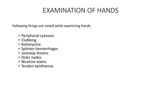
GENERAL PHYSICAL
EXAMINATION
DR. NAEEM SHAHZAD
HOUSE OFFICER
DEPARTMENT OF CARDIOLOGY
BENAZIR BHUTTO HOSPITAL
General physical examination includes:
• General appearance(Built, nutrition, cooperation, decubitus etc).
• Vital signs
• Examination of hands
• Examination of Scalp, face and neck
• Lymph nodes
• Oedema
First impressions
Diagnosis at first sight
From the first moment you set eyes on the patient, you should be forming
impressions of their general state of health. It takes experience and practice
to pick up all the possible clues but much can be gained by combining
common sense with medical knowledge. Ask yourself:
1. Is the patient comfortable or distressed?
2. Is the patient well or ill?
3. Is there a recognizable syndrome or facies?
4. Is the patient well nourished and hydrated?
Many of these features will be noted subconsciously, but you must make
yourself consciously aware of them.
Bed side clues:
In a hospital setting, there may be additional clues as to the patient's
state of health in the objects around them. In other circumstances, look
at objects that they are carrying or are visible in their pockets.
Examples include oxygen tubing, inhalers, GTN spray, insulin injections,
glucose meter, or cigarettes.
Vital signs
It may also be appropriate to assess vital signs at an early stage.
These usually include:
1. Pulse
2. Blood pressure.
3. Respiratory rate.
4. Oxygen saturation.
5. Temperature.
6. Blood glucose.
EXAMINATION OF HANDS
Following things are noted while examining hands.
• Peripheral cyanosis
• Clubbing
• Koilonychia
• Splinter hemorrhages
• Janeway lesions
• Osler nodes
• Nicotine stains
• Tendon xanthomas
Peripheral
cyanosis
Janeway lesions
and Osler nodes
Splinter
hemorrhage
Tendon
xanthomas
Nicotine stains
EXAMINATION OF FACE
Jaundice: seen as a yellow discolouration of the sclera.
Anemia: seen as an unusually pale conjunctiva.
Xanthelasma: yellow, raised lesions found particularly around the
eyes, indicative of a high serum cholesterol.
Corneal arcus: a whitish ring seen overlying the iris. Significant in
patients <40 years but not in older persons.
Mitral facies: rosy cheeks suggestive of mitral stenosis.
Cyanosis: bluish discoloration of lips and tongue.
Dental hygiene: a source of organisms causing infective
endocarditis.
Palate: may be suggestive of Marfan syndrome
Uvula: pulsating uvula may be seen in aortic regurgitation.
Nicotine stomatitis: diffuse white patch on hard palate caused
by tobacco smoking.
Jaundice
Pallor
Mitral facies
High arched
palate
Nicotine
stomatitis
NECK EXAMINATION
1. Carotid pulse examination.
2. Jugular venous pressure.
3. Thyroid gland.
4. Cervical lymph nodes.
Lymph nodes
Inspection
Large nodes are often clearly visible on inspection, particularly if the
enlargement is asymmetrical. If nodes are infected, the overlying skin
may be red and inflamed.
Palpation
Lymph nodes should be palpated using the most sensitive part of your
hands (the fingertips).
Head and neck: the nodes should be palpated with the patient
in an upright position and the examiner standing behind
(similar to the examination of the thyroid gland)
Axillae: To examine the nodes at the right axilla:
1. The patient should be sitting comfortably and you should stand at
their right-hand side.
2. Support their right arm abducted to 90 with your right hand.
3. Examine the axilla with your left hand.
To examine the nodes at the left axilla, perform the opposite
manoeuvre to the above.
Inguinal: with the patient lying supine, palpate their inguinal region
along the inguinal ligament.
There are 2 chains of superficial inguinal lymph nodes (a horizontal
chain which runs just below the inguinal ligament and a vertical chain
which runs along the saphenous vein).
Epitrochlear nodes: place the palm of the right hand under the
patient's slightly flexed right elbow and feel with your fingers in the
groove above and posterior to the medial epicondyle of the humerus.
Popliteal: best examined by passively flexing the knee and exploring
the fossa with the fingers of both hands.
Axilla Epitrochlear nodes Inguinal
Oedema
• Oedema refers to fluid accumulation in the subcutaneous tissues and
implies an imbalance of the Starling forces (increase of intravascular
pressure or reduced intravascular oncotic pressure) causing fluid to
seep into the interstitial space.
• Oedema will occur in hypoproteinaemic states (especially nephrotic
syndrome, malnutrition and malabsorption) and severe cardiac and
renal failure.
Examination
In ambulant patients, palpate the distal shaft of the tibia for oedema by
gently compressing the area for up to 10 seconds with the thumb.
If the oedema is pitting, the skin will show an indention where pressure was
applied which refills slowly.
If oedema is present, note its upper level.
Oedema may also involve the anterior abdominal wall and external genitalia.
When lying down, fluid moves to the new dependent area causing a sacral
pad. This can be checked for by asking the patient to sit forwards, exposing
the lower back and sacral region, and again applying gentle pressure with
your finger-tips.
Pulse examination
The pulse waveform depends on the heart rate, stroke volume,
peripheral resistance (especially in the arterioles), left ventricular
outflow obstruction and the elasticity of peripheral vessels.
When taking a pulse, characterize the information according to:
Site.
Rate.
Rhythm.
Volume.
Character of the pulse .
Radio-radial delay.
Radio-femoral delay.
Bruit.
Site of pulsation:
Radial artery: place the pads of three middle fingers over the right radial artery.
Brachial artery: Feel at the medial side of the cubital fossa, just medial to the
tendenous insertion of the biceps.
Carotid artery: This is the best place to assess the pulse volume and waveform.
Find the larynx, move a couple of centimeters laterally and press backwards medial
to the sternomastoid muscle.
Femoral artery: can be felt midway between the pubic tubercle and the anterior
superior iliac spine.
Popliteal artery: the patient lying flat and knees slightly flexed, press into the
center of the popliteal fossa with tips of the fingers of the left hand and use the
fingers of the right hand to add extra pressure to these.
Posterior tibial artery: Palpate at the ankle just posterior and inferior to the medial
malleolus.
Dorsalis pedis: This runs lateral to the exterior hallucis longus tendon on the
superior surface of the foot between the bases of the 1st and 2nd metatarsals.
Pulse rate:This should be expressed in beats per minute. A rate
<60bpm is called bradycardia whilst tachycardia is a pulse >100bpm.
The most accurate method is to count the pulse for a full minute.
Rhythm: In general, the pulse can be either regular or irregular but
variations exist.
Regular
Irregularly irregular
Regularly irregular
Character/waveform and volume
This is best assessed at the carotid artery. You are feeling for the speed
at which the artery expands and collapses and force with which it does
so.
Pulsus parvus et tardus: a slow rising pulse seen in aortic stenosis.
Collapsing pulse: a collapsing pulse which feels it suddenly hits your
fingers and falls away just as quickly. You could try feeling at the
brachial artery and raising the arm above the patient's heart.
Sometimes referred to as a waterhammer pulse.
Pulsus bisferiens: a waveform with 2 peaks, found where aortic
stenosis and regurgitation co-exist.
Pulsus alternans: an alternating strong and weak pulsation,
synonymous with a severely impaired left ventricle in a heart failure
COLLAPSING PULSE
• It is characterized by rapid upstroke followed by rapid
descent(collapse) of the pulse wave without dicrotic notch.
Detection:
It is best appreciated with the palm of hand palpating radial artery
while arm is lifted above the shoulder.
This may be related to the artery becoming more in line with the
central aorta, allowing direct systolic ejection and diastolic backfkow.
PULSUS PARADOXUS
Pulsus paradoxus refers to a fall in systolic blood pressure > 10 mmHg
during inspiration.
Causes:
• Cardiac temponade
• Massive pulmonary embolism
• Severe obstructive lung disease
• Tension pneumothorax
Blood pressure
Blood pressure (BP) is a measure of the force that the circulating blood
exerts against the arterial wall.
The systolic BP is the maximal pressure that occurs during ventricular
contraction (systole).
During ventricular filling (diastole), arterial pressure is maintained, but
at a lower level, by the elasticity and compliance of the vessel wall. The
lowest value (diastolic BP) occurs immediately before the next cycle.
BP is usually measured by means of a sphygmomanometer cuff. It is
measured in mmHg and recorded as systolic pressure/diastolic
pressure, together with where, and how, the reading was taken,
e.g. BP: 146/92 mmHg, right arm, supine.
Blood pressure should be measured in both arms either in rapid
succession or simultaneously.
Normally the measurements should differ by less than 10 mmHg.
A BP differential of more than 10 mmHg can be associated with
following conditions:
• Aortic dissection
• Subclavian artery disease
• Supravalvular aortic stenosis
• Aortic coarctation
Orthostatic hypotension
It is defined as a fall in systolic pressure >20 mmHg or in diastolic
pressure > 10 mmHg in response to assumption of the upright posture
from a supine position within 3 minutes(may be accompanied by lack
of compensatory tachycardia).
Causes
• Diabetes
• Parkinson’s disease
Jugular venous pressure
The jugular veins connect to the SVC and the right atrium without any
intervening valves. Therefore, changes in pressure in the right atrium
will transmit a pressure wave up these veins which can be seen in the
neck.
It is often the JVP must be measured in the internal jugular vein (IJV).
The EJV had tortuous course means that impulses are not transmitted
as readily or as reliably. It is for this reason that the IJV is used.
The centre of the right atrium lies ~5cm below the sternal angle which
is used as the reference point.
The normal JVP is ~8cm of blood (therefore 3cm above the sternal
angle).
Examination sequel:
1.The patient lying back at 45°, and expose the neck. With the patient tilted back to
45°the upper border of the pulse is just hidden at the base of the neck. This,
therefore, is used as the standard position for JVP measurement.
2. Ask the patient to turn their head away from you (their left) and ensure that the
neck muscles are relaxed.
3. Look for the JVP and measure the vertical distance from the top of the pulsation
to the sternal angle.
4. adding an extra 5cm.
It can sometimes be difficult to distinguish the jugular venous pulse from the carotid pulse
Character of the jugular venous pulsation
The jugular pulsation has 2 main peaks. You should establish the timing of the
peaks in the cardiac cycle by palpating the carotid pulse at the same time.
a wave: caused by atrial contraction. Seen just before the carotid pulse.
c point: slight AV-ring bulge during ventricular contraction.
x decent: atrial relaxation.
v wave: tricuspid closure and atrial filling.
y decent: ventricular filling as tricuspid valve opens.
KUSSMAUL’S SIGN
A rise in venous pressure(or its failure to decrease) with inspiration.
CAUSES:
• Constrictive pericarditis
• Restrictive cardiomyopathy
• Pulmonary embolism
• Right ventricular infarction
• Advanced systolic heart failure
Abdominojugular reflux
The abdominojugular reflux elicit venous hypertension.
Firm and consistent pressure is applied over the upper abdomen,
preferably the right upper quadrant, for atleast 10 seconds.
Positive response is defined as a rise of more than 3 cm in the venous
pressure sustained for at least 15 seconds.
A positive abdominojugular reflux can predict heart failure in patients
with dyspnea as well as pulmonary artery wedge pressure greater than
15 mm Hg.
THANK YOU!

general physical examination .pptx

  • 1.
    GENERAL PHYSICAL EXAMINATION DR. NAEEMSHAHZAD HOUSE OFFICER DEPARTMENT OF CARDIOLOGY BENAZIR BHUTTO HOSPITAL
  • 2.
    General physical examinationincludes: • General appearance(Built, nutrition, cooperation, decubitus etc). • Vital signs • Examination of hands • Examination of Scalp, face and neck • Lymph nodes • Oedema
  • 3.
    First impressions Diagnosis atfirst sight From the first moment you set eyes on the patient, you should be forming impressions of their general state of health. It takes experience and practice to pick up all the possible clues but much can be gained by combining common sense with medical knowledge. Ask yourself: 1. Is the patient comfortable or distressed? 2. Is the patient well or ill? 3. Is there a recognizable syndrome or facies? 4. Is the patient well nourished and hydrated? Many of these features will be noted subconsciously, but you must make yourself consciously aware of them.
  • 4.
    Bed side clues: Ina hospital setting, there may be additional clues as to the patient's state of health in the objects around them. In other circumstances, look at objects that they are carrying or are visible in their pockets. Examples include oxygen tubing, inhalers, GTN spray, insulin injections, glucose meter, or cigarettes.
  • 5.
    Vital signs It mayalso be appropriate to assess vital signs at an early stage. These usually include: 1. Pulse 2. Blood pressure. 3. Respiratory rate. 4. Oxygen saturation. 5. Temperature. 6. Blood glucose.
  • 6.
    EXAMINATION OF HANDS Followingthings are noted while examining hands. • Peripheral cyanosis • Clubbing • Koilonychia • Splinter hemorrhages • Janeway lesions • Osler nodes • Nicotine stains • Tendon xanthomas
  • 7.
  • 8.
  • 11.
    EXAMINATION OF FACE Jaundice:seen as a yellow discolouration of the sclera. Anemia: seen as an unusually pale conjunctiva. Xanthelasma: yellow, raised lesions found particularly around the eyes, indicative of a high serum cholesterol. Corneal arcus: a whitish ring seen overlying the iris. Significant in patients <40 years but not in older persons. Mitral facies: rosy cheeks suggestive of mitral stenosis.
  • 12.
    Cyanosis: bluish discolorationof lips and tongue. Dental hygiene: a source of organisms causing infective endocarditis. Palate: may be suggestive of Marfan syndrome Uvula: pulsating uvula may be seen in aortic regurgitation. Nicotine stomatitis: diffuse white patch on hard palate caused by tobacco smoking.
  • 13.
  • 14.
  • 15.
  • 16.
    NECK EXAMINATION 1. Carotidpulse examination. 2. Jugular venous pressure. 3. Thyroid gland. 4. Cervical lymph nodes.
  • 18.
    Lymph nodes Inspection Large nodesare often clearly visible on inspection, particularly if the enlargement is asymmetrical. If nodes are infected, the overlying skin may be red and inflamed. Palpation Lymph nodes should be palpated using the most sensitive part of your hands (the fingertips).
  • 19.
    Head and neck:the nodes should be palpated with the patient in an upright position and the examiner standing behind (similar to the examination of the thyroid gland)
  • 21.
    Axillae: To examinethe nodes at the right axilla: 1. The patient should be sitting comfortably and you should stand at their right-hand side. 2. Support their right arm abducted to 90 with your right hand. 3. Examine the axilla with your left hand. To examine the nodes at the left axilla, perform the opposite manoeuvre to the above.
  • 22.
    Inguinal: with thepatient lying supine, palpate their inguinal region along the inguinal ligament. There are 2 chains of superficial inguinal lymph nodes (a horizontal chain which runs just below the inguinal ligament and a vertical chain which runs along the saphenous vein). Epitrochlear nodes: place the palm of the right hand under the patient's slightly flexed right elbow and feel with your fingers in the groove above and posterior to the medial epicondyle of the humerus. Popliteal: best examined by passively flexing the knee and exploring the fossa with the fingers of both hands.
  • 23.
  • 24.
    Oedema • Oedema refersto fluid accumulation in the subcutaneous tissues and implies an imbalance of the Starling forces (increase of intravascular pressure or reduced intravascular oncotic pressure) causing fluid to seep into the interstitial space. • Oedema will occur in hypoproteinaemic states (especially nephrotic syndrome, malnutrition and malabsorption) and severe cardiac and renal failure.
  • 25.
    Examination In ambulant patients,palpate the distal shaft of the tibia for oedema by gently compressing the area for up to 10 seconds with the thumb. If the oedema is pitting, the skin will show an indention where pressure was applied which refills slowly. If oedema is present, note its upper level. Oedema may also involve the anterior abdominal wall and external genitalia. When lying down, fluid moves to the new dependent area causing a sacral pad. This can be checked for by asking the patient to sit forwards, exposing the lower back and sacral region, and again applying gentle pressure with your finger-tips.
  • 27.
    Pulse examination The pulsewaveform depends on the heart rate, stroke volume, peripheral resistance (especially in the arterioles), left ventricular outflow obstruction and the elasticity of peripheral vessels. When taking a pulse, characterize the information according to: Site. Rate. Rhythm. Volume. Character of the pulse . Radio-radial delay. Radio-femoral delay. Bruit.
  • 28.
    Site of pulsation: Radialartery: place the pads of three middle fingers over the right radial artery. Brachial artery: Feel at the medial side of the cubital fossa, just medial to the tendenous insertion of the biceps. Carotid artery: This is the best place to assess the pulse volume and waveform. Find the larynx, move a couple of centimeters laterally and press backwards medial to the sternomastoid muscle. Femoral artery: can be felt midway between the pubic tubercle and the anterior superior iliac spine. Popliteal artery: the patient lying flat and knees slightly flexed, press into the center of the popliteal fossa with tips of the fingers of the left hand and use the fingers of the right hand to add extra pressure to these. Posterior tibial artery: Palpate at the ankle just posterior and inferior to the medial malleolus. Dorsalis pedis: This runs lateral to the exterior hallucis longus tendon on the superior surface of the foot between the bases of the 1st and 2nd metatarsals.
  • 30.
    Pulse rate:This shouldbe expressed in beats per minute. A rate <60bpm is called bradycardia whilst tachycardia is a pulse >100bpm. The most accurate method is to count the pulse for a full minute. Rhythm: In general, the pulse can be either regular or irregular but variations exist. Regular Irregularly irregular Regularly irregular
  • 31.
    Character/waveform and volume Thisis best assessed at the carotid artery. You are feeling for the speed at which the artery expands and collapses and force with which it does so. Pulsus parvus et tardus: a slow rising pulse seen in aortic stenosis. Collapsing pulse: a collapsing pulse which feels it suddenly hits your fingers and falls away just as quickly. You could try feeling at the brachial artery and raising the arm above the patient's heart. Sometimes referred to as a waterhammer pulse. Pulsus bisferiens: a waveform with 2 peaks, found where aortic stenosis and regurgitation co-exist. Pulsus alternans: an alternating strong and weak pulsation, synonymous with a severely impaired left ventricle in a heart failure
  • 32.
    COLLAPSING PULSE • Itis characterized by rapid upstroke followed by rapid descent(collapse) of the pulse wave without dicrotic notch. Detection: It is best appreciated with the palm of hand palpating radial artery while arm is lifted above the shoulder. This may be related to the artery becoming more in line with the central aorta, allowing direct systolic ejection and diastolic backfkow.
  • 36.
    PULSUS PARADOXUS Pulsus paradoxusrefers to a fall in systolic blood pressure > 10 mmHg during inspiration. Causes: • Cardiac temponade • Massive pulmonary embolism • Severe obstructive lung disease • Tension pneumothorax
  • 39.
    Blood pressure Blood pressure(BP) is a measure of the force that the circulating blood exerts against the arterial wall. The systolic BP is the maximal pressure that occurs during ventricular contraction (systole). During ventricular filling (diastole), arterial pressure is maintained, but at a lower level, by the elasticity and compliance of the vessel wall. The lowest value (diastolic BP) occurs immediately before the next cycle. BP is usually measured by means of a sphygmomanometer cuff. It is measured in mmHg and recorded as systolic pressure/diastolic pressure, together with where, and how, the reading was taken, e.g. BP: 146/92 mmHg, right arm, supine.
  • 40.
    Blood pressure shouldbe measured in both arms either in rapid succession or simultaneously. Normally the measurements should differ by less than 10 mmHg. A BP differential of more than 10 mmHg can be associated with following conditions: • Aortic dissection • Subclavian artery disease • Supravalvular aortic stenosis • Aortic coarctation
  • 41.
    Orthostatic hypotension It isdefined as a fall in systolic pressure >20 mmHg or in diastolic pressure > 10 mmHg in response to assumption of the upright posture from a supine position within 3 minutes(may be accompanied by lack of compensatory tachycardia). Causes • Diabetes • Parkinson’s disease
  • 43.
    Jugular venous pressure Thejugular veins connect to the SVC and the right atrium without any intervening valves. Therefore, changes in pressure in the right atrium will transmit a pressure wave up these veins which can be seen in the neck. It is often the JVP must be measured in the internal jugular vein (IJV). The EJV had tortuous course means that impulses are not transmitted as readily or as reliably. It is for this reason that the IJV is used. The centre of the right atrium lies ~5cm below the sternal angle which is used as the reference point. The normal JVP is ~8cm of blood (therefore 3cm above the sternal angle).
  • 44.
    Examination sequel: 1.The patientlying back at 45°, and expose the neck. With the patient tilted back to 45°the upper border of the pulse is just hidden at the base of the neck. This, therefore, is used as the standard position for JVP measurement. 2. Ask the patient to turn their head away from you (their left) and ensure that the neck muscles are relaxed. 3. Look for the JVP and measure the vertical distance from the top of the pulsation to the sternal angle. 4. adding an extra 5cm.
  • 45.
    It can sometimesbe difficult to distinguish the jugular venous pulse from the carotid pulse
  • 47.
    Character of thejugular venous pulsation The jugular pulsation has 2 main peaks. You should establish the timing of the peaks in the cardiac cycle by palpating the carotid pulse at the same time. a wave: caused by atrial contraction. Seen just before the carotid pulse. c point: slight AV-ring bulge during ventricular contraction. x decent: atrial relaxation. v wave: tricuspid closure and atrial filling. y decent: ventricular filling as tricuspid valve opens.
  • 48.
    KUSSMAUL’S SIGN A risein venous pressure(or its failure to decrease) with inspiration. CAUSES: • Constrictive pericarditis • Restrictive cardiomyopathy • Pulmonary embolism • Right ventricular infarction • Advanced systolic heart failure
  • 49.
    Abdominojugular reflux The abdominojugularreflux elicit venous hypertension. Firm and consistent pressure is applied over the upper abdomen, preferably the right upper quadrant, for atleast 10 seconds. Positive response is defined as a rise of more than 3 cm in the venous pressure sustained for at least 15 seconds. A positive abdominojugular reflux can predict heart failure in patients with dyspnea as well as pulmonary artery wedge pressure greater than 15 mm Hg.
  • 52.