Gene mapping in phages . How the gens are traced in phages
1.
GENE MAPPING INPHAGES
FINE STRUCTURE MAPPING OF PHAGE GENES –
COMPLEMENTAION MAPPING
2.
GENE MAPPING INPHAGES
• Gene mapping in phages (bacterial viruses) is the process of
determining the order and relative distance of genes on a phage
chromosome.
• It’s similar to how genes are mapped in eukaryotes—but here, we use
recombination between phage DNA molecules.
• When two phages infect the same bacterial cell, their genetic material
can undergo recombination.
• The resulting recombinant phages may carry new combinations of
genetic markers.
• By counting the proportion of recombinant vs non recombinant
progeny, we can calculate recombination frequency (RF) and estimate
gene distances
3.
Hershey and Rotman’sExperiment
• To determine the relative distance between two genes (h and r) on the
chromosome of T2 bacteriophage using recombination frequency.
• The Genes They Studied:
• 1. h (host range gene)
• h⁺: Infects only E. coli B
• h⁻: Infects both E. coli B and B/2
• 2.r (rapid lysis gene)
• r⁺: Forms small, clear plaques
• r⁻: Forms large, fuzzy plaques
4.
• These twogenes control what kind of bacteria the phage can
infect and what kind of plaque they make on a bacterial lawn.
• The Experimental Setup:
• 1. Start with Two Parent Phage Strains:One is h⁺ r⁻
The other is h⁻ r⁺
• 2. Coinfect E. coli B with both strains.
Both phages inject DNA into the same cell.
Inside the cell, recombination between their genomes can occur.
5.
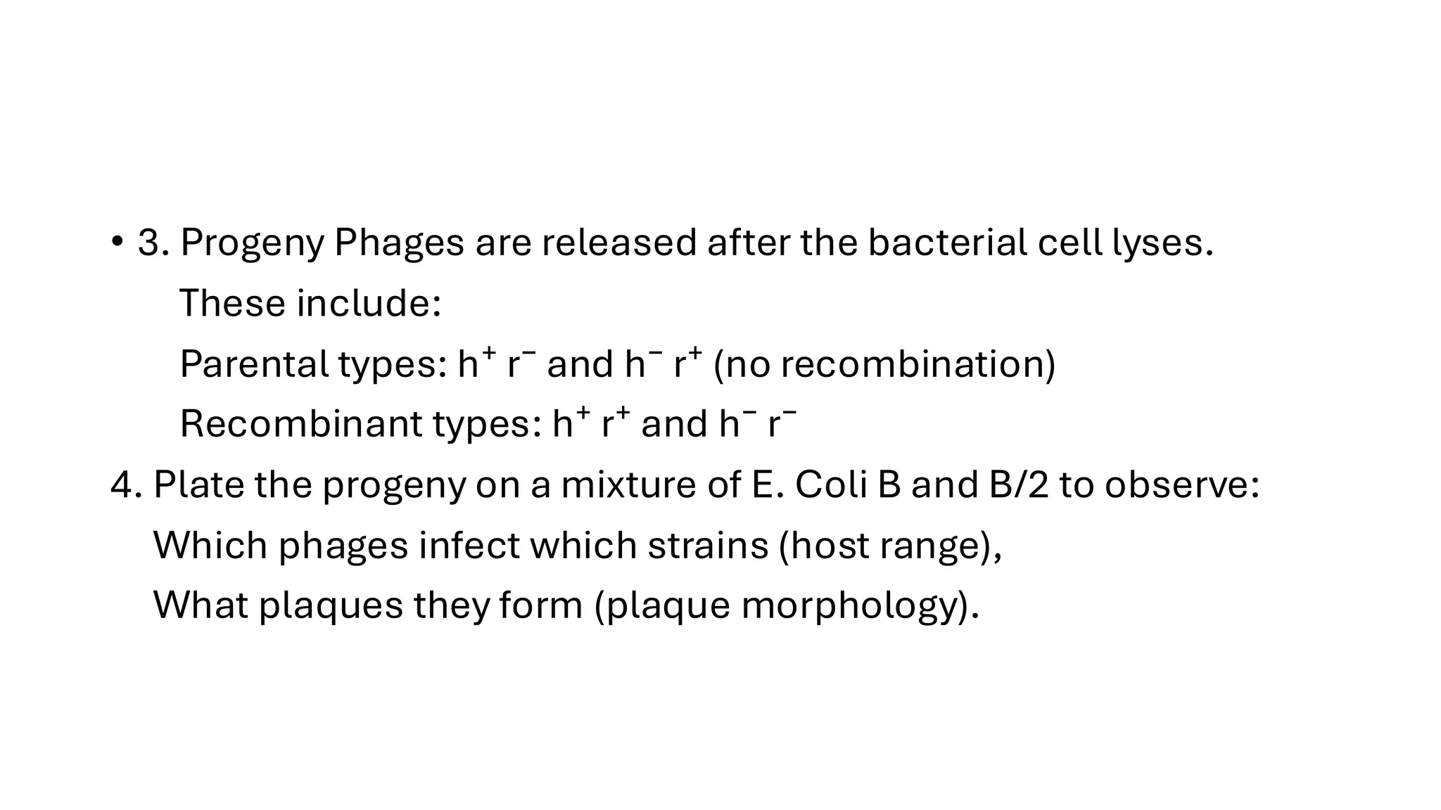
• 3. ProgenyPhages are released after the bacterial cell lyses.
These include:
Parental types: h⁺ r⁻ and h⁻ r⁺ (no recombination)
Recombinant types: h⁺ r⁺ and h⁻ r⁻
4. Plate the progeny on a mixture of E. Coli B and B/2 to observe:
Which phages infect which strains (host range),
What plaques they form (plaque morphology).
6.
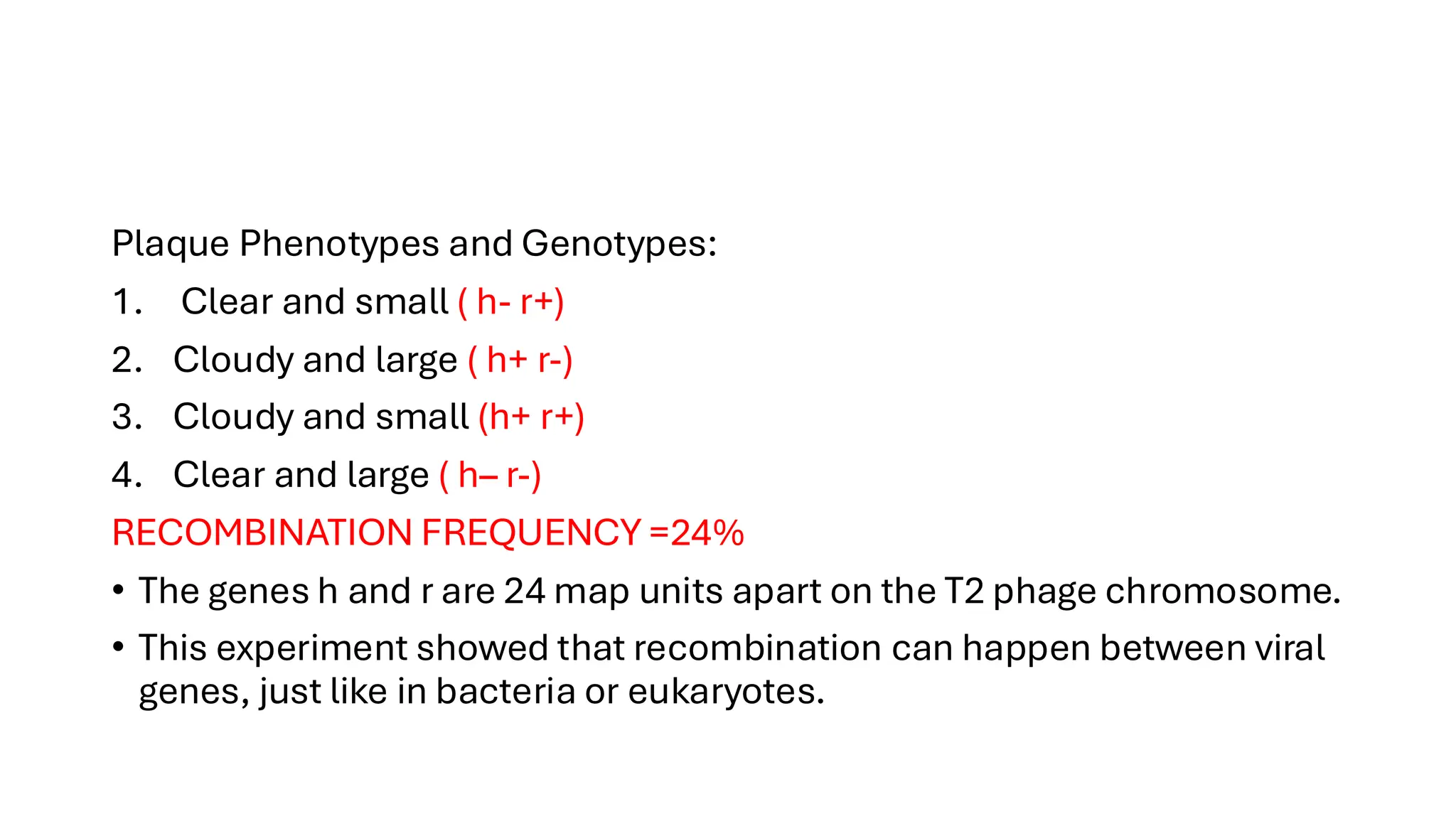
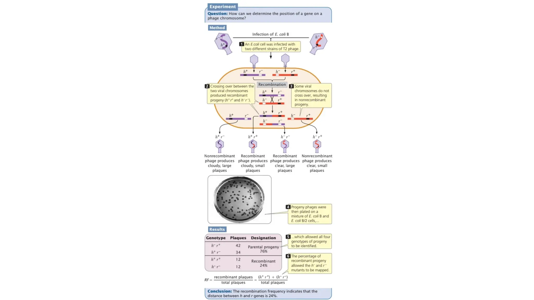
Plaque Phenotypes andGenotypes:
1. Clear and small ( h- r+)
2. Cloudy and large ( h+ r-)
3. Cloudy and small (h+ r+)
4. Clear and large ( h– r-)
RECOMBINATION FREQUENCY =24%
• The genes h and r are 24 map units apart on the T2 phage chromosome.
• This experiment showed that recombination can happen between viral
genes, just like in bacteria or eukaryotes.
7.
• Higher RF→ genes are farther apart.
• Lower RF → genes are closer together.
• This helps create a linear gene map of the phage chromosome.
9.
Fine-Structure Mapping inBacteriophage
Genes
• Fine-structure mapping is a detailed method to locate mutations within
a single gene.
• It helps us figure out how close different mutation sites are — even
within the same gene.
• This was first done in viruses (phages), especially T4 phage by Seymour
Benzer in the 1950s–60s.
• Phages (viruses that infect bacteria) are perfect for this kind of mapping
because:
They produce huge numbers of progeny quickly.
Mutations can be easily identified by plaque morphology.
Mutants of a gene (like the rII gene) can’t grow on certain strains of E.
coli (like K strain), so recombinants are easy to spot.
10.
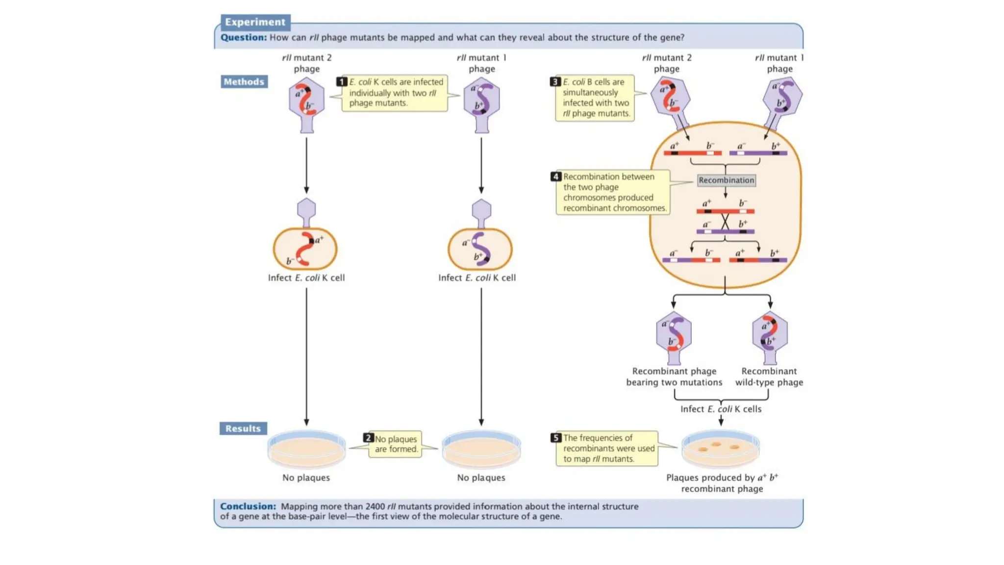
BENZER’S EXPERIMENT
• Benzerused two rII mutants, each with a mutation in a different
part of the same gene. When both mutants infected an E. Coli B
cell:
1. Recombination could happen between their genomes.
2. If recombination restored a functional rII gene, the phage could
now infect E. coli K (where mutants can't grow).
3. These recombinant phages produced plaques on E. coli K. That
means recombination had occurred.
11.
• Benzer collectedthousands of rII mutants with different mutations
in the rII region.
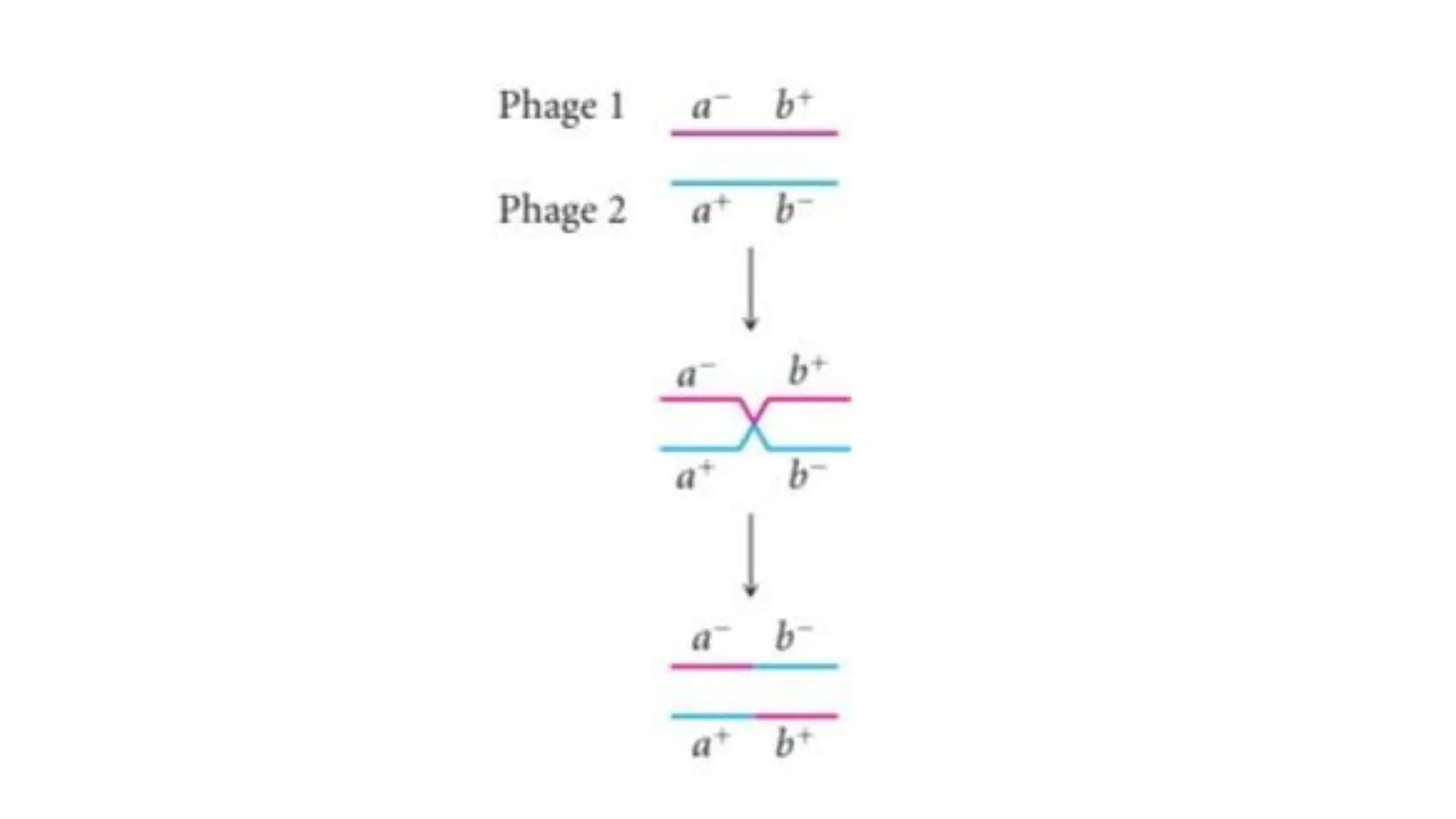
• Two mutants with different mutations (say, a⁻ b⁺ and a⁺ b⁻) were
used to infect E. Coli B strain (where both mutants can grow).
• Inside the bacteria, recombination between phage genomes can
occur.
• Recombinants like a⁺ b⁺ (wild-type) can now grow on E. Coli K cells.
only functional recombinants will form plaques.
• The recombination frequency is calculated by counting how many
plaques form (on E. Coli K). This tells you how far apart the two
mutations are.
12.
• He mappedover 2,400 mutations in the rII gene.
• Found out that genes have a linear structure (mutations can be
ordered).
• Showed that mutations within a single gene can recombine.
15.
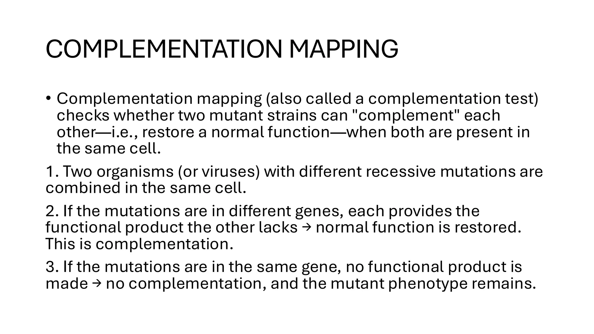
COMPLEMENTATION MAPPING
• Complementationmapping (also called a complementation test)
checks whether two mutant strains can "complement" each
other—i.e., restore a normal function—when both are present in
the same cell.
1. Two organisms (or viruses) with different recessive mutations are
combined in the same cell.
2. If the mutations are in different genes, each provides the
functional product the other lacks → normal function is restored.
This is complementation.
3. If the mutations are in the same gene, no functional product is
made → no complementation, and the mutant phenotype remains.
16.
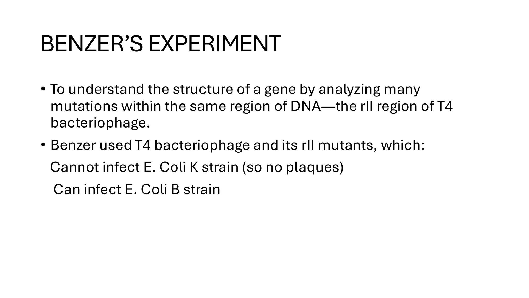
BENZER’S EXPERIMENT
• Tounderstand the structure of a gene by analyzing many
mutations within the same region of DNA—the rII region of T4
bacteriophage.
• Benzer used T4 bacteriophage and its rII mutants, which:
Cannot infect E. Coli K strain (so no plaques)
Can infect E. Coli B strain
17.
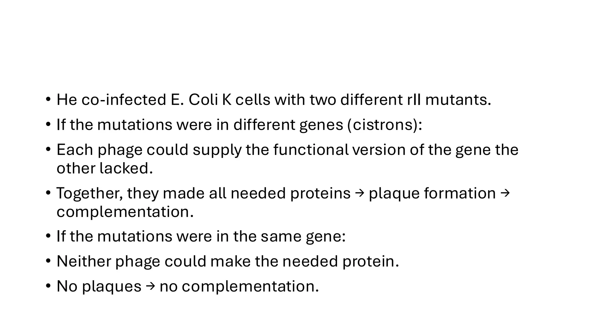
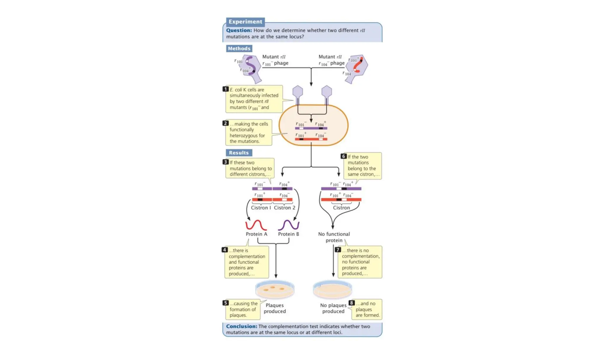
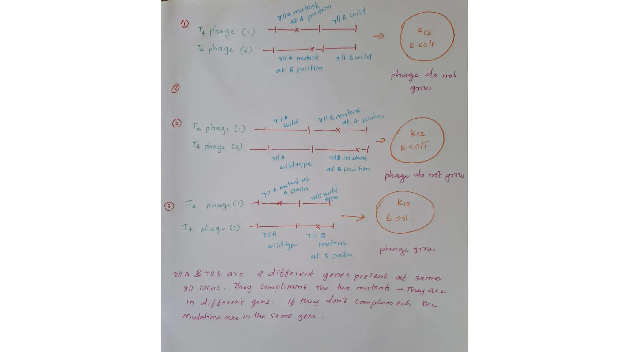
• He co-infectedE. Coli K cells with two different rII mutants.
• If the mutations were in different genes (cistrons):
• Each phage could supply the functional version of the gene the
other lacked.
• Together, they made all needed proteins → plaque formation →
complementation.
• If the mutations were in the same gene:
• Neither phage could make the needed protein.
• No plaques → no complementation.
18.
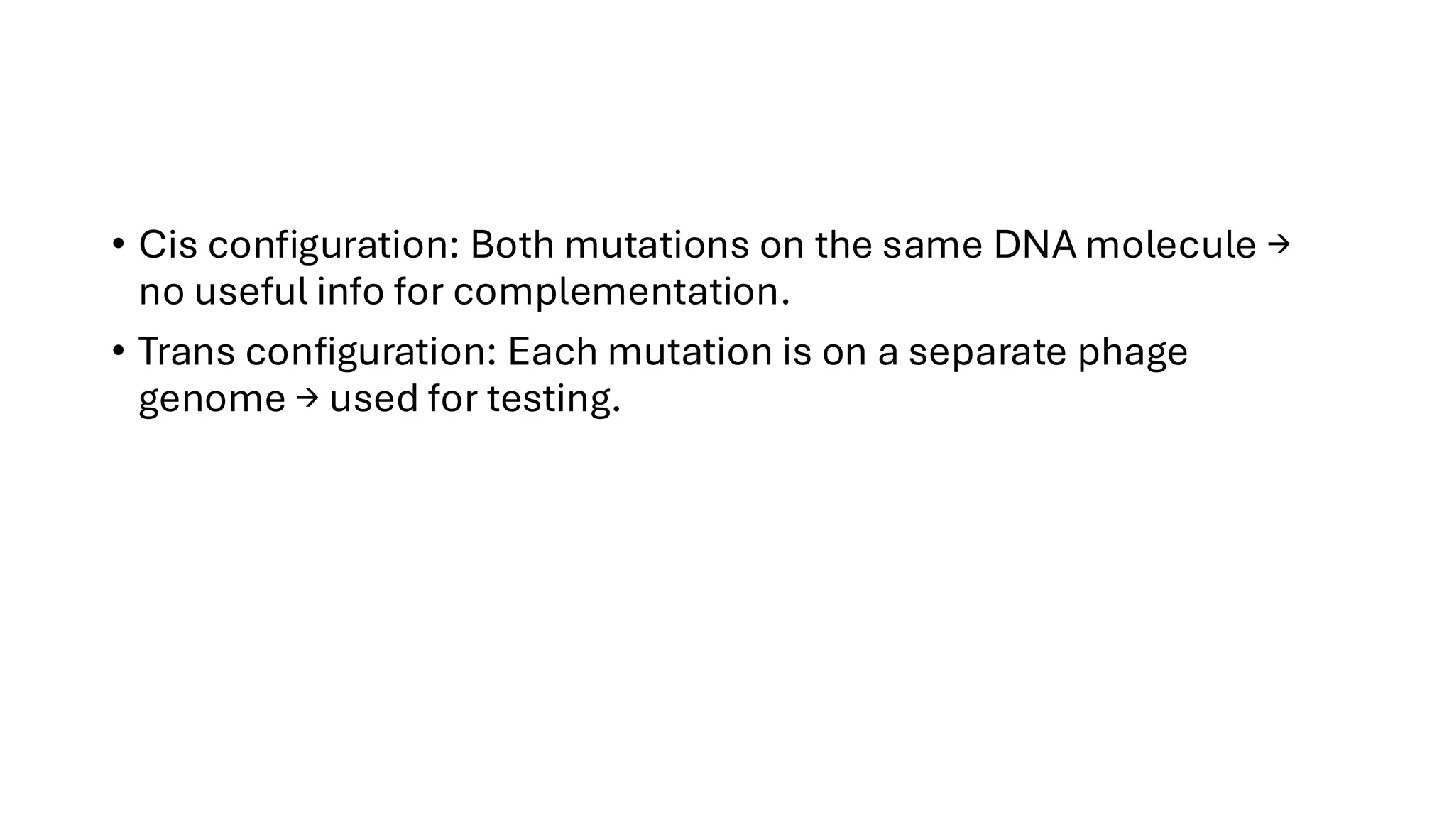
• Cis configuration:Both mutations on the same DNA molecule →
no useful info for complementation.
• Trans configuration: Each mutation is on a separate phage
genome → used for testing.