Acute liver failure is characterized by initial non-specific symptoms that progress to hepatic encephalopathy. It is defined as evidence of coagulopathy and mental alteration occurring within 26 weeks in a patient without pre-existing liver disease. Initial management involves fluid/electrolyte maintenance, nutrition, treatment of the underlying cause, and consideration of liver transplantation if criteria are met. ICU admission is warranted for altered sensorium, respiratory distress, bleeding, or hemodynamic instability. The prognosis depends on the severity and etiology of the liver injury.
The patient is a 4-year-old girl presenting with rapid breathing for 1 week and fever and cough for 1 month. On examination, she is tachypnic, tachycardic, and hypoxic. Investigations show metabolic acidosis, leukocytosis, and chest x-ray findings consistent with pneumonia. She deteriorates and requires intubation for septic shock secondary to severe pneumonia. Management involves fluid resuscitation, antibiotics, and supportive care including ventilation as she progresses to multi-organ dysfunction. Early recognition and goal-directed therapy are emphasized to prevent progression to irreversible shock.
1) Spontaneous bacterial peritonitis (SBP) is an infection of the ascitic fluid without an evident intra-abdominal surgically treatable cause.
2) Abdominal paracentesis should be performed for patients with ascites who present with signs of infection like fever or abdominal pain. The ascitic fluid is examined for white blood cell count and cultured.
3) SBP is diagnosed if the ascitic fluid white blood cell count is above 250 cells/mm3 or has a positive culture. Treatment involves antibiotics like cefotaxime for 5 days along with intravenous albumin to prevent kidney injury.
A 59-year-old man presented to the emergency department with vomiting blood and loose black stool. He had a history of hypertension, hepatitis C, smoking, and alcohol use. Physical examination found distension of the abdomen with positive shifting dullness. Laboratory tests showed signs of liver dysfunction. Endoscopy revealed esophageal varices as the likely cause of bleeding given the patient's risk factors of liver disease. He was admitted and treated with fluid resuscitation, blood transfusion, and balloon tamponade to stop the bleeding from his esophageal varices which developed due to cirrhosis and portal hypertension.
Principles Of Management Of Ascites Combinedsarafurness
The document discusses the causes, management, and treatment of ascites. Ascites is most commonly caused by cirrhosis and portal hypertension. Management involves dietary sodium restriction, diuretics, paracentesis, and TIPS for refractory ascites. For refractory ascites unresponsive to medical management, TIPS has been shown to better control ascites and improve survival rates compared to serial paracentesis, though it carries a higher risk of complications like encephalopathy.
Management of Ascites involves treating the underlying cause, restricting sodium intake, and using diuretics such as spironolactone. Refractory ascites is treated with therapeutic paracentesis or TIPSS. Complications include spontaneous bacterial peritonitis, hepatorenal syndrome, and hepatic hydrothorax. Diagnosis involves ascitic fluid analysis including SAAG to determine if ascites is portal hypertensive or not.
This case involves a 35-year-old female with a history of non-cirrhotic portal fibrosis, splenectomy, recurrent tuberculosis, and recurrent bouts of hematemesis and fever. She presents with cyanosis, clubbing, and shortness of breath. Testing reveals findings consistent with hepatopulmonary syndrome, including low oxygen saturation that decreases further in an upright position. A contrast echocardiogram confirms intrapulmonary vascular dilations. She is diagnosed with severe, longstanding hepatopulmonary syndrome secondary to non-cirrhotic portal hypertension.
This document provides information on acute pancreatitis, including its etiology, pathophysiology, clinical presentation, diagnosis, treatment and management. It notes that acute pancreatitis is characterized by inflammation of the pancreas and abdominal pain, and is most commonly caused by gallstones or alcohol use. The pathophysiology involves premature activation of digestive enzymes within the pancreas, leading to autodigestion. Treatment aims to relieve pain, provide fluid resuscitation and nutritional support, and manage complications through a variety of pharmacological and non-pharmacological therapies.
A 22-year-old male presented with confusion and jaundice. Laboratory results showed abnormal liver function tests and coagulopathy consistent with acute liver failure (ALF). The differential diagnosis for ALF includes drug toxicity, viral hepatitis, and other conditions. The most likely cause in this patient is isoniazid toxicity from his recent tuberculosis treatment, as 1-2% of patients can develop severe liver injury from isoniazid. Management involves supportive care, investigating the underlying cause, and consideration of liver transplantation if criteria are met.
Call prep: emergency nuclear medicine proceduresHerbert Klein
Guidelines for radiology and nuclear medicine procedures taking call on nights and weekends: gastrointestinal bleeding, hepatobiliary and lung scans. It is interactive, e.g. using Keynote presentation software. PowerPoint
Gastroparesis is a syndrome of objectively delayed gastric emptying in the absence of a mechanical obstruction and cardinal symptoms of nausea, vomiting, early satiety, bloating, and/or upper abdominal pain
Hepatic Encephalopathy -Pathophysiology,Evaluation And ManagementSantosh Narayankar
Hepatic encephalopathy is a brain dysfunction caused by liver disease or portosystemic shunting. It presents as a wide range of neurological or psychiatric abnormalities from mild alterations to coma. The prevalence is 30-40% in those with cirrhosis and risk of first episode is 5-25% within 5 years of cirrhosis diagnosis. Recurrence risk after an initial episode is 40% within 1 year. Ammonia, systemic inflammation, manganese, genetics, and oxidative stress may all contribute to pathogenesis. Diagnosis involves clinical exam and testing like serum ammonia levels or neuropsychological tests on phone apps. Management involves treating precipitating factors, lactulose, antibiotics like rifaximin, and
This document provides an overview of chronic pancreatitis, including its definition, epidemiology, pathology, classification, clinical features, diagnosis and treatment. Chronic pancreatitis is defined as permanent and irreversible damage to the pancreas resulting in inflammation, fibrosis and destruction of pancreatic tissue. It has an annual incidence of 3-9 cases per 100,000 people. Alcohol is a major risk factor. Diagnosis involves evaluating pancreatic function and structure through imaging, endoscopy and genetic/serological testing. Treatment focuses on pain management, pancreatic enzyme supplementation and surgery for severe cases.
This document discusses gastroesophageal reflux disease (GERD). It describes GERD as occurring when reflux of stomach contents causes troublesome symptoms or complications. The pathogenesis of GERD involves an imbalance between defensive factors that protect the esophagus and aggressive factors like gastric acid that reflux from the stomach. Defensive factors include the lower esophageal sphincter, esophageal motility, the diaphragm, and stomach function. Transient lower esophageal sphincter relaxations are a major mechanism for acid reflux. Symptoms of GERD include heartburn, regurgitation, dysphagia, chest pain, laryngeal and pulmonary issues. Tests to diagnose GERD include questionnaires
Acute Kidney Injury-case management and discussion
AKI secondary to sepsis secondary to acute bacterial salphingitis vs TB salphingitis
KDIGO 2012 guidelines
AKI, harrison's 19th edition
A 42-year old male patient presented with back pain and vomiting. Medical tests showed changes in the pancreas with calculi in the dilated duct, consistent with a diagnosis of chronic calcific pancreatitis. The patient's condition is likely due to a history of heavy alcohol abuse. He was prescribed medications including antibiotics, painkillers, and proton pump inhibitors to treat his symptoms and the underlying condition.
This document discusses acute liver failure (ALF), defining it as the rapid development of severe liver injury and encephalopathy in someone who previously had a normal liver or well-compensated liver disease. It describes the development of encephalopathy within different time periods to classify ALF as hyper-acute, acute, or subacute. Common causes, clinical presentation, investigations, complications including cerebral edema, and treatment approaches are summarized.
Anaesthesia to patiens with liver disease or a liver transplantscanFOAM
A presentation by Anna Januszkiewicz at the 2017 meeting of the Scandinavian Society of Anaestesiology and Intensive Care Medicine.
All available content from SSAI2017: https://scanfoam.org/ssai2017/
Delivered in collaboration between scanFOAM, SSAI & SFAI.
This document presents the case of a 70-year-old woman admitted to the ICU with hypotension, hypoxia, abdominal tenderness and distension. She developed intra-abdominal hypertension with an intra-abdominal pressure of 26 mmHg. Imaging revealed occlusion of the superior mesenteric artery near its origin with infarction of the right kidney and spleen. Her condition was too poor for surgery, so medical management was initiated for her intra-abdominal hypertension and multiple organ dysfunction.
David Smith, a 68-year-old man with diabetes and heart disease, was admitted to the emergency department after collapsing at home. Upon admission, he was found to have acute kidney injury (AKI) stage 2, with elevated creatinine and decreased urine output. His multiple medical conditions and medications put him at risk for AKI. After fluid resuscitation and medication review, his kidney function and blood pressure improved. However, further treatment with IV contrast and antibiotics for suspected sepsis again placed him at risk for worsening AKI. His creatinine continued to rise, reaching stage 3 AKI. Ultrasound found no obstructions, so the likely cause of David's AKI was his multiple medical issues
OLD and NEW definition of Hepatorenal syndrome , EASL 2018 +AASLD 2012 guidelines , pathophysiology mechanisms , Precipitants of HRS , prevention and treatment of HRS , new drugs for HRS on lane , few evidences .
A 32-year old man with cirrhosis and spontaneous bacterial peritonitis is admitted to the hospital with worsening renal function. His serum creatinine has risen from 1.2 to 4.1 over 5 days and he has become anuric. The likely diagnosis is hepatorenal syndrome (HRS) type 1. The prognosis for this patient is poor, as HRS type 1 has a high mortality rate without treatment. He should be treated with intravenous albumin and a vasoconstrictor such as terlipressin to try to reverse the HRS.
Upper gastrointestinal (UGI) bleeding can originate from the esophagus, stomach, or duodenum. The document discusses the causes, clinical presentations, associated conditions, and endoscopic findings of UGI bleeding. The major causes include peptic ulcers, esophageal or gastric varices, esophagitis, vascular ectasias, tumors, and Dieulafoy's lesions. An evaluation of UGI bleeding involves assessing severity based on history and labs, and identifying potential bleeding sources and comorbidities to guide management. Endoscopy allows visualization of the source and features to determine the risk of rebleeding.
This document defines different types and grades of ascites and outlines guidelines for diagnosing and treating ascites caused by cirrhosis. It describes diagnostic paracentesis to analyze ascitic fluid and determine the cause. Treatment involves dietary salt restriction, diuretics like spironolactone and furosemide, and therapeutic paracentesis with volume expansion to drain large amounts of fluid. The prognosis is poor, with 50% mortality within two years of developing ascites, indicating evaluation for liver transplantation should be considered.
1) Acute pancreatitis has many etiological causes including gallstones, alcohol use, hypertriglyceridemia, medications, infections, trauma, and others.
2) Diagnosis involves meeting two of three criteria including abdominal pain, elevated pancreatic enzymes, and imaging findings characteristic of pancreatitis. Severity is classified as mild, moderately severe, or severe.
3) Management involves aggressive hydration, pain control, monitoring for organ failure, early enteral nutrition, treating infections, and addressing underlying causes such as performing cholecystectomy for gallstone pancreatitis. Infected pancreatic necrosis requires antibiotics and potentially minimally invasive or open surgical debridement.
Acute pancreatitis is an inflammatory condition of the pancreas characterized by abdominal pain and elevated pancreatic enzymes. The most common causes are gallstones and alcohol. In severe cases, pancreatic enzymes activate prematurely and digest the pancreas. This can lead to systemic inflammatory response and organ failure. Diagnosis is based on abdominal symptoms and blood tests showing elevated pancreatic enzymes. Severity is assessed using criteria like Marshall score and need for ICU care. Treatment involves hydration, pain control, treating the underlying cause, preventing infection, and nutrition support. Surgery is usually not needed for sterile pancreatic necrosis but may be for infected necrosis after 4 weeks.
This document discusses acute pancreatitis. It begins with a case presentation of a 30-year-old patient presenting with epigastric pain. It then provides general information on the pancreas and its secretions of bicarbonate and enzymes. It describes the signs, symptoms, lab tests, imaging studies, differential diagnosis, phases, severity, treatment, and recurrence risks of acute pancreatitis. Treatment involves NPO, IV fluids, analgesics, and treating any underlying causes like gallstones.
This is a practical pocket summary for acute liver cell failure which includes the etiology, clinical picture, investigations and management. It is based on the most recent guidelines.
This document provides an overview of acute pancreatitis including its:
- Pathophysiology involving trypsin activation within acinar cells
- Risk factors such as gallstones, alcohol use, anatomical obstructions
- Clinical manifestations like severe abdominal pain
- Diagnosis using blood tests, imaging, and scoring systems to determine severity
- Management approaches depending on severity, including antibiotics for infected necrosis
Acute-Liver-Failure-2012 power point presentationNishanthTR
- Acute liver failure has a high mortality rate between 56-80%. The main role of intensive care is providing multi-organ support.
- The most common cause in the western world is paracetamol toxicity. Hepatic encephalopathy no longer the main cause of death but its detection and management require sophisticated monitoring.
- Hepatorenal failure results from complex circulatory changes due to liver failure. Terlipressin may help treat it. Novel replacement therapies are under development but more studies are still needed on their efficacy.
- The mortality rate for acute liver failure ranges from 56% to 80%. The main role of intensive care therapy is providing multi-organ support.
- Paracetamol toxicity is the most common cause of acute liver failure in the Western world. Hepatic encephalopathy is no longer the main cause of death, but its detection and management require sophisticated monitoring.
- Hepatorenal failure results from complex interactions between splanchnic, renal, and systemic circulation in response to liver failure. Terlipressin has been shown to help treat it.
- Novel hepatic replacement therapies are under development but definitive studies on their efficacy have not been published yet.
This document presents the case of a 14-year-old female patient admitted with recurrent acute pancreatitis. Her episodes are associated with hypertriglyceridemia. Her brother also had a history of similar symptoms. Laboratory tests revealed elevated triglyceride levels in the patient and her family members. She was diagnosed with familial hypertriglyceridemia-induced recurrent acute pancreatitis. Treatment involved managing triglyceride levels and supportive care for the acute attack.
This document provides information on ascites including its definition, causes, diagnosis, and management. Ascites is defined as the accumulation of free fluid in the peritoneal cavity, most often caused by liver cirrhosis (75% of cases), malignancy, or heart failure. Diagnosis involves history, physical exam finding shifting dullness or fluid wave, and abdominal ultrasound or paracentesis. Initial ascites management consists of sodium restriction, diuretics, and large volume paracentesis for refractory ascites.
This document provides information on acute pancreatitis including:
- The anatomy and blood supply of the pancreas.
- Risk factors, pathophysiology, clinical presentation, diagnosis and management of acute pancreatitis including determining severity.
- Choice of antibiotics and analgesics for severe acute pancreatitis, with imipenem and ciprofloxacin/metronidazole recommended for infected pancreatic necrosis.
- Novel pain management strategies like thoracic epidural analgesia and inhibitors of proteinase-activated receptors and transient receptor potential vanilloid-1 showing promise in animal models of acute pancreatitis.
The document discusses various pancreatic disorders including acute pancreatitis, chronic pancreatitis, and pancreatic cancer. It provides details on the epidemiology, anatomy, physiology, etiology, pathophysiology, clinical manifestations, diagnostic studies, medical and surgical management, nursing management, and complications of each disorder. Acute pancreatitis results from inflammation of the pancreas caused by auto-digestion by pancreatic enzymes. Chronic pancreatitis is a progressive inflammatory disease often caused by alcoholism. Pancreatic cancer develops from the uncontrolled growth of pancreatic cells.
Ascites ( Diagnosis and management ).pdfMohamed Wifi
Ascites is an accumulation of fluid in the abdominal cavity. The document summarizes the causes, symptoms, signs, investigations and treatment of ascites. The main causes are increased hydrostatic pressure, decreased colloid osmotic pressure and increased permeability of capillaries. Symptoms include abdominal distension. Treatment involves diuretics, fluid restriction, large volume paracentesis and TIPS for refractory ascites. Complications include spontaneous bacterial peritonitis and hepatorenal syndrome.
Acute pancreatitis is inflammation of the pancreas that ranges from mild to severe. It commonly results from gallstones or alcohol use. Symptoms include abdominal pain and elevated pancreatic enzymes. Severe cases can lead to organ failure and death in up to 30% of patients. Treatment focuses on fluid resuscitation, treating the underlying cause, and managing complications which may include pancreatic necrosis, pseudocysts, or systemic inflammatory response. Prognosis depends on severity with most patients recovering if the attack is mild but severe cases carrying significant morbidity and mortality.
This document reviews the management of ascites in a patient with cirrhosis. Key points include:
1. Paracentesis was performed on the patient which showed ascitic fluid with a SAAG of 2.4, indicating portal hypertension.
2. Initial management involved salt restriction and diuretic therapy. However, the ascites became refractory to treatment.
3. For tense or refractory ascites, serial large volume paracentesis with albumin infusion is the standard treatment when diuretics fail.
Liver diseases can complicate pregnancy in several ways. Some diseases predate pregnancy like cirrhosis, while others are unique to pregnancy such as acute fatty liver of pregnancy (AFLP). AFLP typically presents in the third trimester with nausea, vomiting, and abdominal pain and can progress rapidly to liver failure without treatment. Timely delivery is crucial for managing AFLP and other pregnancy-related liver conditions like preeclampsia. Close monitoring of liver enzymes and symptoms is needed to identify problems and prevent maternal and fetal complications.
1. Diarrhea is defined as an increased frequency, fluidity, and volume of stool. It can be acute (<14 days), persistent (14-30 days), or chronic (>30 days).
2. The document discusses the pathophysiology, etiology, indications for referral to a gastroenterologist, initial evaluation, and recommendations for evaluating patients with chronic diarrhea.
3. The differential diagnosis and workup of inflammatory bowel diseases like Crohn's disease and ulcerative colitis are outlined, including relevant history, physical exam findings, diagnostic studies, endoscopy, histopathology, and radiology findings.
This document discusses pancreatitis, including its anatomy, physiology, etiology, clinical presentation, diagnosis, prognosis, management, and complications. Pancreatitis is defined as inflammation of the pancreas and can be acute or chronic. Acute pancreatitis is commonly caused by gallstones or alcohol and may range from mild to severe, with severe cases involving pancreatic necrosis and multi-organ failure. Diagnosis involves blood tests measuring amylase and lipase along with imaging like CT. Management depends on severity but generally involves hospitalization, IV fluids, pain control, and monitoring for complications.
The document discusses pancreatitis, including:
1. It describes the parts and functions of the pancreas, including exocrine and endocrine functions.
2. It covers the causes, signs, symptoms, investigations, and treatment of acute and chronic pancreatitis. Conservative treatment includes hydration and antibiotics, while surgery may be needed for complications.
3. It lists complications of pancreatitis such as shock, respiratory failure, infections, and pancreatic insufficiency. Chronic pancreatitis involves permanent structural damage and its treatment aims to control pain and manage complications.
George, a 40-year-old male with a history of chronic alcoholism and gallstones, presented with severe abdominal pain after starting sulfasalazine for ulcerative colitis. Lab results showed elevated amylase, lipase, and white blood cell count. The physician's diagnosis was acute pancreatitis, likely caused by sulfasalazine triggering the condition. Due to the severity of symptoms and lab abnormalities, the patient should be admitted to the ICU and given IV fluids, analgesics, and monitored closely for complications of acute pancreatitis.
Similar to Gastroenterology icu management protocol (20)
This presentation was provided by Racquel Jemison, Ph.D., Christina MacLaughlin, Ph.D., and Paulomi Majumder. Ph.D., all of the American Chemical Society, for the second session of NISO's 2024 Training Series "DEIA in the Scholarly Landscape." Session Two: 'Expanding Pathways to Publishing Careers,' was held June 13, 2024.
Philippine Edukasyong Pantahanan at Pangkabuhayan (EPP) CurriculumMJDuyan
(𝐓𝐋𝐄 𝟏𝟎𝟎) (𝐋𝐞𝐬𝐬𝐨𝐧 𝟏)-𝐏𝐫𝐞𝐥𝐢𝐦𝐬
𝐃𝐢𝐬𝐜𝐮𝐬𝐬 𝐭𝐡𝐞 𝐄𝐏𝐏 𝐂𝐮𝐫𝐫𝐢𝐜𝐮𝐥𝐮𝐦 𝐢𝐧 𝐭𝐡𝐞 𝐏𝐡𝐢𝐥𝐢𝐩𝐩𝐢𝐧𝐞𝐬:
- Understand the goals and objectives of the Edukasyong Pantahanan at Pangkabuhayan (EPP) curriculum, recognizing its importance in fostering practical life skills and values among students. Students will also be able to identify the key components and subjects covered, such as agriculture, home economics, industrial arts, and information and communication technology.
𝐄𝐱𝐩𝐥𝐚𝐢𝐧 𝐭𝐡𝐞 𝐍𝐚𝐭𝐮𝐫𝐞 𝐚𝐧𝐝 𝐒𝐜𝐨𝐩𝐞 𝐨𝐟 𝐚𝐧 𝐄𝐧𝐭𝐫𝐞𝐩𝐫𝐞𝐧𝐞𝐮𝐫:
-Define entrepreneurship, distinguishing it from general business activities by emphasizing its focus on innovation, risk-taking, and value creation. Students will describe the characteristics and traits of successful entrepreneurs, including their roles and responsibilities, and discuss the broader economic and social impacts of entrepreneurial activities on both local and global scales.
🔥🔥🔥🔥🔥🔥🔥🔥🔥
إضغ بين إيديكم من أقوى الملازم التي صممتها
ملزمة تشريح الجهاز الهيكلي (نظري 3)
💀💀💀💀💀💀💀💀💀💀
تتميز هذهِ الملزمة بعِدة مُميزات :
1- مُترجمة ترجمة تُناسب جميع المستويات
2- تحتوي على 78 رسم توضيحي لكل كلمة موجودة بالملزمة (لكل كلمة !!!!)
#فهم_ماكو_درخ
3- دقة الكتابة والصور عالية جداً جداً جداً
4- هُنالك بعض المعلومات تم توضيحها بشكل تفصيلي جداً (تُعتبر لدى الطالب أو الطالبة بإنها معلومات مُبهمة ومع ذلك تم توضيح هذهِ المعلومات المُبهمة بشكل تفصيلي جداً
5- الملزمة تشرح نفسها ب نفسها بس تكلك تعال اقراني
6- تحتوي الملزمة في اول سلايد على خارطة تتضمن جميع تفرُعات معلومات الجهاز الهيكلي المذكورة في هذهِ الملزمة
واخيراً هذهِ الملزمة حلالٌ عليكم وإتمنى منكم إن تدعولي بالخير والصحة والعافية فقط
كل التوفيق زملائي وزميلاتي ، زميلكم محمد الذهبي 💊💊
🔥🔥🔥🔥🔥🔥🔥🔥🔥
Leveraging Generative AI to Drive Nonprofit InnovationTechSoup
In this webinar, participants learned how to utilize Generative AI to streamline operations and elevate member engagement. Amazon Web Service experts provided a customer specific use cases and dived into low/no-code tools that are quick and easy to deploy through Amazon Web Service (AWS.)
Andreas Schleicher presents PISA 2022 Volume III - Creative Thinking - 18 Jun...EduSkills OECD
Andreas Schleicher, Director of Education and Skills at the OECD presents at the launch of PISA 2022 Volume III - Creative Minds, Creative Schools on 18 June 2024.
Level 3 NCEA - NZ: A Nation In the Making 1872 - 1900 SML.pptHenry Hollis
The History of NZ 1870-1900.
Making of a Nation.
From the NZ Wars to Liberals,
Richard Seddon, George Grey,
Social Laboratory, New Zealand,
Confiscations, Kotahitanga, Kingitanga, Parliament, Suffrage, Repudiation, Economic Change, Agriculture, Gold Mining, Timber, Flax, Sheep, Dairying,
What is Digital Literacy? A guest blog from Andy McLaughlin, University of Ab...
Gastroenterology icu management protocol
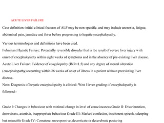
1. ACUTE LIVER FAILURE
Case definition: initial clinical features of ALF may be non-specific, and may include anorexia, fatigue,
abdominal pain, jaundice and fever before progressing to hepatic encephalopathy.
Various terminologies and definitions have been used.
Fulminant Hepatic Failure: Potentially reversible disorder that is the result of severe liver injury with
onset of encephalopathy within eight weeks of symptoms and in the absence of pre-existing liver disease.
Acute Liver Failure: Evidence of coagulopathy (INR>1.5) and any degree of mental alteration
(encephalopathy) occurring within 26 weeks of onset of illness in a patient without preexisting liver
disease.
Note: Diagnosis of hepatic encephalopathy is clinical; West Haven grading of encephalopathy is
followed:-
Grade I: Changes in behaviour with minimal change in level of consciousness Grade II: Disorientation,
drowsiness, asterixis, inappropriate behaviour Grade III: Marked confusion, incoherent speech, ssleeping
but arousable Grade IV: Comatose, unresponsive, decorticate or decerebrate posturing
2.
3. Criteria for ICU admissionPatient with any one of the following:
Altered sensorium: Patients in grade I and perhaps, grade II encephalopathy,
could be managed in a ward. However, rapidly worsening encephalopathy or
grade III/IV encephalopathy warrants ICU admission.
Respiratory distress, i.e., respiratory rate >30/m.
Any evidence of gastrointestinal bleeding
Any hemodynamic instability
4. Investigations
CBC, LFT, PT,INR, Creatinine, Electrolytes, Sugar, USG upper abdomen, blood typing,bloood culture, .Blood smear for
malaria, serology for Dengue and Widal test in selected cases
HbsAg. IgM-anti HBc if HbsAg positive, IgM-antiHEV. If viral markers negative then ANA, SMA, serum ceruloplasmin,
examination for KF rings. In ICU setting: ABG, arterial ammonia.
General Measures:
1. Patients with altered mentation should be admitted to ICU.
2. Avoid stimulation, avoid sedation.
3. Nurse with head end elevation to 30®
4. Consider intubation if grade III/IV encephalopathy.
5. Fluid and electrolyte maintenance. Any fluid may be used.
6. Enteral nutrition preferred till grade 1 /2 encephalopathy. Protein
intake 1G/Kg.
5. treatmenttolerated, dextrose infusion should be started
. Invasive hemodynamic monitoring in the form of intra-arterial blood pressure and central venous pressure
monitoring.
3% saline infusion @ 30 ml/hour till serum Na 145 mEq/L, then maintain 10- 15 ml/hour (aim serum Na 145-
155 mEq/L). Monitor serum sodium 12 hourly. Rehydration is required in many patients as they may be
dehydrated at admission due to vomiting and anorexia.
Inj Vitamin K 10 mg IV single dose.Fresh-frozen plasma transfusion is required for bleeding manifestations, as
and when required. Platelet transfusion is required for platelet counts <10,000/mm3 or invasive procedures.
Intravenous ranitidine or proton pump inhibitors.
Inj Mannitol bolus 0.5-1g/Kg if signs of cerebral edema: systolic hypertension, bradycardia, irregular
respiration or unequal pupils or posturing Use mannitol only if plasma Osmolality is < 320 mosmol/L.
Bowel decontamination with lactulose and antibiotic for gut sterilization (ampicillin / metronidazole /
rifaximin)
All patients in grade 3 or 4 hepatic encephalopathy should be intubated electively for airway
protection. In addition, respiratory support and mechanical ventilation should be provided for
those with inadequate respiratory effort.In ventilated patients, sedation should be instituted to avoid
coughing and bucking on endotracheal tube. which sedation?
The role of antibiotics is not definitive. However, in grades 3 and 4 encephalopathy, prophylactic antibiotics
6. 5. Treatment directed at aetiology:
a.Acetaminophen: N-Acetyl Cystine, Inj N-acetyl cysteine
150mg/Kg over 1 hour, 12.5mg/Kg/hour over next 4 hours and
then 6.25mg/Kg/hour over 67 hours.
b.HSV: Acyclovir: Skin lesions in 50% only
c.Amanita phalloides: Penicillin G 1 million units/kg/day
d.HELLP/AFLP: Terminate pregnancy
7. Avoid
1. Fresh frozen plasma (FFP) as it interferes with assessment for liver transplantation. Use only if invasive procedure
planned.
2. Platelet transfusion with platelets >10,000/cmm unless invasive procedure planned
3. Protein restriction to <1G/Kg
4. Branched chain amino acids
5.Avoid sedation in grades 1 and 2 encephalopathy.
6. Inj L-Ornithine L-Aspartate
7. Prophylactic anticonvulsants
8. Hypothermia
8. liver transplantation
Apply modified King’s College Hospital (KCH) criteria:-
Prothrombin time >100s or INR >6.5 Or, any 3 of the following:-
Age < 10 years or > 40 years
Jaundice-Hepatic Encephalopathy interval > 7 days
S bilirubin > 17.5 mg /dl Prothrombin time >50s or INR > 3.5 NonA-NonE Hepatitis
If answer to step 1 is yes, proceed to step 2. If answer to step1 is no, keep looking for indication for
liver transplantation as the negative predictive value of KCH criteria is low. Give due consider ation
to MELD score (>30) and arterial ammonia (>124).
Step 2: If answer to step 1 is yes, is there a contraindication to transplantation? Contraindications
include sepsis and posturing due to severe cerebral edema. Get in touch with the nearest liver
transplant centre for advise before deciding aginst transplantation.
Step 3: If answer to step 2 is no, can the family afford liver transplantation?
Step 4: If answer to step 3 is yes, is there a suitable donor?
Test blood group of patient and willing near relatives aged 18 to 50 years. If ABO compatible donor
is available, counsel on success and risk of liver transplantation and donor surgery.
9. INTESTINAL OBSTRUCTION
Acute intestinal obstruction occurs when there is an interruption in the forward
flow of intestinal contents. This interruption can occur at any point along the length
of the gastrointestinal tract. The clinical presentation varies depending on the
severity, duration, site and type of intestinal obstruction..The classical clinical
tetrad of presentation is colicky abdominal pain, nausea and vomiting, abdominal
distention, and progressive obstipation.
Mechanical intestinal obstruction
adynamic intestinal obstruction - paralytic ileus
10. Proximal intestinal obstruction typically produces epigastricpain that occurs every 3 to 4
minutes, with frequent bilious vomiting.
Distalintestinal obstruction typically produces peri-umbilical pain that occurs every 15 to
20 minutes, with infrequent feculent emesis.Abdominal distension is more marked in the
distal obstruction
Closed-loop obstruction often presents with pain outof proportion to the abdominal signs
because of concurrent mesentericischemia.
visible peristalsis. Air-filled intestinal loops produce abdominal tympany. Palpation
reveals mild generalized abdominal tenderness. Peritoneal signs, such as rebound
tenderness and guarding, are typically absent, unless intestinal necrosis or perforation
supervenes. Intestinal sounds are initially high-pitched (tinkling) and hyperactive, with
audible rushes or borborygmi corresponding to paroxysms of abdominal pain, but become
progressively hypoactive and softer, and then disappear because of intestinal fatigue.
The development of rigors, high fever, or systemic toxicity suggests thatthe obstruction
may be complicated by intestinal necrosis or perforation
11. history of previous abdominal surgery especially intestinal surgery
abdominal scars, clinically obvious external incarcerated hernia (femoral, inguinal,
umbilical, or incisional), per-rectal examination (fecal impaction, or a rectal mass),
and digital examination of the stoma to rule out stomal obstruction in patients with
colostomy or ileostomy
Presence of hepatomegaly, splenomegaly, a palpableabdominal mass, or
periumbilical (Sister Mary Joseph nodule), inguinal, and right
supraclavicular (Virchow node) lymphadenopathy may suggest presence of a
malignant obstructive lesion in the intestine with systemic dissemination.
12. Plain upright abdominal radiograph
Large air fluid levels occurring more or less at the same level on the radiograph are
suggestive of paralytic ileus whereas air fluid levels at different levels are more likely in a
dynamic obstruction.
the supine abdominal film in patients with small intestinal obstruction shows dilatation of
multiple loops of small intestine, with a paucity of air in the large intestine, the large
intestinal obstruction shows dilatation of the colon, with decompressed small intestine in the
setting of a competent ileocecal valve.
Presence of free air under the diaphragms immediately confirms intestinal
perforation.Although this finding diminishes in significance if it is found on an X- ray done
in the early postoperative period following an abdominal surgery.
Plain abdominal film can appear normal in early obstruction and in high gut obstruction
(jejunal or duodenal obstruction) and thus may be misleading.
. Hemogram, electrolytes, RFT, LFT, Blood gases, Serum lactate levels, X-ray:
13.
14. The basic principal of investigation is to find out site, cause, extent and etiology of the intestinal
obstructive disease.
Small intestinalevaluation: Barium meal follow through (BMFT), Enteroclysis or CT
enteroclysis
Large intestinal evaluation: Preferably colonoscopy and if not available barium enema
Histological examination of the biopsies for pathological diagnosis
Microbiological tests such as PCR and culture for tuberculosis in appropriate clinical setting
. with surgery (adhesiolysis,Whenever resection is warranted, the resected specimen should be
evaluated both histologically and microbiologically to determine the etiology. In some patients,
inflammatory lesions such as tuberculosis or Crohn’s disease, malignant lesions such as
adenocarcinomas/gastrointestinal stromal tumors or lymphomas, a specific treatment will be
required for the management of the disease postresection.)
15. TREATMENT
correction of abnormalities in the fluid and electrolyte imbalances, nasogastric suction and gastric decompression, and bowel rest, prevention of infection,
analgesics, and specific treatment for cause of obstruction.
Monitoring of clinical and hemodynamic status Pulse, Blood pressure
Respiratory rate Abdominal girth Intestinal sound Abdominal tenderness
Fluid and electrolyte balance
Intravenous fluid: Isotonic crystalloids
Arterial blood gas: Correction of acid base abnormalities
Correction of electrolyte imbalance, especially hypokalemia
Measurement of urine output
Symptomatic treatment
Oral administration of water-soluble contrast agent (Urograffin)(see below)*
Analgesics such as intravenous anti-cholinergics or NSAIDS
Anti-emetics: Metoclopromide, ondansetron Antibiotics:
Nutritional support:
o If intestinal obstruction is prolonged (for example ,in patients with post operative
state)and the patients cannot be fed enterally, they should be given total parenteral nutrition. Generally speaking, a previously healthy adult can tolerate 5-7
days of fasting or bowel rest without any significant clinical debility. Beyond this period appropriate parenteral nutritional support is advisable.
With conservative management, resolution generally occurs within 24 to 48 hours. Beyond this time frame, the risk of complications, including vascular
16.
17. INFLAMMATORY BOWEL DISEASE
case definition:
o Ulcerative colitis:
Ulcerative colitis is a chronic disorder of unknown etiology in which a part or the
whole of the mucosa of the colon becomes diffusely inflamed and ulcerated. Rectum
is involved in a vast majority of the cases.
o Crohn’s Disease:
A chronic granulomatous disease which can affect any part of GI tract in a
discontinuous, asymmetric manner. Unlike Ulcerative colitis which is a mucosal
disease, Crohn’s disease is a transmural disease.
o IBD unclassified:
Categorization is not possible after clinical, radiological, endoscopic and histological
features
o Indeterminate colitis:
18.
19. diagnosis:
History
The most important features are the chronic duration of symptoms and the frequent
remissions and relapses which characterize IBD and help in distinguishing from other
infectious diseases affecting the large and small bowel.
Ulcerative colitis
1. Diarrhea : Large bowel diarrhea which is daytime or nocturnal
2. LGI bleed : Blood may be mixed with stool and at times separate from the stools
3. Rectal symptoms: tenesmus, urgency , frequency of stools
4. Abdominal pain and fever in case of severe disease
5. Symptoms are limited to large bowel
20. Symptoms depend upon the site of involvement. In CD , any part of bowel from esophagus to anal canal may be
involved .
Small Intestinal involvement / Ileocolonic involvement :
1. Abdominal Pain
2. Symptoms suggestive of recurrent partial intestinal obstruction may be present
3. Chronic diarrhea
4. Fever, anorexia, weight loss
Large bowel involvement :
1. Chronic diarrhea
2. Hematochezia
3. Peri anal disease
4. Fever, weight loss
5. Abdominal pain
Upper GI involvement
1. Dysphagia, odynophagia
21. Extraintestinal manifestations (EIM) :Arthritis is the most common EIM and is
observed in 15-20% of cases. Other extraintestinal complications include
ankylosing spondylitis, pyodermagangrenosum, erythema nodosum, iritis,
uveitis, episcleritis, and primary sclerosing cholangitis.
Complications include :
1. Hemorrhage: profuse bleeding from ulcers in UC. Bleeding is less common
in CD. Massive bleeding in CD is more often seen from ileal ulceration than
colitis.
2. Strictures, bowel perforation and Intra-abdominal abscesses in CD. 3. Fistula
and perianal disease in CD
4. Colorectal cancer: Significantly increased risk of colon cancer in UC after 8
years of diagnosis; the risk is lower in CD as compared to UC.
22. Investigations:
Haemoglobin , ESR,CRP , serum albumin , Human immunodeficiency virus (HIV) ,Perinuclear antineutrophil cytoplasmic
antibody (p-ANCA) and anti-Saccharomyces cerevisiae antibodies (ASCA): need not be done ,Celiac serology
Monteux skin test Chest X ray
Sigmoidoscopy(An endoscopic examination is necessary a) to establish the diagnosis b) to assess severity of disease c) to take
targeted mucosal biopsies . In cases with severe activity , a full length colonoscopy is not indicated as there is a high risk of
perforation )
Histology (Multiple mucosal biopsies should be taken from inflamed areas. Features suggestive of ulcerative colitis include
crypt architectural destruction, cryptabscesses, goblet cell depletion, paneth cell metaplasia, basal plasmocytosis. In Crohn’s
disease non caseating granulomas may be seen in 30% of cases )
Imaging
Abdominal X ray: In severe UC or CD where perforation or toxic mega colon is suspected. In CD, if intestinal obstruction is
suspected.
Contrast enhanced computed tomography ( Enteroclysis/Enterography) : In cases with CD i) to evaluate small intestinal
involvement ii) to differentiate from intestinal tuberculosis .Barium meal follow through: If facilities for CT (enteroclysis) are
not present.
suspected case of Crohn’s disease should always be referred to a
23.
24. management
goals of treatment are to:
Maintain steroid-free remissions (decreasing the frequency and severity of recurrences and reliance on steroids)
Prevent complications hospitalization and surgery
25. Referral criteria: Criteria for surgery referral :
Toxic megacolon
Perforation peritonitis
Severe bleed
Refractory to medical therapy
Stricturing Crohn’s disease
26. LOWER GASTROINTESTINAL BLEED
Case definition:
Any bleeding beyond ligament of Treitz is called as lower GI bleed. Mid GI bleed is any bleed between ampulla
of Vater and ileocaecal valve. The source of bleed is colonic in 80-90% of the cases and small intestinal in 1-
10% of the casesIt may present as acute lower GI bleed or may present as occult GI bleed leading to chronic GI
blood loss.
Clinical Diagnosis:
History should include following points, which may aid in clinical diagnosis:
. a) Age: Elderly are more predisposed to cancer colon, diverticular disease and ischaemic colitis.
Pediatric age group is predisposed to bleed from rectal polyp or Meckel’s diverticulum.
. b) History of fever : suggests infectious disease as a cause
. c) H/o of altered bowel habits for a long duration suggests inflammatory bowel disease or
intestinal tuberculosis
. d) H/o constipation with bleed suggests haemorrhoidal bleed or solitary rectal ulcer syndrome
. e) Constitutional symptoms – anorexia, pain abdomen, weight loss
. f) History suggestive of partial intestinal or colonic obstruction
. g) H/o of drugs – NSAIDS, anticoagulants
. h) H/o of radiation, mainly to pelvic region in past
. i) Past H/o of similar or less threatening LGI Bleed
. j) Past H/o of endoscopic intervention e.g. polypectomy
Assess for
a) Type of blood per rectum - red, maroon or malaenic which depends upon source and rapidity of blood loss
b) Whether blood is mixed with stools, separate from stool or only blood is seen. Blood if separate from the
stools suggests haemorrhoidal bleed or solitary rectal ulcer syndrome.
27. . Investigations:
• Assess for signs of blood volume loss-Pulse rate, BP
Assess for aetiology of bleed
Skin telengiectesia, Lymph nodes, abdominal tenderness, abdominal lum
Digital rectal examination – to look for anorectal causes and stool colour
Proctoscopy to rule out haemorrhoids and evaluate rectal mucosa
• Assessment of other organ systems mainly cardiovascular and pulmonary
28. Active LGI bleed:
UGI endoscopy
Colonoscopy
Angiography
Radio nuclide scan
Exploratory Laparotomy with intra-operative endoscopy
Occult/Obscure GI bleed:
Stool for occult blood
Stool for parasites
UGI endoscopy
Colonoscopy
Capsule endoscopy/Double balloon enteroscopy
Computed tomography (Enteroclysis)
29. treatment
• Resuscitation and assessment go hand in hand
• Two large bore i.v.cannula or a central venous line if acute LGI bleed
• Start crystalloids and plasma expanders, arrange packed RBC
• Senßd samples for CBC, electrolytes, urea, coagulation profile and sample for
grouping and
cross match
• Ryle’s tube (RT) placement and check for RT aspirate