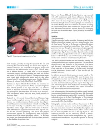
SMALL
ANIMALS
SMALL ANIMALS
© 2007 The Authors Australian Veterinary Journal Volume 85, No 3, March 2007
Journal compilation © 2007 Australian Veterinary Association
113
Blackwell Publishing Asia
Investigation and clinical application of a novel
axial pattern flap for nasal and facial reconstruction
in the dog
G YATES, B LANDON and G EDWARDS
The University of Melbourne Veterinary Clinical Centre, Werribee VIC 3030
gyates@unimelb.edu.au
Objective To describe the vascular supply to a facial skin flap
based at the commissure of the lip in the dog and report on its
use in four dogs.
Study design Experimental and prospective clinical study.
Animals Five canine cadavers and four client-owned dogs.
Methods In the cadavers, the ventral aspect of the zygomatic
arch, the ventral margin of the caudal mandible and the wing of
the atlas were marked as anatomical boundaries of a skin flap
that was elevated from the subcutaneous tissues to the level of
the medial canthus of the eye. Methylene blue dye and barium
sulphate solution were independently infused through a common
carotid (three dogs) or facial artery (two dogs) catheter.
Distribution of dye throughout the harvested skin was assessed
subjectively. After contrast infusion the flap was excised and
radiographed.The technique was used to reconstruct large facial
or nasal defects in four dogs after tumour or skin lesion excision.
Results Cadaver dissections and contrast studies clearly
demonstrated three direct cutaneous arteries, the superior and
inferior labial arteries and the angularis oris artery, arborising
within the base of the flap. A separate direct cutaneous branch
of the angularis oris artery was identified. An arterial plexus
was identified within the distal flap, within which this artery
communicates with the transverse facial artery and a
cutaneous branch of the masseteric artery. Dye infusion
caused discolouration of the elevated skin and vasculature
within the flap. The flap survived in all clinical cases with
marginal distal necrosis in one dog.
Conclusions The complex facial flap described is perfused
by three direct cutaneous arteries and functions reliably in
clinical cases.
Key words: Axial pattern flap, angularis oris, labial artery,
reconstructive surgery
Aust Vet J 2007;85:113–118 doi: 10.1111/j.1751-0813.2007.00111.x
T
he management of large defects involving the facial and
nasal regions of the dog is complicated by the limited
availability of skin for tension free closure. Many local
reconstructive techniques, including buccal rotation1,2
and advance-
ment flaps,1–3
upper labial pull-down,1
single pedicle advancement4
or transposition flaps,4
have been reported to achieve cosmetic
primary closure. Of these, only the transposition and advancement
flaps4
are reported to provide sufficient skin for reconstruction of
large defects involving the rostral nasal skin and nasal planum.
The incorporation of a direct cutaneous artery and vein into the
base of skin flaps enables single-stage transfer of large areas of
skin.10
Reconstruction of large nasal and facial wounds, however,
has been limited by a paucity of reported regional direct cutane-
ous vascular pedicles capable of supporting a sufficiently sized
skin flap to achieve primary closure, with the superficial tem-
poral,5,6
caudal auricular7–9
and superficial cervical10
axial pattern
flaps being relevant to facial reconstruction.
A caudodorsally oriented transposition flap based on the com-
missure of the lip has been previously described for labial and
buccal reconstruction.1
The survival of this flap was explained by
the existence of a rich vascular supply, which is intrinsic to the
labial and buccal soft tissues. With the requirement for increased
flap length in this region it has been recommended to broaden
the flap base to increase the probability of incorporating a direct
cutaneous artery.1
The vascular anatomy of the facial and buccal regions of the dog
has been described.12
The buccal tissues at the commissure of the
lip are supplied by the superior and inferior labial arteries and
the angularis oris artery, terminal branches of the facial artery.12
The angularis oris artery supplies the obicularis oris and buccina-
tor muscles, as well as the adjacent skin and mucosa.12
The buc-
cal mucosal vascular supply has been effectively exploited in
palatal reconstruction with the creation of an angularis oris axial
pattern buccal flap.13
The cutaneous distribution and potential
to support a cutaneous axial pattern flap has not been reported.
The objectives of this study were to describe the arterial vascul-
arity of the previously reported transposition flap1
with the
hypothesis that it is an axial pattern flap, receiving direct cutane-
ous perfusion from the angularis oris artery, define its anatomic
boundaries, and to determine its clinical utility for facial and
nasal reconstruction in the dog.
Materials and methods
Experimental study
Four adult Greyhound cadavers and one adult Jack Russell
Terrier cadaver were used for the experimental component of
avj_111.fm Page 113 Wednesday, February 14, 2007 4:50 PM
SMALL
ANIMALS
SMALL ANIMALS
Australian Veterinary Journal Volume 85, No 3, March 2007 © 2007 The Authors
Journal compilation © 2007 Australian Veterinary Association
114
the study. Each of these dogs was euthanased for reasons
unassociated with this study. The dogs were positioned in right
lateral recumbency and the hair was clipped from the left buccal
and lateral neck regions. The ventral aspect of the zygomatic
arch, the ventral margin of the ramus of the mandible and
the wing of the atlas were used as anatomical landmarks. A
caudodorsally oriented skin flap based on the commissure of the
lip was delineated with a permanent marker (Figure 1) and
elevated deep to the platysma muscle. Meticulous dissection was
conducted at the flap base to preserve cutaneous vasculature.
In three dogs, the ipsilateral common carotid artery was
catheterised with an 18G catheter (Optiva – Medex Medical Ltd,
Rossendale, Great Britain) and a 2/0 ligature (Silk, Ethicon,
Johnson & Johnson Ltd, North Ryde, NSW) placed encircling
the catheter and artery to prevent retrograde flow and
inadvertent dislodgement of the catheter. Five mL of 1%
methylene blue dye (Amyl Media Pty, Ltd, Melbourne, VIC) was
infused into the carotid catheter to facilitate visualisation of the
cutaneous vascular supply and to define the arterial supply of the
skin flap after elevation. In two dogs, the ipsilateral facial artery
was isolated and catheterised with a 24G catheter (Optiva,
Medex Medical Ltd, Rossendale, Great Britain) in an effort to
improve the distribution of radiographic contrast within the
flap’s vasculature.
Barium sulphate (Liquibar, 62.5% w/w barium sulphate, MCI
Forrest Aust. Pty Ltd, Melbourne, VIC) was infused via the
arterial catheter. The flap was excised at its base at the mucocut-
aneous junction of the labial commissure, including its
communications with the facial artery, and radiographed.
Clinical study
The flap was applied to the reconstruction of large facial or nasal
defects in four dogs.
Patient 1: An 8-year-old desexed male Labrador Retriever was
presented with a 4 cm diameter fibrosarcoma on the right rostral
nasal region adjacent to the nasal planum. A caudodorsally ori-
ented skin flap centred on the commissure of the lip was devel-
oped (Figure 2). The ventral margin of the flap was defined by
the ventral border of the caudal mandibular ramus and the dorsal
margin equidistant from the labial commissure dorsally. The
dorsal and ventral boundaries of the flap were continued parallel
to each other, along an axis paralleling the mandible, to the level
of the horizontal ear canal. The flap was harvested and the donor
site closed primarily without tension. The mass was excised with
Figure 1. Facial flap margins were marked, according to anatomic land-
marks, before elevation in cadaver studies.
Figure 2. Nasal fibrosarcoma. Pre-operative planning, including excisional
margins and flap position and dimensions.
Figure 3. The fibrosarcoma was excised with wide margins including the
underlying maxillary and incisive bone.
avj_111.fm Page 114 Wednesday, February 14, 2007 4:50 PM
SMALL
ANIMALS
SMALL ANIMALS
© 2007 The Authors Australian Veterinary Journal Volume 85, No 3, March 2007
Journal compilation © 2007 Australian Veterinary Association
115
wide margins, partially excising the ipsilateral alar fold and
including the subjacent maxillary and incisive bone (Figure 3).
The buccal mucosa was advanced to the gingival margin and
sutured with 3/0 polydioxanone suture (PDS 2, Ethicon, John-
son & Johnson Medical Ltd, North Ryde, NSW) in a simple
continuous pattern. A bridging incision was made and the flap
was rotated to fill the defect (Figure 4). The subcutaneous tissues
were closed with polydioxanone suture and the skin edges
apposed with 3/0 monofilament nylon suture (Ethilon, Ethicon,
Johnson & Johnson Medical Ltd, North Ryde NSW).
Patient 2: A 6-year-old male Border Collie was presented for
excision of a chronic proliferative dermal lesion resulting from
focal adnexal dysplasia of the right nasal skin. The extensive
nature of the lesion necessitated marginal excision. A skin flap
was developed as described above and rotated to fill the defect.
The subcutaneous tissues and skin were closed primarily as
described above.
Patient 3: In a 10-year-old German Short-haired Pointer, a large
caudal maxillary anaplastic sarcoma was excised by caudal maxil-
lectomy and partial orbitectomy. In this case the size and posi-
tion of the excision necessitated a more ventrally based flap. The
dorsal extent of the flap was at the level of the commissure of the
lip and the ventral margin below the ventral ramus of the man-
dible. Otherwise the length and orientation of the flap were sim-
ilar to that described previously. The buccal and gingival
mucosae were apposed and closed primarily with 3/0 polydio-
xanone suture in a simple interrupted pattern. The skin flap
was rotated into the recipient bed and the defect closed primarily
as described for the previous cases.
Patient 4: A 7-year-old female Golden Retriever was presented
with a 1.5 cm diameter, grade 2 mast cell tumour affecting the
muzzle ventrolateral to the nasal planum. After staging, excision
of the mass with wide margins was planned. A similar flap was
harvested, although in this case the flap length was continued to
the level of the wing of the atlas. The mass was excised, including
the ventrolateral portion of the external nare and part of the alar
fold. The flap was transferred into the defect with a bridging
incision and the wounds were closed primarily as described
previously.
Results
Experimental study
Vascular anatomical studies identified the superior and inferior
labial arteries and the angularis oris artery as they branched from
the facial artery. The base of the flap was vascularised by direct
cutaneous arteries arising from each of these three vessels. They
penetrated into and through the platysma muscle joining a rich
anastomosing cutaneous vascular network which could be seen
throughout the flap. A dorsoventral line drawn from the medial
canthus of the eye perpendicular to the axis of the mandibular
body was identified as a safe rostral limit to avoid iatrogenic
trauma to the cutaneous vasculature during dissection.
Two direct cutaneous arteries were also identified entering the
distal regions of the flap in a rostral orientation. The most dorsal
of these arteries was identified as the transverse facial artery
and the other a cutaneous branch of the masseteric artery.
Close inspection of the vasculature of the flap clearly identified
arterial anastomoses between each of these arteries and the
angularis oris artery.
In addition, a separate direct cutaneous arterial branch of the
angularis oris artery was identified. This artery branched from
the angularis oris artery near its division from the facial artery
and reflected caudally, continuing to the level of the horizontal
ear canal. It did not supply the proximal regions of the flap how-
ever, remaining separate from the flap until mid-way along its
length. It is this vessel which forms direct arterial anastomoses
with the secondary and tertiary angiosomes.
Dye infusion through the carotid artery catheter rapidly resulted
in a generalised blue discoloration of the raised flap. Dye could
be seen within branching vessels, which extended to the middle
and distal regions of the flap. Discolouration of the remaining
transverse and cutaneous masseteric vasculature could be readily
appreciated within the distal flap.
Radiographic contrast studies confirmed the presence of three
predominant direct cutaneous arteries. As terminal branches of
the facial artery, these vessels were identified as the angularis oris
and superior and inferior labial arteries. These arteries could
be seen branching within the base of the flap (Figure 5). The
technique performed was not sufficiently sensitive to demon-
strate the extent of the vasculature throughout the flap. Opacific-
ation of vasculature beyond the proximal portion of the flap
was poor.
Figure 4. The postoperative appearance of the flap.
avj_111.fm Page 115 Wednesday, February 14, 2007 4:50 PM
SMALL
ANIMALS
SMALL ANIMALS
Australian Veterinary Journal Volume 85, No 3, March 2007 © 2007 The Authors
Journal compilation © 2007 Australian Veterinary Association
116
Clinical study
The facial axial pattern flap used remained viable in all clinical
cases. Minor marginal flap necrosis (less than 5% of the flap area)
and dehiscence was observed in patient 4, in which the flap had
been extended to the level of the wing of the atlas. In this case
the flap margin was debrided and the wound, adjacent to the
external nares, healed by secondary intention. Otherwise, wound
healing complications were not encountered.
In patients 1, 3 and 4 the extent of the excision created direct
communication between the nasal cavity and the dead space
beneath the flap. Unrestricted air flow from the nasal cavity
resulted in billowing of the flap during expiration. Although cos-
metically displeasing this movement was not associated with
postoperative complications and had resolved by the 10th post-
operative day in all cases, by which time the flap was palpably
firmly adherent to the underlying tissues.
Patient 3 was euthanased after 3 months for reasons unassociated
with the progress of wound healing and the cosmetic appearance
of the reconstruction. Follow-up examination of patients 1, 2
and 4 found no evidence of lesion recurrence after 17, 12 and
9 months respectively. The owner of patient 1, in which the
flap was used to reconstruct the lateral aspect of the nasal plan-
um, reported that it was necessary to trim the hair periodically
from the end of the flap as it grew into the external nare, causing
mild irritation and sneezing.
The flap was well tolerated in all cases and resulted in an accept-
able postoperative cosmetic appearance.
Discussion
After exploring the anatomy of the angularis oris and labial
arteries it was evident that three major direct cutaneous branches
reflect caudally, anastomosing with each other and communicating
with secondary and tertiary angiosomes. This vasculature can be
incorporated into a broad-based, long axial pattern flap suitable
for cosmetic reconstruction of defects of the ipsilateral face
and nose. The observed versatility and durability of this flap
is attributed to the multiplicity of its arterial vascularity and
their contribution to, and communication within, an extensive
subdermal network.
The identification of two large direct cutaneous arteries, the
transverse facial and a cutaneous branch of the masseteric artery,
entering the distal regions of the flap, are further explanations for
its resilience. Anastomoses between these vessels and the angula-
ris oris artery were clearly demonstrated by cadaver dissection
and dye studies and are consistent with a previous report of com-
munication between a cutaneous branch of the masseteric artery
and the superior labial artery.12
The cutaneous distributions of
the transverse facial and cutaneous branch of the masseteric
arteries represent secondary and tertiary angiosomes within
the flap described and the authors theorise that the flap may be
harvested in the opposite direction, being based caudally on
these vessels. This possibility has not been investigated in this
study however.
Anatomical dissection and radiographic perfusion studies are
reliable, objective methods of examining cutaneous vascular-
ity.6,10,11,14,15
Although the angularis oris and dorsal and ventral
labial arteries course cranially, supplying the soft tissues of the
labia, cheek and regions of the nose, cadaver dissections consist-
ently demonstrated these vessels supplying and ramifying within
the flap base. The existence of a separate direct cutaneous branch
of the angularis oris artery is evidence of regional differences in
the perfusion of this flap (Figure 6). The flap base receives abun-
dant arterial supply from the angularis oris and labial arteries.
The distal regions of the flap, however, appear to be vascularised
by this separate cutaneous branch of the angularis oris artery and
its anastomoses with the transverse facial artery and a cutaneous
branch of the masseteric artery. The anatomy of this cutaneous
vessel is believed to be an additional factor contributing to the
functional length of this flap and its observed durability. It is
reasonable to expect that this vessel would be more easily
affected by twisting or kinking, or if tension was created within
the flap during transposition.
Radiographic contrast studies have been widely used to define
vascular angiosomes within skin flaps.6,10,11,14,15
Positive contrast
radiography10,11,14,15
and subtraction angiography6,15
have been
described. The use of barium sulphate for skin flap angiography
has been reported11,14
and in this study the cutaneous branches of
the angularis oris and dorsal and ventral branches of the labial
arteries were clearly demonstrated within the flap base using this
method. There was poor opacification, however, of these vessels
beyond the proximal third of the flap. This observation is
explained by poor filling of the microvasculature due to the
Figure 5. Positive contrast angiogram demonstrating the extensive
branching vasculature extending into the base of the flap. The flap has
been secured with two needles, seen at the lower and left margins of the
flap in the image. The facial artery (small arrow head) branches into the
inferior labial (double arrow), angularis oris (arrow) and superior labial
(large arrow head) arteries. The separate direct cutaneous branch of the
angularis oris (large arrow) is less well defined.
avj_111.fm Page 116 Wednesday, February 14, 2007 4:50 PM
SMALL
ANIMALS
SMALL ANIMALS
© 2007 The Authors Australian Veterinary Journal Volume 85, No 3, March 2007
Journal compilation © 2007 Australian Veterinary Association
117
viscosity of the contrast liquid infused. Barium sulphate solution
was infused at the recommended concentration for reconstitu-
tion without further dilution. Although the use of a viscous con-
trast agent limited its penetration into the smaller arteries, pilot
studies with less viscous agents, including dilute barium sulphate
and water soluble contrast medium (Omnipaque 350), rapidly
resulted in loss of infusion pressure by leakage of the agent from
the ends of small vessels that had been traumatised during the
dissection. This resulted in poor contrast distribution and insuf-
ficient opacification. The limited penetration of contrast along
the length of the flap precluded objective radiographic definition
of the perfusion of the distal regions of the flap. It does, however,
indicate that the ventral aspect of the zygomatic arch and the
ventral aspect of the ramus of the mandible are appropriate
dorsal and ventral flap boundaries, as substantial branching
vasculature was identified extending close to these margins.
Clinically, the flap consistently survived when the level of the
vertical ear canal was used as its caudal boundary. Although the
wing of the atlas was used as a caudal margin for the experimen-
tal study, it was not necessary to extend the flap beyond this level
to reconstruct the nasal defects described in the clinical study.
Distal necrosis of the flap occurred in patient 4, in which the flap
was incorrectly extended to the level of the wing of the atlas.
Despite pre-planning of the flap it was realised after harvesting
that it was unnecessarily long. As this was the only case in the
present series using such a caudal margin, it cannot be determined
whether the necrosis observed was a consequence of technical
error or of exceeding the limits of the flap’s vascular supply.
Meticulous atraumatic technique is recommended to optimise
flap survival if extension of the flap to the wing of the atlas is
planned, however the authors suggest that extending the flap
beyond the level of the vertical ear canal is unnecessary for
reconstruction of ipsilateral rostral nasal defects.
No other intra-operative or postoperative complications were
attributed to the use of this skin flap. Elevation of the skin flap
described exposes the dorsal, ventral and auriculopalpebral
branches of the facial nerve, branches of the auriculotemporal
nerve, the parotid salivary duct and the facial vein. Careful tech-
nique is necessary to avoid trauma to these structures. Although
clinical complications arising from trauma to these structures
were not recognised, cutaneous sensation within the transposed
flap was not assessed. Accordingly, the influence of elevation and
transposition of this flap on its sensory innervation is unknown.
In all cases the donor site could be closed without tension and
healing progressed uneventfully. When tumour excision
included maxillectomy or excision of the nasal bone, air flow
from the nasal cavity caused billowing of the flap during expira-
tion. In all patients this had abated by the 10th postoperative
day, at which time the flap was firmly fixed to subcutaneous tis-
sue. Passive wound drainage or a nasal catheter may be useful to
limit billowing of the flap and facilitate adherence, however their
value has not been investigated. The skin flap was well tolerated
and resulted in acceptable cosmetic appearance. For patient 1, in
which the flap was used to reconstruct areas of the nasal planum,
periodic trimming of the hair from the distal regions of the flap
was necessary to avoid irritation and occasional sneezing.
Conclusion
The complex facial flap described, based at the commissure of
the lip, is vascularised by the angularis oris and labial arteries. It
is a durable and versatile axial pattern flap that was consistently
effective in the cosmetic reconstruction of large facial and nasal
defects in these dogs. Although further clinical studies are
indicated to explore the limits of this flap’s length and whether
similar results can be achieved when only one or two of these
direct cutaneous arteries are included in the flap base, the use of
the ventral aspect of the zygomatic arch, the ventral aspect of the
mandibular ramus and extending the flap to the vertical ear canal
can be expected to yield consistently successful results. This flap
represents another alternative in the expanding field of facial and
oral reconstruction.
References
1. Pavletic MM. Atlas of Small Animal Reconstructive Surgery. 2nd edn. Saun-
ders, Philadelphia, 1999:298–327.
2. Swaim SF, Henderson RA. Small Animal Wound Management. 2nd edn.
Williams and Wilkins, Baltimore, 1997:191–233.
3. Welsh JA, Swaim SF. Nasal and facial reconstruction in a dog following
severe trauma. J Am Anim Hosp Assoc 2003;39:407–415.
4. Pavletic MM. Pedicle Grafts. In: Slatter D, editor. Textbook of Small Animal
Surgery. Vol 1. 3rd edn. Saunders, Philadelphia, 2003:292–321.
5. Fahie MA, Smith BJ, Ballard JB, Moon ML, Smith MM. Regional peripheral
vascular supply based on the superficial temporal artery in dogs and cats.
Anat Histol Embryol 1998;27:205–208.
Figure 6. Schematic diagram of the flap’s vasculature showing the facial
artery (small arrowhead) branching into the inferior labial (double arrow),
angularis oris (arrow) and superior labial (triple arrow) arteries. The
separate direct cutaneous branch of the angularis oris artery (double
small arrowhead) anastomoses with the transverse facial artery (large
arrowhead) and a cutaneous branch of the masseteric artery (double large
arrowhead).
avj_111.fm Page 117 Wednesday, February 14, 2007 4:50 PM
SMALL
ANIMALS
SMALL ANIMALS
Australian Veterinary Journal Volume 85, No 3, March 2007 © 2007 The Authors
Journal compilation © 2007 Australian Veterinary Association
118
6. Fahie MA, Smith MM. Axial pattern flap based on the cutaneous branch of the
superficial temporal artery in dogs: an experimental study and case report. Vet
Surg 1999;28:141–147.
7. Smith MM, Payne JT, Moon ML, Freeman LE. Axial pattern flap based on the
caudal auricular artery in dogs. Am J Vet Res 1999;52:922–925.
8. Moses PA, Hunt GB. Use of a caudal auricular axial pattern flap in a dog.
Aust Vet Pract 2001;31:155.
9. Stiles J, Townsend W, Willis M, Moore PA, Smith E. Use of a caudal auricular
axial pattern flap in three cats and one dog following orbital exenteration.
Vet Ophthalmol 2003;6:121–126.
10. Dundas JM, Fowler JD, Shmon CL, Clapson JB. Modification of the superficial
cervical axial pattern skin flap for oral reconstruction. Vet Surg 2005;34:206–213.
11. Pavletic MM. Canine axial pattern flaps, using the omocervical, thoracodorsal,
and deep circumflex iliac direct cutaneous arteries. Am J Vet Res 1981;
42:391–406.
12. Evans HE. The heart and arteries. In: Evans HE, editor. Miller’s Anatomy of
the Dog. 3rd edn. Saunders, Phildelphia, 1993:610–614.
13. Bryant KJ, Moore K, McAnulty JF. Angularis oris axial buccal flap for recon-
struction of recurrent fistulae of the palate. Vet Surg 2003:32:113–119.
14. Anderson DM, Charlesworth TC, White RAS. A novel axial pattern skin flap
based on the lateral thoracic artery in the dog. Vet Comp Orth Traumatol
2004;17:73–77.
15. Gregory CR, Gourley IM, Koblik PD, Patz JD. Experimental definition of
latissimus dorsi, gracilis and rectus abdominus musculocutaneous flaps in the
dog. Am J Vet Res 1988;49:878–884.
(Accepted for publication 27 October 2006)
Blackwell Publishing Asia
avj_111.fm Page 118 Wednesday, February 14, 2007 4:50 PM

Flap facial.pdf

  • 1.
    SMALL ANIMALS SMALL ANIMALS © 2007The Authors Australian Veterinary Journal Volume 85, No 3, March 2007 Journal compilation © 2007 Australian Veterinary Association 113 Blackwell Publishing Asia Investigation and clinical application of a novel axial pattern flap for nasal and facial reconstruction in the dog G YATES, B LANDON and G EDWARDS The University of Melbourne Veterinary Clinical Centre, Werribee VIC 3030 gyates@unimelb.edu.au Objective To describe the vascular supply to a facial skin flap based at the commissure of the lip in the dog and report on its use in four dogs. Study design Experimental and prospective clinical study. Animals Five canine cadavers and four client-owned dogs. Methods In the cadavers, the ventral aspect of the zygomatic arch, the ventral margin of the caudal mandible and the wing of the atlas were marked as anatomical boundaries of a skin flap that was elevated from the subcutaneous tissues to the level of the medial canthus of the eye. Methylene blue dye and barium sulphate solution were independently infused through a common carotid (three dogs) or facial artery (two dogs) catheter. Distribution of dye throughout the harvested skin was assessed subjectively. After contrast infusion the flap was excised and radiographed.The technique was used to reconstruct large facial or nasal defects in four dogs after tumour or skin lesion excision. Results Cadaver dissections and contrast studies clearly demonstrated three direct cutaneous arteries, the superior and inferior labial arteries and the angularis oris artery, arborising within the base of the flap. A separate direct cutaneous branch of the angularis oris artery was identified. An arterial plexus was identified within the distal flap, within which this artery communicates with the transverse facial artery and a cutaneous branch of the masseteric artery. Dye infusion caused discolouration of the elevated skin and vasculature within the flap. The flap survived in all clinical cases with marginal distal necrosis in one dog. Conclusions The complex facial flap described is perfused by three direct cutaneous arteries and functions reliably in clinical cases. Key words: Axial pattern flap, angularis oris, labial artery, reconstructive surgery Aust Vet J 2007;85:113–118 doi: 10.1111/j.1751-0813.2007.00111.x T he management of large defects involving the facial and nasal regions of the dog is complicated by the limited availability of skin for tension free closure. Many local reconstructive techniques, including buccal rotation1,2 and advance- ment flaps,1–3 upper labial pull-down,1 single pedicle advancement4 or transposition flaps,4 have been reported to achieve cosmetic primary closure. Of these, only the transposition and advancement flaps4 are reported to provide sufficient skin for reconstruction of large defects involving the rostral nasal skin and nasal planum. The incorporation of a direct cutaneous artery and vein into the base of skin flaps enables single-stage transfer of large areas of skin.10 Reconstruction of large nasal and facial wounds, however, has been limited by a paucity of reported regional direct cutane- ous vascular pedicles capable of supporting a sufficiently sized skin flap to achieve primary closure, with the superficial tem- poral,5,6 caudal auricular7–9 and superficial cervical10 axial pattern flaps being relevant to facial reconstruction. A caudodorsally oriented transposition flap based on the com- missure of the lip has been previously described for labial and buccal reconstruction.1 The survival of this flap was explained by the existence of a rich vascular supply, which is intrinsic to the labial and buccal soft tissues. With the requirement for increased flap length in this region it has been recommended to broaden the flap base to increase the probability of incorporating a direct cutaneous artery.1 The vascular anatomy of the facial and buccal regions of the dog has been described.12 The buccal tissues at the commissure of the lip are supplied by the superior and inferior labial arteries and the angularis oris artery, terminal branches of the facial artery.12 The angularis oris artery supplies the obicularis oris and buccina- tor muscles, as well as the adjacent skin and mucosa.12 The buc- cal mucosal vascular supply has been effectively exploited in palatal reconstruction with the creation of an angularis oris axial pattern buccal flap.13 The cutaneous distribution and potential to support a cutaneous axial pattern flap has not been reported. The objectives of this study were to describe the arterial vascul- arity of the previously reported transposition flap1 with the hypothesis that it is an axial pattern flap, receiving direct cutane- ous perfusion from the angularis oris artery, define its anatomic boundaries, and to determine its clinical utility for facial and nasal reconstruction in the dog. Materials and methods Experimental study Four adult Greyhound cadavers and one adult Jack Russell Terrier cadaver were used for the experimental component of avj_111.fm Page 113 Wednesday, February 14, 2007 4:50 PM
  • 2.
    SMALL ANIMALS SMALL ANIMALS Australian VeterinaryJournal Volume 85, No 3, March 2007 © 2007 The Authors Journal compilation © 2007 Australian Veterinary Association 114 the study. Each of these dogs was euthanased for reasons unassociated with this study. The dogs were positioned in right lateral recumbency and the hair was clipped from the left buccal and lateral neck regions. The ventral aspect of the zygomatic arch, the ventral margin of the ramus of the mandible and the wing of the atlas were used as anatomical landmarks. A caudodorsally oriented skin flap based on the commissure of the lip was delineated with a permanent marker (Figure 1) and elevated deep to the platysma muscle. Meticulous dissection was conducted at the flap base to preserve cutaneous vasculature. In three dogs, the ipsilateral common carotid artery was catheterised with an 18G catheter (Optiva – Medex Medical Ltd, Rossendale, Great Britain) and a 2/0 ligature (Silk, Ethicon, Johnson & Johnson Ltd, North Ryde, NSW) placed encircling the catheter and artery to prevent retrograde flow and inadvertent dislodgement of the catheter. Five mL of 1% methylene blue dye (Amyl Media Pty, Ltd, Melbourne, VIC) was infused into the carotid catheter to facilitate visualisation of the cutaneous vascular supply and to define the arterial supply of the skin flap after elevation. In two dogs, the ipsilateral facial artery was isolated and catheterised with a 24G catheter (Optiva, Medex Medical Ltd, Rossendale, Great Britain) in an effort to improve the distribution of radiographic contrast within the flap’s vasculature. Barium sulphate (Liquibar, 62.5% w/w barium sulphate, MCI Forrest Aust. Pty Ltd, Melbourne, VIC) was infused via the arterial catheter. The flap was excised at its base at the mucocut- aneous junction of the labial commissure, including its communications with the facial artery, and radiographed. Clinical study The flap was applied to the reconstruction of large facial or nasal defects in four dogs. Patient 1: An 8-year-old desexed male Labrador Retriever was presented with a 4 cm diameter fibrosarcoma on the right rostral nasal region adjacent to the nasal planum. A caudodorsally ori- ented skin flap centred on the commissure of the lip was devel- oped (Figure 2). The ventral margin of the flap was defined by the ventral border of the caudal mandibular ramus and the dorsal margin equidistant from the labial commissure dorsally. The dorsal and ventral boundaries of the flap were continued parallel to each other, along an axis paralleling the mandible, to the level of the horizontal ear canal. The flap was harvested and the donor site closed primarily without tension. The mass was excised with Figure 1. Facial flap margins were marked, according to anatomic land- marks, before elevation in cadaver studies. Figure 2. Nasal fibrosarcoma. Pre-operative planning, including excisional margins and flap position and dimensions. Figure 3. The fibrosarcoma was excised with wide margins including the underlying maxillary and incisive bone. avj_111.fm Page 114 Wednesday, February 14, 2007 4:50 PM
  • 3.
    SMALL ANIMALS SMALL ANIMALS © 2007The Authors Australian Veterinary Journal Volume 85, No 3, March 2007 Journal compilation © 2007 Australian Veterinary Association 115 wide margins, partially excising the ipsilateral alar fold and including the subjacent maxillary and incisive bone (Figure 3). The buccal mucosa was advanced to the gingival margin and sutured with 3/0 polydioxanone suture (PDS 2, Ethicon, John- son & Johnson Medical Ltd, North Ryde, NSW) in a simple continuous pattern. A bridging incision was made and the flap was rotated to fill the defect (Figure 4). The subcutaneous tissues were closed with polydioxanone suture and the skin edges apposed with 3/0 monofilament nylon suture (Ethilon, Ethicon, Johnson & Johnson Medical Ltd, North Ryde NSW). Patient 2: A 6-year-old male Border Collie was presented for excision of a chronic proliferative dermal lesion resulting from focal adnexal dysplasia of the right nasal skin. The extensive nature of the lesion necessitated marginal excision. A skin flap was developed as described above and rotated to fill the defect. The subcutaneous tissues and skin were closed primarily as described above. Patient 3: In a 10-year-old German Short-haired Pointer, a large caudal maxillary anaplastic sarcoma was excised by caudal maxil- lectomy and partial orbitectomy. In this case the size and posi- tion of the excision necessitated a more ventrally based flap. The dorsal extent of the flap was at the level of the commissure of the lip and the ventral margin below the ventral ramus of the man- dible. Otherwise the length and orientation of the flap were sim- ilar to that described previously. The buccal and gingival mucosae were apposed and closed primarily with 3/0 polydio- xanone suture in a simple interrupted pattern. The skin flap was rotated into the recipient bed and the defect closed primarily as described for the previous cases. Patient 4: A 7-year-old female Golden Retriever was presented with a 1.5 cm diameter, grade 2 mast cell tumour affecting the muzzle ventrolateral to the nasal planum. After staging, excision of the mass with wide margins was planned. A similar flap was harvested, although in this case the flap length was continued to the level of the wing of the atlas. The mass was excised, including the ventrolateral portion of the external nare and part of the alar fold. The flap was transferred into the defect with a bridging incision and the wounds were closed primarily as described previously. Results Experimental study Vascular anatomical studies identified the superior and inferior labial arteries and the angularis oris artery as they branched from the facial artery. The base of the flap was vascularised by direct cutaneous arteries arising from each of these three vessels. They penetrated into and through the platysma muscle joining a rich anastomosing cutaneous vascular network which could be seen throughout the flap. A dorsoventral line drawn from the medial canthus of the eye perpendicular to the axis of the mandibular body was identified as a safe rostral limit to avoid iatrogenic trauma to the cutaneous vasculature during dissection. Two direct cutaneous arteries were also identified entering the distal regions of the flap in a rostral orientation. The most dorsal of these arteries was identified as the transverse facial artery and the other a cutaneous branch of the masseteric artery. Close inspection of the vasculature of the flap clearly identified arterial anastomoses between each of these arteries and the angularis oris artery. In addition, a separate direct cutaneous arterial branch of the angularis oris artery was identified. This artery branched from the angularis oris artery near its division from the facial artery and reflected caudally, continuing to the level of the horizontal ear canal. It did not supply the proximal regions of the flap how- ever, remaining separate from the flap until mid-way along its length. It is this vessel which forms direct arterial anastomoses with the secondary and tertiary angiosomes. Dye infusion through the carotid artery catheter rapidly resulted in a generalised blue discoloration of the raised flap. Dye could be seen within branching vessels, which extended to the middle and distal regions of the flap. Discolouration of the remaining transverse and cutaneous masseteric vasculature could be readily appreciated within the distal flap. Radiographic contrast studies confirmed the presence of three predominant direct cutaneous arteries. As terminal branches of the facial artery, these vessels were identified as the angularis oris and superior and inferior labial arteries. These arteries could be seen branching within the base of the flap (Figure 5). The technique performed was not sufficiently sensitive to demon- strate the extent of the vasculature throughout the flap. Opacific- ation of vasculature beyond the proximal portion of the flap was poor. Figure 4. The postoperative appearance of the flap. avj_111.fm Page 115 Wednesday, February 14, 2007 4:50 PM
  • 4.
    SMALL ANIMALS SMALL ANIMALS Australian VeterinaryJournal Volume 85, No 3, March 2007 © 2007 The Authors Journal compilation © 2007 Australian Veterinary Association 116 Clinical study The facial axial pattern flap used remained viable in all clinical cases. Minor marginal flap necrosis (less than 5% of the flap area) and dehiscence was observed in patient 4, in which the flap had been extended to the level of the wing of the atlas. In this case the flap margin was debrided and the wound, adjacent to the external nares, healed by secondary intention. Otherwise, wound healing complications were not encountered. In patients 1, 3 and 4 the extent of the excision created direct communication between the nasal cavity and the dead space beneath the flap. Unrestricted air flow from the nasal cavity resulted in billowing of the flap during expiration. Although cos- metically displeasing this movement was not associated with postoperative complications and had resolved by the 10th post- operative day in all cases, by which time the flap was palpably firmly adherent to the underlying tissues. Patient 3 was euthanased after 3 months for reasons unassociated with the progress of wound healing and the cosmetic appearance of the reconstruction. Follow-up examination of patients 1, 2 and 4 found no evidence of lesion recurrence after 17, 12 and 9 months respectively. The owner of patient 1, in which the flap was used to reconstruct the lateral aspect of the nasal plan- um, reported that it was necessary to trim the hair periodically from the end of the flap as it grew into the external nare, causing mild irritation and sneezing. The flap was well tolerated in all cases and resulted in an accept- able postoperative cosmetic appearance. Discussion After exploring the anatomy of the angularis oris and labial arteries it was evident that three major direct cutaneous branches reflect caudally, anastomosing with each other and communicating with secondary and tertiary angiosomes. This vasculature can be incorporated into a broad-based, long axial pattern flap suitable for cosmetic reconstruction of defects of the ipsilateral face and nose. The observed versatility and durability of this flap is attributed to the multiplicity of its arterial vascularity and their contribution to, and communication within, an extensive subdermal network. The identification of two large direct cutaneous arteries, the transverse facial and a cutaneous branch of the masseteric artery, entering the distal regions of the flap, are further explanations for its resilience. Anastomoses between these vessels and the angula- ris oris artery were clearly demonstrated by cadaver dissection and dye studies and are consistent with a previous report of com- munication between a cutaneous branch of the masseteric artery and the superior labial artery.12 The cutaneous distributions of the transverse facial and cutaneous branch of the masseteric arteries represent secondary and tertiary angiosomes within the flap described and the authors theorise that the flap may be harvested in the opposite direction, being based caudally on these vessels. This possibility has not been investigated in this study however. Anatomical dissection and radiographic perfusion studies are reliable, objective methods of examining cutaneous vascular- ity.6,10,11,14,15 Although the angularis oris and dorsal and ventral labial arteries course cranially, supplying the soft tissues of the labia, cheek and regions of the nose, cadaver dissections consist- ently demonstrated these vessels supplying and ramifying within the flap base. The existence of a separate direct cutaneous branch of the angularis oris artery is evidence of regional differences in the perfusion of this flap (Figure 6). The flap base receives abun- dant arterial supply from the angularis oris and labial arteries. The distal regions of the flap, however, appear to be vascularised by this separate cutaneous branch of the angularis oris artery and its anastomoses with the transverse facial artery and a cutaneous branch of the masseteric artery. The anatomy of this cutaneous vessel is believed to be an additional factor contributing to the functional length of this flap and its observed durability. It is reasonable to expect that this vessel would be more easily affected by twisting or kinking, or if tension was created within the flap during transposition. Radiographic contrast studies have been widely used to define vascular angiosomes within skin flaps.6,10,11,14,15 Positive contrast radiography10,11,14,15 and subtraction angiography6,15 have been described. The use of barium sulphate for skin flap angiography has been reported11,14 and in this study the cutaneous branches of the angularis oris and dorsal and ventral branches of the labial arteries were clearly demonstrated within the flap base using this method. There was poor opacification, however, of these vessels beyond the proximal third of the flap. This observation is explained by poor filling of the microvasculature due to the Figure 5. Positive contrast angiogram demonstrating the extensive branching vasculature extending into the base of the flap. The flap has been secured with two needles, seen at the lower and left margins of the flap in the image. The facial artery (small arrow head) branches into the inferior labial (double arrow), angularis oris (arrow) and superior labial (large arrow head) arteries. The separate direct cutaneous branch of the angularis oris (large arrow) is less well defined. avj_111.fm Page 116 Wednesday, February 14, 2007 4:50 PM
  • 5.
    SMALL ANIMALS SMALL ANIMALS © 2007The Authors Australian Veterinary Journal Volume 85, No 3, March 2007 Journal compilation © 2007 Australian Veterinary Association 117 viscosity of the contrast liquid infused. Barium sulphate solution was infused at the recommended concentration for reconstitu- tion without further dilution. Although the use of a viscous con- trast agent limited its penetration into the smaller arteries, pilot studies with less viscous agents, including dilute barium sulphate and water soluble contrast medium (Omnipaque 350), rapidly resulted in loss of infusion pressure by leakage of the agent from the ends of small vessels that had been traumatised during the dissection. This resulted in poor contrast distribution and insuf- ficient opacification. The limited penetration of contrast along the length of the flap precluded objective radiographic definition of the perfusion of the distal regions of the flap. It does, however, indicate that the ventral aspect of the zygomatic arch and the ventral aspect of the ramus of the mandible are appropriate dorsal and ventral flap boundaries, as substantial branching vasculature was identified extending close to these margins. Clinically, the flap consistently survived when the level of the vertical ear canal was used as its caudal boundary. Although the wing of the atlas was used as a caudal margin for the experimen- tal study, it was not necessary to extend the flap beyond this level to reconstruct the nasal defects described in the clinical study. Distal necrosis of the flap occurred in patient 4, in which the flap was incorrectly extended to the level of the wing of the atlas. Despite pre-planning of the flap it was realised after harvesting that it was unnecessarily long. As this was the only case in the present series using such a caudal margin, it cannot be determined whether the necrosis observed was a consequence of technical error or of exceeding the limits of the flap’s vascular supply. Meticulous atraumatic technique is recommended to optimise flap survival if extension of the flap to the wing of the atlas is planned, however the authors suggest that extending the flap beyond the level of the vertical ear canal is unnecessary for reconstruction of ipsilateral rostral nasal defects. No other intra-operative or postoperative complications were attributed to the use of this skin flap. Elevation of the skin flap described exposes the dorsal, ventral and auriculopalpebral branches of the facial nerve, branches of the auriculotemporal nerve, the parotid salivary duct and the facial vein. Careful tech- nique is necessary to avoid trauma to these structures. Although clinical complications arising from trauma to these structures were not recognised, cutaneous sensation within the transposed flap was not assessed. Accordingly, the influence of elevation and transposition of this flap on its sensory innervation is unknown. In all cases the donor site could be closed without tension and healing progressed uneventfully. When tumour excision included maxillectomy or excision of the nasal bone, air flow from the nasal cavity caused billowing of the flap during expira- tion. In all patients this had abated by the 10th postoperative day, at which time the flap was firmly fixed to subcutaneous tis- sue. Passive wound drainage or a nasal catheter may be useful to limit billowing of the flap and facilitate adherence, however their value has not been investigated. The skin flap was well tolerated and resulted in acceptable cosmetic appearance. For patient 1, in which the flap was used to reconstruct areas of the nasal planum, periodic trimming of the hair from the distal regions of the flap was necessary to avoid irritation and occasional sneezing. Conclusion The complex facial flap described, based at the commissure of the lip, is vascularised by the angularis oris and labial arteries. It is a durable and versatile axial pattern flap that was consistently effective in the cosmetic reconstruction of large facial and nasal defects in these dogs. Although further clinical studies are indicated to explore the limits of this flap’s length and whether similar results can be achieved when only one or two of these direct cutaneous arteries are included in the flap base, the use of the ventral aspect of the zygomatic arch, the ventral aspect of the mandibular ramus and extending the flap to the vertical ear canal can be expected to yield consistently successful results. This flap represents another alternative in the expanding field of facial and oral reconstruction. References 1. Pavletic MM. Atlas of Small Animal Reconstructive Surgery. 2nd edn. Saun- ders, Philadelphia, 1999:298–327. 2. Swaim SF, Henderson RA. Small Animal Wound Management. 2nd edn. Williams and Wilkins, Baltimore, 1997:191–233. 3. Welsh JA, Swaim SF. Nasal and facial reconstruction in a dog following severe trauma. J Am Anim Hosp Assoc 2003;39:407–415. 4. Pavletic MM. Pedicle Grafts. In: Slatter D, editor. Textbook of Small Animal Surgery. Vol 1. 3rd edn. Saunders, Philadelphia, 2003:292–321. 5. Fahie MA, Smith BJ, Ballard JB, Moon ML, Smith MM. Regional peripheral vascular supply based on the superficial temporal artery in dogs and cats. Anat Histol Embryol 1998;27:205–208. Figure 6. Schematic diagram of the flap’s vasculature showing the facial artery (small arrowhead) branching into the inferior labial (double arrow), angularis oris (arrow) and superior labial (triple arrow) arteries. The separate direct cutaneous branch of the angularis oris artery (double small arrowhead) anastomoses with the transverse facial artery (large arrowhead) and a cutaneous branch of the masseteric artery (double large arrowhead). avj_111.fm Page 117 Wednesday, February 14, 2007 4:50 PM
  • 6.
    SMALL ANIMALS SMALL ANIMALS Australian VeterinaryJournal Volume 85, No 3, March 2007 © 2007 The Authors Journal compilation © 2007 Australian Veterinary Association 118 6. Fahie MA, Smith MM. Axial pattern flap based on the cutaneous branch of the superficial temporal artery in dogs: an experimental study and case report. Vet Surg 1999;28:141–147. 7. Smith MM, Payne JT, Moon ML, Freeman LE. Axial pattern flap based on the caudal auricular artery in dogs. Am J Vet Res 1999;52:922–925. 8. Moses PA, Hunt GB. Use of a caudal auricular axial pattern flap in a dog. Aust Vet Pract 2001;31:155. 9. Stiles J, Townsend W, Willis M, Moore PA, Smith E. Use of a caudal auricular axial pattern flap in three cats and one dog following orbital exenteration. Vet Ophthalmol 2003;6:121–126. 10. Dundas JM, Fowler JD, Shmon CL, Clapson JB. Modification of the superficial cervical axial pattern skin flap for oral reconstruction. Vet Surg 2005;34:206–213. 11. Pavletic MM. Canine axial pattern flaps, using the omocervical, thoracodorsal, and deep circumflex iliac direct cutaneous arteries. Am J Vet Res 1981; 42:391–406. 12. Evans HE. The heart and arteries. In: Evans HE, editor. Miller’s Anatomy of the Dog. 3rd edn. Saunders, Phildelphia, 1993:610–614. 13. Bryant KJ, Moore K, McAnulty JF. Angularis oris axial buccal flap for recon- struction of recurrent fistulae of the palate. Vet Surg 2003:32:113–119. 14. Anderson DM, Charlesworth TC, White RAS. A novel axial pattern skin flap based on the lateral thoracic artery in the dog. Vet Comp Orth Traumatol 2004;17:73–77. 15. Gregory CR, Gourley IM, Koblik PD, Patz JD. Experimental definition of latissimus dorsi, gracilis and rectus abdominus musculocutaneous flaps in the dog. Am J Vet Res 1988;49:878–884. (Accepted for publication 27 October 2006) Blackwell Publishing Asia avj_111.fm Page 118 Wednesday, February 14, 2007 4:50 PM