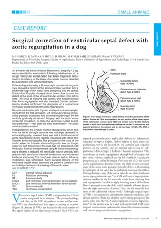
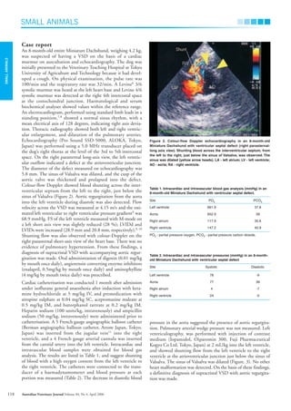
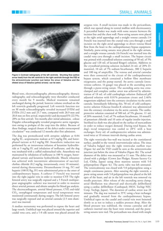
This document describes a case study of a Miniature Dachshund that was diagnosed with a ventricular septal defect (VSD) and aortic regurgitation. Echocardiography revealed a defect between the ventricles and a thickened aortic valve prolapsing into the defect. Cardiac catheterization confirmed a supracristal VSD with aortic regurgitation. Despite medication, the dog's left ventricular dimensions worsened over time. The dog ultimately underwent surgery to close the VSD using cardiopulmonary bypass, which improved the condition and controlled further valve deterioration.
Trio of Rheumatic Mitral Stenosis, Right Posterior Septal Accessory Pathway a...Ramachandra Barik
A 57-year-old male presented with recurrent palpitations. He was diagnosed with rheumatic mitral stenosis, right posterior septal accessory pathway and atrial flutter. An electrophysiological study after percutaneous balloon mitral valvotomy showed that the palpitations were due to atrial flutter with right bundle branch aberrancy. The right posterior septal pathway was a bystander because it had a higher refractory period than the atrioventricular node.
Transposition of the great arteries is a serious but rare heart defect present at birth (congenital), in which the two main arteries leaving the heart are reversed (transposed). The condition is also called dextro-transposition of the great arteries.
Fourth Recurrence of Aortic Annular Dehiscence Following AVR for Aortic Regur...Apollo Hospitals
It is a report of an unusual case of recurrent periprosthetic aortic regurgitation which occurred four times in the same patient over a span of 26 years. After pre-operative stabilization, the patient was subjected to a fifth open heart surgery to replace the 4th prosthetic aortic valve by an upsized 5th prosthetic valve. Since endocarditis was excluded by repeated blood culture and paucity of vegetations, a working diagnosis of Behcet's disease was arrived upon. The fifth prosthetic aortic valve was implanted using a special suturing technique as described by Azumo, et al in 2009, which involves a sub-annular reinforcement by a Teflon ring. The patient was discharged after an uneventful recovery and is well on regular follow-up.
Adjusting primitives for graph : SHORT REPORT / NOTESSubhajit Sahu
Graph algorithms, like PageRank Compressed Sparse Row (CSR) is an adjacency-list based graph representation that is
Multiply with different modes (map)
1. Performance of sequential execution based vs OpenMP based vector multiply.
2. Comparing various launch configs for CUDA based vector multiply.
Sum with different storage types (reduce)
1. Performance of vector element sum using float vs bfloat16 as the storage type.
Sum with different modes (reduce)
1. Performance of sequential execution based vs OpenMP based vector element sum.
2. Performance of memcpy vs in-place based CUDA based vector element sum.
3. Comparing various launch configs for CUDA based vector element sum (memcpy).
4. Comparing various launch configs for CUDA based vector element sum (in-place).
Sum with in-place strategies of CUDA mode (reduce)
1. Comparing various launch configs for CUDA based vector element sum (in-place).
Techniques to optimize the pagerank algorithm usually fall in two categories. One is to try reducing the work per iteration, and the other is to try reducing the number of iterations. These goals are often at odds with one another. Skipping computation on vertices which have already converged has the potential to save iteration time. Skipping in-identical vertices, with the same in-links, helps reduce duplicate computations and thus could help reduce iteration time. Road networks often have chains which can be short-circuited before pagerank computation to improve performance. Final ranks of chain nodes can be easily calculated. This could reduce both the iteration time, and the number of iterations. If a graph has no dangling nodes, pagerank of each strongly connected component can be computed in topological order. This could help reduce the iteration time, no. of iterations, and also enable multi-iteration concurrency in pagerank computation. The combination of all of the above methods is the STICD algorithm. [sticd] For dynamic graphs, unchanged components whose ranks are unaffected can be skipped altogether.
Opendatabay - Open Data Marketplace.pptxOpendatabay
Opendatabay.com unlocks the power of data for everyone. Open Data Marketplace fosters a collaborative hub for data enthusiasts to explore, share, and contribute to a vast collection of datasets.
First ever open hub for data enthusiasts to collaborate and innovate. A platform to explore, share, and contribute to a vast collection of datasets. Through robust quality control and innovative technologies like blockchain verification, opendatabay ensures the authenticity and reliability of datasets, empowering users to make data-driven decisions with confidence. Leverage cutting-edge AI technologies to enhance the data exploration, analysis, and discovery experience.
From intelligent search and recommendations to automated data productisation and quotation, Opendatabay AI-driven features streamline the data workflow. Finding the data you need shouldn't be a complex. Opendatabay simplifies the data acquisition process with an intuitive interface and robust search tools. Effortlessly explore, discover, and access the data you need, allowing you to focus on extracting valuable insights. Opendatabay breaks new ground with a dedicated, AI-generated, synthetic datasets.
Leverage these privacy-preserving datasets for training and testing AI models without compromising sensitive information. Opendatabay prioritizes transparency by providing detailed metadata, provenance information, and usage guidelines for each dataset, ensuring users have a comprehensive understanding of the data they're working with. By leveraging a powerful combination of distributed ledger technology and rigorous third-party audits Opendatabay ensures the authenticity and reliability of every dataset. Security is at the core of Opendatabay. Marketplace implements stringent security measures, including encryption, access controls, and regular vulnerability assessments, to safeguard your data and protect your privacy.
Ch03-Managing the Object-Oriented Information Systems Project a.pdf
reparación septal.pdf
1. SMALL ANIMALS
SMALL
ANIMALS
117
Australian Veterinary Journal Volume 84, No 4, April 2006
termed perimembranous trabecular defects or infracristal,
subaortic, or type 2 Kirklin. Defects related to both aortic and
pulmonary valves are located in the anterior and superior
portion of the septum and are termed supracristal or sub-
pulmonary defects (type 1 Kirklin).3
Because supracristal VSD
contributes to aortic regurgitation through lack of support for
the valve, volume overload on the left ventricle is gradually
progressive, as it suffers the impact of not only the VSD, but also of
aortic regurgitation. During systole, the right or non-coronary
cusp of the aortic valve prolapses into the defect, with resulting
hypertrophy of the valve due to turbulent shunting blood flow.
During diastole, cusps of the aortic valve do not close firmly and
aortic regurgitation occurs.4
In VSD with aortic regurgitation,
volume overload in the left ventricle results from shunting flow
and regurgitation. In a left to right shunting, much of the shunt
flow is pumped across the defect with variable volumes ejected
into the right ventricular chamber. Thus, the left ventricle does
most of the additional volume work.5
Although surgical repair
of VSD is not commonly attempted in veterinary medicine, early
closure has been effective in children in preventing prolapse of the
aortic valve into the VSD and progression of aortic regurgita-
tion.4
In the present case of a dog with supracristal VSD, early
surgical treatment was performed to reduce the volume overload
and control progression of aortic regurgitation.
An 8-month-old entire Miniature Dachshund, weighing 4.2 kg,
was presented for examination following delvelopment of a
cough. Ventricular septal defect had been diagnosed tenta-
tively in its infancy on the basis of a cardiac murmur detected
by auscultation and echocardiography.
Echocardiography using a B mode right parasternal long-axis
view showed a defect at the atrioventricular junction and a
thickened cusp of the aortic valve prolapsing into the defect.
Colour-flow Doppler showed shunt blood flow across the
defect at the level of the atrioventricular junction, from left to
right. The sinus of Valsalva was dilated, with turbulent blood
flow. Aortic regurgitation was also observed. Cardiac catheter-
isation studies confirmed the diagnosis of a supracristal
ventricular septal defect with aortic regurgitation.
Despite medication with digoxin, enalapril and aminophylin,
started from the first admission, left ventricular internal dimen-
sions gradually increased, and fractional shortening of the left
ventricle gradually decreased. Surgery, with the aid of extra-
corporeal circulation, to close the ventricular septal defect,
was performed 1 year after the initial examination. The aortic
valve was left untreated.
Postoperatively, the systolic murmur disappeared. Shunt flow
from the left to the right ventricle was no longer observed on
echocardiography, however there was still a small amount of
aortic regurgitation during diastole visualised with colour-flow
Doppler echocardiography. The prolapse of the cusp of the
aortic valve on B-mode echocardiography was no longer
observed and thickening of the cusp had not progressed. Left
ventricular function measurement using M mode echocardiog-
raphy showed a reduced left ventricular volume overload with
reduced left ventricular internal dimensions and increased
fractional shortening.The cough was relieved and no follow-up
medication was scheduled. Early surgical closure of the
ventricular septal defect improved the patient’s condition and
controlled prolapse and thickening of the aortic valve.
Aust Vet J 2006;84:117-121
CVP Central venous pressure
FS Fractional shortening
IM Intramuscularly
LVIDd Left ventricular internal dimension at end diastole
LVIDs Left ventricular internal dimension at end systole
SC Subcutaneously
VSD Ventricular septal defect
V
entricular septal defect (VSD) is responsible for 7 % of
canine congenital heart diseases.1
The pathophysiolog-
ical effect of the VSD depends on its size and location,
and VSDs are classified into four types according to location
(Figure 1). Almost all VSDs are located in the mid portion of
the upper region of the ventricular septum.2
Such VSDs are
M SHIMIZU, R TANAKA, K HOSHI, H HIRAO, M KOBAYASHI, S SHIMAMURA and Y YAMANE
Department of Veterinary Surgery, Faculty of Agriculture, Tokyo University of Agriculture and Technology, 3-5-8 Saiwai-cho,
Fuchu-shi, Tokyo 183-0054, Japan
Surgical correction of ventricular septal defect with
aortic regurgitation in a dog
CASE REPORT
Figure 1. Four types ventricular septal defects according to location of the
defect.. Almost all VSDs are located in the mid portion of the upper region
of the ventricular septum. Such VSDs are termed type 2 Kirklin. Defects
related to both aortic and pulmonary valves are located in the anterior and
superior portion of the septum and are termed type 1 Kirklin. The VSD in
the present case was type 1 Kirklin.
AVJApril06N.qxd 24/03/2006 10:27 AM Page 117
2. pressure in the aorta suggested the presence of aortic regurgita-
tion. Pulmonary arterial wedge pressure was not measured. Left
ventriculography, was performed with injection of contrast
medium (Iopamidol, Oiparomin 300, Fuji Pharmaceutical
Kogyo Co Ltd, Tokyo, Japan) at 2 mL/kg into the left ventricle,
and showed shunting flow from the left ventricle to the right
ventricle at the atrioventricular junction just below the sinus of
Valsalva. The sinus of Valsalva was dilated (Figure. 3). No other
heart malformation was detected. On the basis of these findings,
a definitive diagnosis of supracristal VSD with aortic regurgita-
tion was made.
Case report
An 8-month-old entire Miniature Dachshund, weighing 4.2 kg,
was suspected of having a VSD on the basis of a cardiac
murmur on auscultation and echocardiography. The dog was
initially presented to the Veterinary Teaching Hospital at Tokyo
University of Agriculture and Technology because it had devel-
oped a cough. On physical examination, the pulse rate was
100/min and the respiratory rate was 32/min. A Levine6
3/6
systolic murmur was heard at the left heart base and Levine 4/6
systolic murmur was detected at the right 4th intercostal space
at the costochondral junction. Haematological and serum
biochemical analyses showed values within the reference range.
An electrocardiogram, performed using standard limb leads in a
standing position,7,8
showed a normal sinus rhythm, with a
mean electrical axis of 128 degrees, indicating right axis devia-
tion. Thoracic radiography showed both left and right ventric-
ular enlargement, and dilatation of the pulmonary arteries.
Echocardiography (Pro Sound SSD-5000, ALOKA, Tokyo,
Japan) was performed using a 5.0 MHz transducer placed on
the dog’s right thorax at the level of the 3rd to 5th intercostal
space. On the right parasternal long-axis view, the left ventric-
ular outflow indicated a defect at the atrioventricular junction.
The diameter of the defect measured on echocardiography was
5.8 mm. The sinus of Valsalva was dilated, and the cusp of the
aortic valve was thickened and prolapsed into the defect.
Colour-flow Doppler showed blood shunting across the inter-
ventricular septum from the left to the right, just below the
sinus of Valsalva (Figure 2). Aortic regurgitation from the aorta
into the left ventricle during diastole was also detected. Flow
velocity across the VSD was measured at 4.15 m/s and the esti-
mated left ventricular to right ventricular pressure gradient9
was
68.9 mmHg. FS of the left ventricle measured with M-mode on
a left short axis view was slightly reduced (28 %); LVIDd and
LVIDs were increased (28.9 mm and 20.8 mm, respectively).9, 10
Shunting flow was also observed with colour-Doppler on the
right parasternal short-axis view of the heart base. There was no
evidence of pulmonary hypertension. From these findings, a
diagnosis of supracristal VSD with accompanying aortic regur-
gitation was made. Oral administration of digoxin (0.01 mg/kg
by mouth once daily), angiotensin converting enzyme inhibitors
(enalapril, 0.5mg/kg by mouth once daily) and aminophylline
(4 mg/kg by mouth twice daily) was prescribed.
Cardiac catheterisation was conducted 1 month after admission
under isoflurane general anaesthesia after induction with keta-
mine hydrochloride at 5 mg/kg IV, and premedication with
atropine sulphate at 0.04 mg/kg SC, acepromazine maleate at
0.5 mg/kg IM, and butorphanol tartrate at 0.2 mg/kg IM.
Heparin sodium (100 units/kg, intravenously) and ampicillin
sodium (50 mg/kg, intravenously) were administered prior to
catheterisation. A 5 French gauge angiographic balloon catheter
(Berman angiographic balloon catheter, Arrow Japan, Tokyo,
Japan) was inserted from the jugular vein11
into the right
ventricle, and a 4 French gauge arterial cannula was inserted
from the carotid artery into the left ventricle. Intracardiac and
intravascular blood samples were obtained for blood gas
analysis. The results are listed in Table 1, and suggest shunting
of blood with a high oxygen content from the left ventricle to
the right ventricle. The catheters were connected to the trans-
ducer of a haemadynamometer and blood pressure at each
portion was measured (Table 2). The decrease in diastolic blood
SMALL ANIMALS
118 Australian Veterinary Journal Volume 84, No 4, April 2006
SMALL
ANIMALS
Figure 2. Colour-flow Doppler echocardiography in an 8-month-old
Miniature Dachshund with ventricular septal defect (right parasternal-
long axis view). Shunting blood across the interventricular septum, from
the left to the right, just below the sinus of Valsalva, was observed. The
sinus was dilated (yellow arrow heads). LA - left atrium; LV - left ventricle;
AO - aorta; RA - right ventricle.
Table 1. Intracardiac and intravascular blood gas analysis (mmHg) in an
8-month-old Miniature Dachshund with ventricular septal defect.
Site PO2 PCO2
Left ventricle 661.9 37.8
Aorta 652.9 39
Right atrium 117.9 35.6
Right ventricle 147.2 40.9
PO2 - partial pressure oxygen, PCO2 - partial pressure carbon dioxide.
Table 2. Intracardiac and intravascular pressures (mmHg) in an 8-month-
old Miniature Dachshund with ventricular septal defect
Site Systolic Diastolic
Left ventricle 78 -9
Aorta 77 39
Right atrium 4 -7
Right ventricle 24 -5
AVJApril06N.qxd 24/03/2006 10:27 AM Page 118
3. Blood tests, electrocardiography, phonocardiography, thoracic
radiography, and echocardiography were thereafter conducted
every month for 9 months. Medical treatment remained
unchanged during the period, however volume overload on the
left ventricle gradually progressed. Left ventricle function test
on M mode echocardiography revealed increased LVIDd and
LVIDs (33.2 mm and 25.7 mm, compared with 28.9 mm and
20.8 mm on first arrival, respectively) and decreased FS (22.5%;
28% on first arrival). Ten months after initial admission, colour
Doppler echocardiography revealed progressive aortic regurgita-
tion owing to prolapse of the cusp into the defect. Because of
these findings, surgical closure of the defect under extracorporal
circulation12
was conducted 12 months after first admission.
The dog was premedicated with atropine sulphate at 0.04
mg/kg SC, acepromazine maleate at 0.5 mg/kg IM, and butor-
phanol tartrate at 0.2 mg/kg IM. Anaesthetic induction was
performed by an intravenous infusion of ketamine hydrochlo-
ride at 5 mg/kg IV, and inhalation of isoflurane, and the dog
was intubated with a cuffed endotracheal tube. Anaesthesia was
maintained by inhalation of isoflurane in 100 % oxygen, butor-
phanol tartrate and ketamine hydrochloride. Muscle relaxation
was achieved with intermittent administration of succinyl-
choline chloride (0.2 mg/kg, intravenously, every 10 minutes),
started during thoracotomy. Dexamethasone (4 mg, IV) and
heparin (100 IU/kg, IV) were administered prior to the onset of
cardiopulmonary bypass. A catheter (7 French) was inserted
into the right jugular vein in order to monitor CVP. The right
femoral artery was surgically exposed and a 5 French multipur-
pose angiographic catheter was inserted in order to monitor
direct arterial pressure and obtain samples for blood gas analysis.
The electrocardiogram, arterial blood pressure, CVP, end-tidal
CO2, oesophageal temperature and rectal temperature were
monitored continuously during surgery. The left femoral artery
was surgically exposed and an arterial cannula (2.5 mm diam-
eter) was inserted.
A median sternotomy was performed to expose the heart and
large vessels. Umbilical tape was placed around the superior and
caudal vena cava, and a 1-0 silk suture was placed around the
azygous vein. A small incision was made in the pericardium,
which was opened along its ventral midline with electrocautery.
A percardial basket was made with some sutures between the
incision line and the chest wall. Purse-string sutures were placed
at the right atrial appendage and a straight venous cannula (14
French) was inserted into the cranial vena cava through a small
incision on the right atrial appendage to divert venous blood
flow from the heart to the cardiopulmonary bypass equipment.
Similarly, purse-string sutures were placed in the right atrium,
and a straight venous cannula (16 French) was inserted into the
caudal vena cava through a small incision. The bypass circuit
was primed with crystalloid solution consisting of 50 mL of 5%
glucose and 120 mL of lactated Ringer’s solution. Additives to
the crystalloid priming solution included 24 mL of 20% D-
mannitol, 10 mL of 7% sodium bicarbonate, 300 U heparin
and 100 mg of ampicillin. The venous and arterial cannulae
were then connected to the circuit of the cardiopulmonary
bypass system, which contained a hollow fibre membrane
oxygenator, and the pump started. An aortic cannula (intra-
venous catheter, 16 gauge) was placed at the ascending aorta
through a purse-string suture. The ascending aorta was cross-
clamped and complete cardiac arrest was achieved by adminis-
tration of 18 mL of cold cardioplegia solution (balanced pH
adjusted solution with 0.81% potassium citrate and 2.46%
magnesium sulphate) into the coronary circulation via the aortic
cannula. Immediately following this, 50 mL of cold cardiopro-
tective solution (Glucose-Insulin-K solution) was administered
into the coronary circulation via the aortic cannula. The cardio-
protective solution consisted of 500 mL of 5% glucose, 11 mL
of 20% mannitol, 5 mL of 7% sodium bicarbonate, 10 mmol/L
of potassium chloride and 10 units of regular insulin injection.
Sterilised ice slush was also placed on the surface of myocardium
in order to reduce oxygen demand. During total bypass, the
dog’s rectal temprature was cooled to 28°C with a heat
exchanger. Forty mL of cardioprotective solution was adminis-
tered twice at 10 minute intervals during cardiac arrest.
The right ventricular free-wall was incised at the craniolateral
surface, parallel to the ventral interventricular sulcus. The sinus
of Valsalva bulged into the right ventricular outflow tract
(Figure 4a) and the VSD could be seen at the atrioventricular
junction just below the sinus of Valsalva (Figure 4b). The actual
diameter of the defect was measured as 11 mm. The defect was
closed with a pledget (Crown Jun Pledget, Kouno factory Co
Ltd, Chiba, Japan) using three mattress sutures with 5-0
polypropylene (Figure 4c). The aortic valve was left untreated.
The ventriculotomy was closed with 5-0 polypropylene using a
simple continuous pattern. After suturing the right ventricle, a
purse string suture with 5-0 polypropylene was placed at the left
apex of the heart, and air in the left ventricle was removed via
needle puncture. After release of the ascending aorta clamp,
atrial fibrillation was observed and electrically defibrillated
using a cardiac defibrillator (Cardiopack 3M33, Tochigi NEC
Corp, Tochigi, Japan). The duration of cardiac arrest was 28
minutes. The dog was warmed to 35°C using a heat exchanger
and gradually weaned from the cardiopulmonary bypass.
Umbilical tapes on the caudal and cranial vein were loosened
slowly so as not to induce a sudden pressure drop. After the
blood pressure was stabilised, the venous cannulae and all
catheters inserted into the heart were removed, and the purse-
string sutures were tied. The pericardium was closed with simple
SMALL ANIMALS
SMALL
ANIMALS
119
Australian Veterinary Journal Volume 84, No 4, April 2006
Figure 3. Contrast radiography of the left ventricle. Shunting flow (yellow
arrow head) from the left ventricle to the right ventricle through the VSD at
the atrioventricular junction just below the sinus of Valsalva and the
dilated sinus of Valsalva (yellow arrows) can be seen.
AVJApril06N.qxd 24/03/2006 10:27 AM Page 119
4. interrupted sutures using 5-0 polypropylene at 1 to 2 cm inter-
vals. Thoracic drainage tubes were placed in the left and right
chest walls and the thorax was closed routinely. The cannula in
the aorta was removed and the incision sutured with 6-0
polypropylene using a simple continuous pattern.
After surgery, the cardiac murmur was inaudible. Ventricular
premature complexes occurred the next morning and were
corrected with lignocaine injection (2 mg/kg bolus, followed 30
min later by an additional 1 mg/kg). Ventricular premature
complexes became sporadic, and disappeared in 2 days without
additional medication.
On echocardiography 16 days after surgery, closure of the defect
was confirmed on the left ventricular long axis view and the
pledget could be seen at the defect site. Prolapse of the aortic
valve was no longer observed. On colour-flow Doppler, shunt
flow had disappeared. Because aortic regurgitation was relieved
on colour Doppler, no follow up medication was scheduled.
On echocardiography 14 months after surgery, LVIDd and
LVIDs had returned to the normal range (22.7 mm and 15.1
mm, respectively). This improvement in left ventricular func-
tion seemed ascribable to the decrease in volume load following
VSD closure. Although aortic regurgitation was still evident on
colour-flow Doppler, the cough was no longer observed.
Discussion
The association of VSD and aortic regurgitation is well recog-
nised in human patients, with a reported prevalence of 3% to
8%.13
In patients with a defect located at the atrioventricular
junction just below the sinus of Valsalva, VSD contributes to
aortic regurgitation.4,14
In the early systolic phase, the anatomi-
cally unsupported aortic valve and sinus of Valsalva are drawn
into the defect by shunting of blood through the VSD. In the
mid systolic phase, the blood ejected from the left ventricle
pushes the unsupported sinus outward and makes a large bulge
in the sinus of Valsalva. In the diastolic phase, the free margins
of the three aortic cusps close, providing complete closure of the
valve as a result of intra-aortic pressure. However, the free
margin of the prolapsed cusp is then forced down and becomes
elongated due to intra-aortic pressure, finally separating from
another valves, thus resulting in incomplete aortic valve
closure.4
Once aortic regurgitation is established, the jet causes
thickening and rolling of the cusps of the aortic valve. This
aggravates aortic regurgitation and predisposes to bacterial
endocarditis. The severity of aortic regurgitation determines the
prognosis. Because aortic regurgitation with VSD is usually
progressive, early surgical treatment is recommended.15,16
The prevalence of VSD with aortic regurgitation in dogs is
unknown, and the prognosis has been reported as poor.13
In
dogs VSD is not routinely closed surgically. In the present case,
early surgical treatment was indicated because the animal had
volume overload, partially due to aortic regurgitation, and
because it was thought that early occlusion of the VSD could
stop prolapse of the aortic valve into the VSD, minimising
progression of aortic regurgitation.
VSD closure is a surgical challenge, especially in toy breed dogs.
Various operations have been conducted as treatments, some of
which are palliative, because a direct approach to the VSD is
difficult.17
While surgical closure of VSD has been reported
previously, most were in large dogs (15 to 25 kg in weight),18,19
SMALL ANIMALS
120
SMALL
ANIMALS
Figure 4. Intracardiac findings on the right ventricle outflow incision
(cranial side is on the upper side) and hand-drawn diagram of suture
pattern a. On the right ventricle outflow incision, the bulging sinus of Valsalva
can be observed. Sterilised ice slush was placed on the surface of the
myocardium in order to reduce oxygen demand. b. The sinus of Valsalva was
retracted, and the VSD was found at the atrioventricular junction just below the
sinus of Valsalva. The diameter of the defect was 11 mm. c. The defect was
closed with a pledget using three mattress sutures with 5-0 polypropylene.
a
b
c
pledget
aorta
pulmonary artery
ventricular septal defect
5-0 polypropylene
suture
AVJApril06N.qxd 24/03/2006 10:27 AM Page 120
5. using an artificial heart and lung circuit designed for human
use. We have developed a technique for cardiopulmonary bypass
for use in small animals.
The approach to surgical repair of VSD with aortic regurgita-
tion is controversial in human medicine. Repair or replacement
of the aortic valve is conducted in many cases,20
however, this
technique is not established for veterinary use. In cases with
mild aortic regurgitation and mild deformity of the aortic valve,
surgical correction can be effective, with simple closure of the
VSD and elimination of aortic regurgitation,21-23
especially in
cases with VSD located high in the septum such as the present
case. Moreover, when the VSD is located high in the septum,
the defect is separated from the conduction pathway and the
risk of postoperative right bundle branch block is relatively low.
In the present case, the operation was conducted without
significant complications. After surgery, the cardiac murmur
was not audible and shunting blood was not seen on colour-
flow Doppler echocardiography. VSD closure in the present case
had two advantages: relieving volume overload resulting from
VSD, and interrupting the progression of aortic regurgitation.
Although aortic regurgitation is difficult to treat surgically, early
VSD closure can be beneficial to control its progression and
minimise the risk of bacterial endocarditis.
Acknowledgments
We thank Senko medical instrument Mfg, Co, Ltd for assis-
tance of extracorporal circulation.
References
1. Sisson DD, Thomas WP, Bonagura JD. Congenital heart disease. In: Ettinger
SJ, editors. Textbook of Veterinary Internal Medicine: Disease of the Dog and
Cat. 5th edn. Saunders, Philadelphia, 2000:737-787.
2. Goodwin J. Congenital heart disease. In: Tilley LP, editors. Manual of Canine
and Feline Cardiology. Saunders, Philadelphia, 2001:273-293.
3. Hijazi ZM, Hakim F, Haweleh AA et al. Catheter closure of perimembranous
ventricular septal defects using the new Amplatzer membranous VSD occluder:
initial clinical experience. Catheter Cardiovasc Interv 2002;56:508-515.
4. Tatsuno K, Konno S, Ando M, Sakakibara S. Pathogenetic mechanisms of
prolapsing aortic valve and aortic regurgitation associated with ventricular
septal defect. Anatomical, angiographic, and surgical considerations. Circulation
1973;48:1028-1037.
5. Bonagura JD, Lehmkuhl LB. Congenital Heart Diseae. In: Fox PR, Sisson D,
Moise NS, editors. Textbook of Canine and Feline Cardiology: Principles and
Clinical Practice. 2nd edn. Saunders, Philadelphia, 1999:471-535.
6. Sisson D, Ettinger SJ. The physical examination. In: Fox PR, Sisson D, Moise
NS, editors. Textbook of Canine and Feline Cardiology: Principles and Clinical
Practice. 2nd edn. Saunders, Philadelphia, 1999:46-64.
7. Goodwin J. Electrocardiography. In: Tilley LP, Goodwin J, editors. Manual of
Canine and Feline Cardiology. 3rd edn. Saunders, Philadelphia, 2001:43-70.
8. Miller MS, Tilley LP, Smith FWK, Fox PR. Electrocardiography. In: Fox PR,
Sisson D, Moise NS, editors. Textbook of Canine and Feline Cardiology.
Principles and Clinical Practice. 2nd edn. Saunders, Philadelphia, 1999:67-105.
9. Moise NS, Fox PR. Echocardiography and Doppler imaging. In: Fox PR,
Sisson D, Moise NS, editors. Textbook of Canine and Feline Cardiology:
Principles and Clinical Practice. 2nd edn. Saunders, Philadelphia, 1999:130-
171.
10. Boon JA. Echocardiographic reference values. In: Cann CC, editor. Manual
of Veterinary Echocardiography. 1st edn. Williams & Wilkins, Maryland, 1998:
453-473.
11. Thomas WP, Sisson D. Cardiac catheterization and angiocardiography. In:
Fox PR, Sisson D, Moise NS, editors. Textbook of Canine and Feline Cardiology.
Principles and Clinical Practice. 2nd edn. Saunders, Philadelphia, 1999:173-
192.
12. Tanaka R, Hoshi K, Shimizu M et al. Surgical correction of cor triatriatum
dexter in a dog under extracorporeal circulation. J Small Anim Pract
2003;44:370-373.
13. Sisson D, Luethy M, Thomas WP. Ventricular septal defect accompanied by
aortic regurgitation in five dogs. J Am Anim Hosp Assoc 1991;27:441-448.
14. Van Praagh R, McNamara JJ. Anatomic types of ventricular septal defect
with aortic insufficiency. Diagnostic and surgical considerations. Am Heart J
1968; 75: 604-619.
15. Robinson G, Fell SC, Jacobson BE. Ventricular septal defect with aortic
insufficiency. A method of management. J Thorac Cardiovasc Surg 1962; 43:
785-791.
16. Lun K, Li H, Leung MP et al. Analysis of indications for surgical closure of
subarterial ventricular septal defect without associated aortic cusp prolapse and
aortic regurgitation. Am J Cardiol 2001; 87:1266-1270.
17. Eyster GE, Whipple RD, Anderson LK, Evans AT, O’Handley P. Pulmonary
artery banding for ventricular septal defect in dogs and cats. J Am Vet Med
Assoc 1977; 170:434-438.
18. Braden TD, Appleford MD, Hartsfield SM. Correction of a ventricular septal
defect in a dog. J Am Vet Med Assoc 1972;161:507-512.
19. Breznock EM, Hilwig RW, Vasko JS, Hamlin RL. Surgical correction of an
interventricular septal defect in the dog. J Am Vet Med Assoc 1970;157:1343-
1353.
20. Somerville J, Brandao A, Ross DN. Aortic regurgitation with ventricular
septal defect. Surgical management and clinical features. Circulation
1970;41:317-330.
21. Chung KJ, Manning JA. Ventricular septal defect associated with aortic
insufficiency: medical and surgical management. Am Heart J 1974;87:435-438.
22. Kawashima Y, Danno M, Shimizu Y, Matsuda H, Miyamoto T. Ventricular
septal defect associated with aortic insufficiency: anatomic classification and
method of operation. Circulation 1973;47:1057-1064.
23. Keane JF, Plauth WH Jr, Nadas AS. Ventricular septal defect with aortic
regurgitation. Circulation 1977;56:I72-77.
(Accepted for publication 10 October 2005)
SMALL ANIMALS
SMALL
ANIMALS
121
Australian Veterinary Journal Volume 84, No 4, April 2006
AVJApril06N.qxd 24/03/2006 10:34 AM Page 121