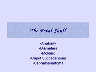
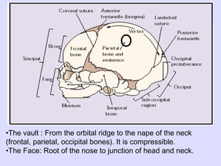
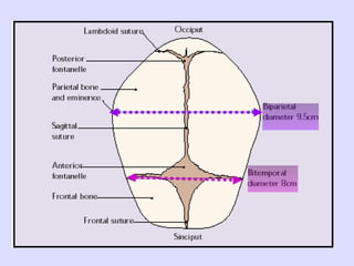
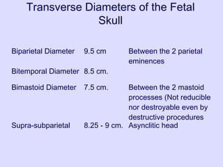
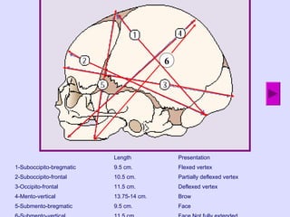
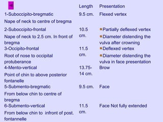
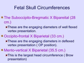
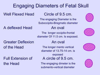
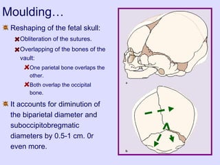
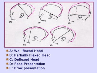
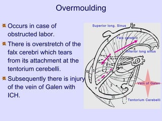
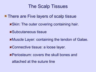
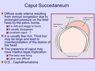
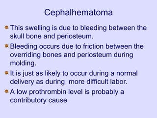
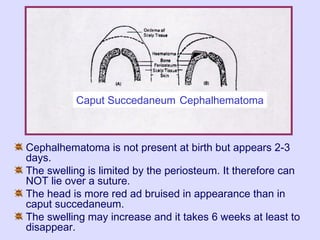
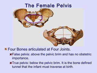
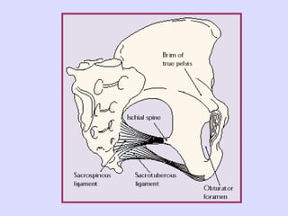
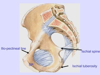
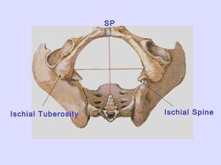
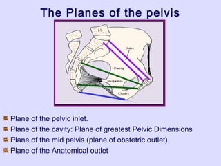
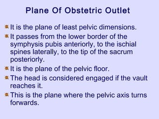
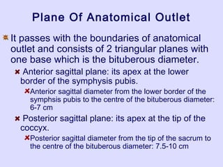
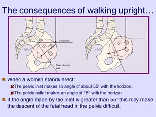
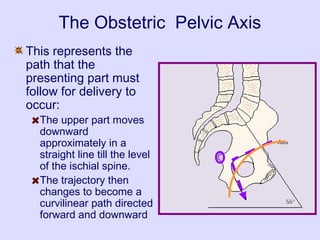
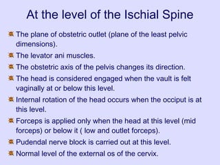
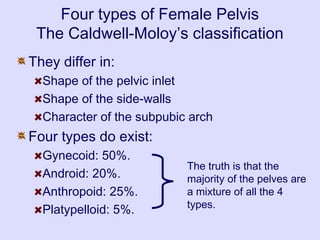
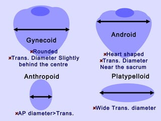
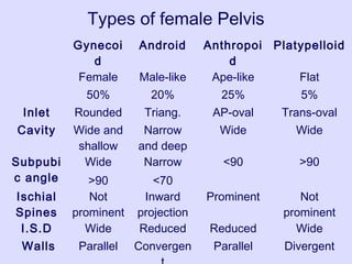
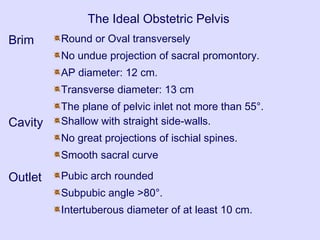
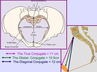
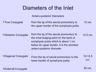
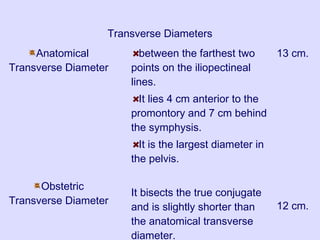
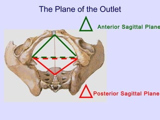
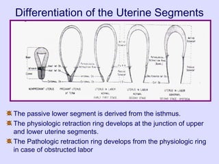
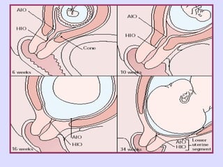
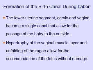
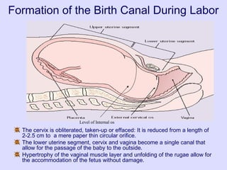
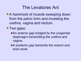
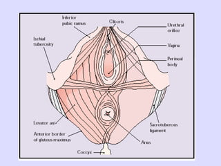
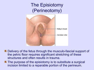
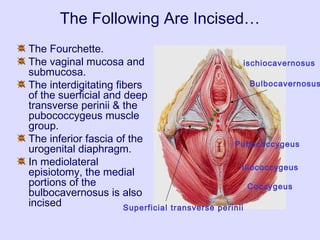
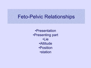
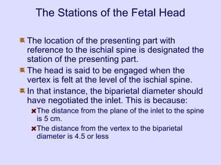
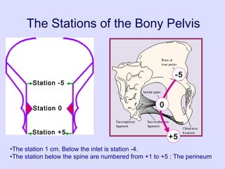
Obstetric Anatomy
Ahmed Mukhtar
Consultant and Lecturer of Obstetrics andConsultant and Lecturer of Obstetrics and
Gynecology, Faculty ofGynecology, Faculty of
MEDICINE, Zagazig University.MEDICINE, Zagazig University.
The Fetal Skull
•Anatomy
•Diameters
•Molding
•Caput Succedaneum
•Cephalhematoma
•The vault : From the orbital ridge to the nape of the neck
(frontal, parietal, occipital bones). It is compressible.
•The Face: Root of the nose to junction of head and neck.
Transverse Diameters of the Fetal
Skull
Biparietal Diameter 9.5 cm Between the 2 parietal
eminences
Bitemporal Diameter 8.5 cm.
Bimastoid Diameter 7.5 cm. Between the 2 mastoid
processes (Not reducible
nor destroyable even by
destructive procedures
Supra-subparietal 8.25 - 9 cm. Asynclitic head
Length Presentation
1-Suboccipito-bregmatic 9.5 cm. Flexed vertex
2-Suboccipito-frontal 10.5 cm. Partially deflexed vertex
3-Occipito-frontal 11.5 cm. Deflexed vertex
4-Mento-vertical 13.75-14 cm. Brow
5-Submento-bregmatic 9.5 cm. Face
6
Length Presentation
1-Suboccipito-bregmatic
Nape of neck to centre of bregma
9.5 cm. Flexed vertex
2-Suboccipito-frontal
Nape of neck to 2.5 cm. In front of
bregma
10.5
cm.
Partially deflexed vertex
Diameter distending the
vulva after crowning
3-Occipito-frontal
Root of nose to occipital
protuberance
11.5
cm.
Deflexed vertex
Diameter distending the
vulva in face presentation
4-Mento-vertical
Point of chin to above posterior
fontanelle
13.75-
14 cm.
Brow
5-Submento-bregmatic
From below chin to centre of
bregma
9.5 cm. Face
6-Submento-vertical
From below chin to infront of post.
fontannelle
11.5
cm.
Face Not fully extended
Fetal Skull Circumferences
The Suboccipito-Bregmatic X Bipareital (28
cm.)
These are the engaging diameters of well flexed
vertex presentation.
Occipito-frontal X Biparietal (33 cm.)
These are the engaging diameters in deflexed
vertex presentation ( OP position).
Mento-vertical X Biparietal (35.5 cm.)
This is the largest head circumference ( Brow
presentation)
Engaging Diameters of Fetal Skull
Well Flexed Head Circle of 9.5 cm.
The engaging Diameter is the
Suboccipito-Bregmatic diameter
A deflexed Head An oval
The longer occipito-frontal
diameter Of 11.5 cm. Is exposed.
Greater Deflexion
of the Head
An oval
The longer mento vertical
diameter of 13.75-14 cm. is
exposed
Full Extension of
the Head
A circle of 9.5 cm.
The engaging dimeter is the
submento-vertical diameter
Moulding…
Reshaping of the fetal skull:
Obliteration of the sutures.
Overlapping of the bones of the
vault:
One parietal bone overlaps the
other.
Both overlap the occipital
bone.
It accounts for diminution of
the biparietal diameter and
suboccipitobregmatic
diameters by 0.5-1 cm. 0r
even more.
A: Well flexed Head
B: Partially Flexed Head
C: Deflexed Head
D: Face Presentation
E: Brow presentation
Superior long. Sinus
Inferior long sinus
Falx cerebri
Vein of Galen
Tentorium Cerebelli
Overmoulding
Occurs in case of
obstructed labor.
There is overstretch of the
falx cerebri which tears
from its attachment at the
tentorium cerebelli.
Subsequently there is injury
of the vein of Galen with
ICH.
The Scalp Tissues
There are Five layers of scalp tissue
Skin: The outer covering containing hair.
Subcutaneous tissue
Muscle Layer: containing the tendon of Galae.
Connective tissue: a loose layer.
Periosteum: covers the skull bones and
attached at the suture line
Caput Succedaneum
Diffuse scalp edema resulting
from venous congestion due to
prolonged pressure on the fetal
head by the pelvic bones.
It is soft and boggy to touch
It usually disappears
Localized caput…?
It is usually few mm. Thick but
may be large and lead to
misinterpretation of the station of
the head.
The presence of caput may
have medico-legal implication:
The baby was living
Labor was difficult
D.D…Cephalhematoma
Cephalhematoma
This swelling is due to bleeding between the
skull bone and periosteum.
Bleeding occurs due to friction between the
overriding bones and periosteum during
molding.
It is just as likely to occur during a normal
delivery as during more difficult labor.
A low prothrombin level is probably a
contributory cause
Cephalhematoma is not present at birth but appears 2-3
days.
The swelling is limited by the periosteum. It therefore can
NOT lie over a suture.
The head is more red ad bruised in appearance than in
caput succedaneum.
The swelling may increase and it takes 6 weeks at least to
disappear.
Caput Succedaneum Cephalhematoma
The Female Pelvis
•Anatomy
•Pelvic Diameters
•Pelvic Types
The Female Pelvis
Four Bones articulated at Four Joints.
False pelvis: above the pelvic brim and has no obstetric
importance.
True pelvis: below the pelvic brim. It is the bone defined
tunnel that the infant must traverse at birth.
Ischial spine
Ischial tuberosity
Ilio-pectineal line
SP
Ischial Tuberosity Ischial Spine
SP
The Planes of the pelvis
Plane of the pelvic inlet.
Plane of the cavity: Plane of greatest Pelvic Dimensions
Plane of the mid pelvis (plane of obstetric outlet)
Plane of the Anatomical outlet
Plane Of The Pelvic Inlet
passing with the boundaries of pelvic
brim and making an angle of 55o with
the horizon (angle of pelvic inclination).
Plane of the Pelvic Cavity
It is the plane of greatest pelvic dimensions.
It passes between the middle of the posterior
surface of the symphysis pubis and the junction
between 2nd and 3rd sacral vertebrae. Laterally, it
passes to the centre of the acetabulum and the
upper part of the greater sciatic notch.
It is a round plane with diameter of 12.5 cm.
Internal rotation of the head occurs when the
biparietal diameter occupies this wide pelvic plane
while the occiput is on the pelvic floor i.e. at the
plane of the least pelvic dimensions.
Plane Of Obstetric Outlet
It is the plane of least pelvic dimensions.
It passes from the lower border of the
symphysis pubis anteriorly, to the ischial
spines laterally, to the tip of the sacrum
posteriorly.
It is the plane of the pelvic floor.
The head is considered engaged if the vault
reaches it.
This is the plane where the pelvic axis turns
forwards.
Plane Of Anatomical Outlet
It passes with the boundaries of anatomical
outlet and consists of 2 triangular planes with
one base which is the bituberous diameter.
Anterior sagittal plane: its apex at the lower
border of the symphysis pubis.
Anterior sagittal diameter from the lower border of the
symphsis pubis to the centre of the bituberous diameter:
6-7 cm
Posterior sagittal plane: its apex at the tip of the
coccyx.
Posterior sagittal diameter from the tip of the sacrum to
the centre of the bituberous diameter: 7.5-10 cm
The consequences of walking upright…
When a women stands erect:
The pelvic inlet makes an angle of about 55° with the horizon.
The pelvic outlet makes an angle of 15° with the horizon
If the angle made by the inlet is greater than 55° this may make
the descent of the fetal head in the pelvis difficult.
The Obstetric Pelvic Axis
This represents the
path that the
presenting part must
follow for delivery to
occur:
The upper part moves
downward
approximately in a
straight line till the level
of the ischial spine.
The trajectory then
changes to become a
curvilinear path directed
forward and downward
At the level of the Ischial Spine
The plane of obstetric outlet (plane of the least pelvic
dimensions).
The levator ani muscles.
The obstetric axis of the pelvis changes its direction.
The head is considered engaged when the vault is felt
vaginally at or below this level.
Internal rotation of the head occurs when the occiput is at
this level.
Forceps is applied only when the head at this level (mid
forceps) or below it ( low and outlet forceps).
Pudendal nerve block is carried out at this level.
Normal level of the external os of the cervix.
Four types of Female Pelvis
The Caldwell-Moloy’s classification
They differ in:
Shape of the pelvic inlet
Shape of the side-walls
Character of the subpubic arch
Four types do exist:
Gynecoid: 50%.
Android: 20%.
Anthropoid: 25%.
Platypelloid: 5%.
The truth is that the
majority of the pelves are
a mixture of all the 4
types.
Gynecoid
Android
Anthropoid Platypelloid
Rounded
Trans. Diameter Slightly
behind the centre
Heart shaped
Trans. Diameter
Near the sacrum
AP diameter>Trans.
Wide Trans. diameter
Types of female Pelvis
Gynecoi
d
Android Anthropoi
d
Platypelloid
Female Male-like Ape-like Flat
50% 20% 25% 5%
Inlet Rounded Triang. AP-oval Trans-oval
Cavity Wide and
shallow
Narrow
and deep
Wide Wide
Subpubi
c angle
Wide
>90
Narrow
<70
<90 >90
Ischial
Spines
Not
prominent
Inward
projection
Prominent Not
prominent
I.S.D Wide Reduced Reduced Wide
Walls Parallel Convergen Parallel Divergent
The Ideal Obstetric Pelvis
Brim Round or Oval transversely
No undue projection of sacral promontory.
AP diameter: 12 cm.
Transverse diameter: 13 cm
The plane of pelvic inlet not more than 55°.
Cavity Shallow with straight side-walls.
No great projections of ischial spines.
Smooth sacral curve
Outlet Pubic arch rounded
Subpubic angle >80°.
Intertuberous diameter of at least 10 cm.
The True Conjugate = 11 cm
The Obstet. Conjugate = 10.5cm
The Diagonal Conjugate = 12 cm
Diameters of the Inlet
Antero-posterior Diameters
True Conjugate
Obstetric Conjugate
Diagonal Conjugate
External Conjugate
from the tip of the sacral promontory to
the upper border of the symphysis pubis.
from the tip of the sacral promontory to
the most bulging point on the back of
symphysis pubis which is about 1 cm
below its upper border. It is the shortest
antero-posterior diameter
From the tip of sacral promontory to the
lower border of symphysis pubis.
12 cm.
10.5 cm.
12-12.5
cm.
20 cm.
Transverse Diameters
Anatomical
Transverse Diameter
Obstetric
Transverse Diameter
between the farthest two
points on the iliopectineal
lines.
It lies 4 cm anterior to the
promontory and 7 cm behind
the symphysis.
It is the largest diameter in
the pelvis.
It bisects the true conjugate
and is slightly shorter than
the anatomical transverse
diameter.
13 cm.
12 cm.
Oblique Diameters
Right and left
oblique diameters
Right and left
Sacro-cotyloid
diameters
From the right Sacro-iliac
joint to the left ilio-pectineal
eminence and vice-versa.
From the right ilio-
pectineal eminence to the
promontory of the sacrum
(rt.)
12 cm.
9-9.5
cm.
Interspinous diam. = 10.5 cm.
Obstet. Ant. Post diam= 13 cm.
Anato. Ant. Post diam= 11 cm.
Diameters of the Outlet
Antero-Posterior Diameters
Anatomical
antero-posterior
diameter
Obstetric
antero-posterior
diameter
From the tip of the coccyx
to the lower border of
symphysis pubis.
From the tip of the sacrum
to the lower border of
symphysis pubis as the
coccyx moves backwards
during the second stage of
labour.
11cm
13 cm
Transverse Diameters
Anatomical
Transverse
Diameter
(Bituberous)
Obstetric
Transverse
Diameter
(interspinous)
Extends between the inner
aspects of the ischial
tuberosities.
Extends between the tips
of the ischial spines. It is the
smallest diameter of the
pelvis.
11cm
10.5 cm.
Tom’s Dictum: If the sum of the Bituberous diameter and Post.
Sagittal diameter is less than 15, the pelvic outlet is
contracted . This is an indication of performing a Cesarean
section.
The Plane of the Outlet
Anterior Sagittal Plane
Posterior Sagittal Plane
Pelvic Soft Tissues
•The Formation Of The Lower Uterine Segment
•The Levatores Ani
•The Perineal Muscles
•Formation of the birth canal during labor
•The Episiotomy
The formation of the lower uterine segment
It is the part between the vesico-uterine fold
of peritoneum superiorly and the cervix
inferiorly.
It develops as early as the 16th
week by
incorporating the upper part of the cervix in
the lower part of the uterus to accommodate
for the presenting part of the fetus.
Differentiation of the Uterine Segments
The passive lower segment is derived from the isthmus.
The physiologic retraction ring develops at the junction of upper
and lower uterine segments.
The Pathologic retraction ring develops from the physiologic ring
in case of obstructed labor
Formation of the Birth Canal During Labor
The lower uterine segment, cervix and vagina
become a single canal that allow for the
passage of the baby to the outside.
Hypertrophy of the vaginal muscle layer and
unfolding of the rugae allow for the
accommodation of the fetus without damage.
Level of Internal os
The cervix is obliterated, taken-up or effaced: It is reduced from a length of
2-2.5 cm to a mere paper thin circular orifice.
The lower uterine segment, cervix and vagina become a single canal that
allow for the passage of the baby to the outside.
Hypertrophy of the vaginal muscle layer and unfolding of the rugae allow for
the accommodation of the fetus without damage.
Formation of the Birth Canal During Labor
The Levatores Ani
A hammock of muscle sweeping down
from the pelvic brim and investing the
urethra, vagina and rectum.
Two gaps:
An anterior gap bridged by the urogenital
diaphragm transmitting the urethra and
vagina.
A posterior gap transmits the rectum and
anal canal.
The resistance and shape of the pelvic floor
play an important role in facilitating rotation
and flexion of the presenting part.
As the presenting part descends:
The anterior portion of the pelvic floor is pressed
outwards against the SP.
The posterior part becomes stretched into a thin-
walled tube.
The perineal body stretches and thins from 5 cm.
To 0.05 cm. and is displaced downward.
The Episiotomy
(Perineotomy)
Delivery of the fetus through the musculo-fascial support of
the pelvic floor requires significant stretching of these
structures and often results in trauma.
The purpose of the episiotomy is to substitute a surgical
incision limited to a reparable portion of the perineum.
The Following Are Incised…
The Fourchette.
The vaginal mucosa and
submucosa.
The interdigitating fibers
of the suerficial and deep
transverse perinii & the
pubococcygeus muscle
group.
The inferior fascia of the
urogenital diaphragm.
In mediolateral
episiotomy, the medial
portions of the
bulbocavernosus is also
incised
ischiocavernosus
Bulbocavernosus
Pubococcygeus
Superficial transverse perinii
Iliococcygeus
Coccygeus
Feto-Pelvic Relationships
•Presentation
•Presenting part
•Lie
•Attitude
•Position
•station
As the journey progresses…
The fetal head descends along the pelvic
axis.
It must rotate to accommodate the
appropriate diameters of the head to the
pelvic diameters.
The reference points during this journey:
The ischial spine is the pelvic reference point
The presenting part is the fetal reference point.
Fetal Presentation & The Presenting Part
Fetal Presentation:
Is the fetal pole that presents at the pelvic
inlet:
Cephalic: Head First
Breech: Feet or Buttocks
Shoulder: back or abdomen
The Presenting part:
Is the part of the fetus first touched by the
examining fingers during pelvic examination.
The Fetal Lie
Refers to the relationship between the fetal
longitudinal axis and that of the mother.
Position
It refers to the relationships of a designated point on the
presenting part “Denominator” to the walls of maternal
pelvis.
P
LT
A
RT
RA
RP LP
LA
As the fetal head descends through the birth
canal, the suboccipito-bregmatic diameter
successively occupies the :
Transverse diameter of the inlet.
Oblique diameter of the cavity.
AP diameter of the outlet
What is the predominant fetal head position?
During labor, in 90% of vertex
presentation, The head assumes either a
LOA or a ROP position
The sagittal suture occupies the Right
Oblique diameter of the pelvis.
The right oblique diameter of the pelvis goes
from the left iliopectineal eminence to the
Right sacroiliac joint.
Why should the head rotate?
The larger transverse diameter of the
pelvis is more posterior.
However the presence of the sacral
promontory pushes the head anteriorly
towards a smaller transverse diameter.
The head will therefore rotate to take
advantage of the greater oblique diameter
at that level
Why the LOA or the ROP are favored
over the LOP or ROA?
The presence of the sigmoid colon in the post left
quadrant of the pelvic inlet pushes the head
anteriorly towards the pubis.
The sagittal suture is tending to occupy the wider
Right oblique diameter rather then the left oblique
diameter which is encroached upon by the sigmoid
colon.
Thus a LOA or a ROP positions are favored in 90%
of cases.
The Stations of the Fetal Head
The location of the presenting part with
reference to the ischial spine is designated the
station of the presenting part.
The head is said to be engaged when the
vertex is felt at the level of the ischial spine.
In that instance, the biparietal diameter should
have negotiated the inlet. This is because:
The distance from the plane of the inlet to the spine
is 5 cm.
The distance from the vertex to the biparietal
diameter is 4.5 or less
The Stations of the Bony Pelvis
Station 0
Station -5
Station +5
-5
0
+5
•The station 1 cm. Below the inlet is station -4.
•The station below the spine are numbered from +1 to +5 : The perineum
The Fetal Head Has Five Fifths…
0 : Head Not Palpable
1 : Sinciput felt – Occiput Not Felt
2 : Sinciput felt – Occiput Just Felt
3 : Sinciput easily felt – Occiput
Felt
4 : Sinciput High – Occiput easily
Felt
5 : Complete above pelvic brim
fifthabove
-5
0
+5

Fetal skull ahmed mukhtar

  • 1.
    Obstetric Anatomy Ahmed Mukhtar Consultantand Lecturer of Obstetrics andConsultant and Lecturer of Obstetrics and Gynecology, Faculty ofGynecology, Faculty of MEDICINE, Zagazig University.MEDICINE, Zagazig University.
  • 2.
  • 3.
    •The vault :From the orbital ridge to the nape of the neck (frontal, parietal, occipital bones). It is compressible. •The Face: Root of the nose to junction of head and neck.
  • 5.
    Transverse Diameters ofthe Fetal Skull Biparietal Diameter 9.5 cm Between the 2 parietal eminences Bitemporal Diameter 8.5 cm. Bimastoid Diameter 7.5 cm. Between the 2 mastoid processes (Not reducible nor destroyable even by destructive procedures Supra-subparietal 8.25 - 9 cm. Asynclitic head
  • 6.
    Length Presentation 1-Suboccipito-bregmatic 9.5cm. Flexed vertex 2-Suboccipito-frontal 10.5 cm. Partially deflexed vertex 3-Occipito-frontal 11.5 cm. Deflexed vertex 4-Mento-vertical 13.75-14 cm. Brow 5-Submento-bregmatic 9.5 cm. Face 6
  • 7.
    Length Presentation 1-Suboccipito-bregmatic Nape ofneck to centre of bregma 9.5 cm. Flexed vertex 2-Suboccipito-frontal Nape of neck to 2.5 cm. In front of bregma 10.5 cm. Partially deflexed vertex Diameter distending the vulva after crowning 3-Occipito-frontal Root of nose to occipital protuberance 11.5 cm. Deflexed vertex Diameter distending the vulva in face presentation 4-Mento-vertical Point of chin to above posterior fontanelle 13.75- 14 cm. Brow 5-Submento-bregmatic From below chin to centre of bregma 9.5 cm. Face 6-Submento-vertical From below chin to infront of post. fontannelle 11.5 cm. Face Not fully extended
  • 8.
    Fetal Skull Circumferences TheSuboccipito-Bregmatic X Bipareital (28 cm.) These are the engaging diameters of well flexed vertex presentation. Occipito-frontal X Biparietal (33 cm.) These are the engaging diameters in deflexed vertex presentation ( OP position). Mento-vertical X Biparietal (35.5 cm.) This is the largest head circumference ( Brow presentation)
  • 9.
    Engaging Diameters ofFetal Skull Well Flexed Head Circle of 9.5 cm. The engaging Diameter is the Suboccipito-Bregmatic diameter A deflexed Head An oval The longer occipito-frontal diameter Of 11.5 cm. Is exposed. Greater Deflexion of the Head An oval The longer mento vertical diameter of 13.75-14 cm. is exposed Full Extension of the Head A circle of 9.5 cm. The engaging dimeter is the submento-vertical diameter
  • 10.
    Moulding… Reshaping of thefetal skull: Obliteration of the sutures. Overlapping of the bones of the vault: One parietal bone overlaps the other. Both overlap the occipital bone. It accounts for diminution of the biparietal diameter and suboccipitobregmatic diameters by 0.5-1 cm. 0r even more.
  • 11.
    A: Well flexedHead B: Partially Flexed Head C: Deflexed Head D: Face Presentation E: Brow presentation
  • 12.
    Superior long. Sinus Inferiorlong sinus Falx cerebri Vein of Galen Tentorium Cerebelli Overmoulding Occurs in case of obstructed labor. There is overstretch of the falx cerebri which tears from its attachment at the tentorium cerebelli. Subsequently there is injury of the vein of Galen with ICH.
  • 13.
    The Scalp Tissues Thereare Five layers of scalp tissue Skin: The outer covering containing hair. Subcutaneous tissue Muscle Layer: containing the tendon of Galae. Connective tissue: a loose layer. Periosteum: covers the skull bones and attached at the suture line
  • 14.
    Caput Succedaneum Diffuse scalpedema resulting from venous congestion due to prolonged pressure on the fetal head by the pelvic bones. It is soft and boggy to touch It usually disappears Localized caput…? It is usually few mm. Thick but may be large and lead to misinterpretation of the station of the head. The presence of caput may have medico-legal implication: The baby was living Labor was difficult D.D…Cephalhematoma
  • 15.
    Cephalhematoma This swelling isdue to bleeding between the skull bone and periosteum. Bleeding occurs due to friction between the overriding bones and periosteum during molding. It is just as likely to occur during a normal delivery as during more difficult labor. A low prothrombin level is probably a contributory cause
  • 16.
    Cephalhematoma is notpresent at birth but appears 2-3 days. The swelling is limited by the periosteum. It therefore can NOT lie over a suture. The head is more red ad bruised in appearance than in caput succedaneum. The swelling may increase and it takes 6 weeks at least to disappear. Caput Succedaneum Cephalhematoma
  • 17.
    The Female Pelvis •Anatomy •PelvicDiameters •Pelvic Types
  • 18.
    The Female Pelvis FourBones articulated at Four Joints. False pelvis: above the pelvic brim and has no obstetric importance. True pelvis: below the pelvic brim. It is the bone defined tunnel that the infant must traverse at birth.
  • 20.
  • 21.
  • 22.
    The Planes ofthe pelvis Plane of the pelvic inlet. Plane of the cavity: Plane of greatest Pelvic Dimensions Plane of the mid pelvis (plane of obstetric outlet) Plane of the Anatomical outlet
  • 23.
    Plane Of ThePelvic Inlet passing with the boundaries of pelvic brim and making an angle of 55o with the horizon (angle of pelvic inclination).
  • 24.
    Plane of thePelvic Cavity It is the plane of greatest pelvic dimensions. It passes between the middle of the posterior surface of the symphysis pubis and the junction between 2nd and 3rd sacral vertebrae. Laterally, it passes to the centre of the acetabulum and the upper part of the greater sciatic notch. It is a round plane with diameter of 12.5 cm. Internal rotation of the head occurs when the biparietal diameter occupies this wide pelvic plane while the occiput is on the pelvic floor i.e. at the plane of the least pelvic dimensions.
  • 25.
    Plane Of ObstetricOutlet It is the plane of least pelvic dimensions. It passes from the lower border of the symphysis pubis anteriorly, to the ischial spines laterally, to the tip of the sacrum posteriorly. It is the plane of the pelvic floor. The head is considered engaged if the vault reaches it. This is the plane where the pelvic axis turns forwards.
  • 26.
    Plane Of AnatomicalOutlet It passes with the boundaries of anatomical outlet and consists of 2 triangular planes with one base which is the bituberous diameter. Anterior sagittal plane: its apex at the lower border of the symphysis pubis. Anterior sagittal diameter from the lower border of the symphsis pubis to the centre of the bituberous diameter: 6-7 cm Posterior sagittal plane: its apex at the tip of the coccyx. Posterior sagittal diameter from the tip of the sacrum to the centre of the bituberous diameter: 7.5-10 cm
  • 27.
    The consequences ofwalking upright… When a women stands erect: The pelvic inlet makes an angle of about 55° with the horizon. The pelvic outlet makes an angle of 15° with the horizon If the angle made by the inlet is greater than 55° this may make the descent of the fetal head in the pelvis difficult.
  • 28.
    The Obstetric PelvicAxis This represents the path that the presenting part must follow for delivery to occur: The upper part moves downward approximately in a straight line till the level of the ischial spine. The trajectory then changes to become a curvilinear path directed forward and downward
  • 29.
    At the levelof the Ischial Spine The plane of obstetric outlet (plane of the least pelvic dimensions). The levator ani muscles. The obstetric axis of the pelvis changes its direction. The head is considered engaged when the vault is felt vaginally at or below this level. Internal rotation of the head occurs when the occiput is at this level. Forceps is applied only when the head at this level (mid forceps) or below it ( low and outlet forceps). Pudendal nerve block is carried out at this level. Normal level of the external os of the cervix.
  • 30.
    Four types ofFemale Pelvis The Caldwell-Moloy’s classification They differ in: Shape of the pelvic inlet Shape of the side-walls Character of the subpubic arch Four types do exist: Gynecoid: 50%. Android: 20%. Anthropoid: 25%. Platypelloid: 5%. The truth is that the majority of the pelves are a mixture of all the 4 types.
  • 31.
    Gynecoid Android Anthropoid Platypelloid Rounded Trans. DiameterSlightly behind the centre Heart shaped Trans. Diameter Near the sacrum AP diameter>Trans. Wide Trans. diameter
  • 32.
    Types of femalePelvis Gynecoi d Android Anthropoi d Platypelloid Female Male-like Ape-like Flat 50% 20% 25% 5% Inlet Rounded Triang. AP-oval Trans-oval Cavity Wide and shallow Narrow and deep Wide Wide Subpubi c angle Wide >90 Narrow <70 <90 >90 Ischial Spines Not prominent Inward projection Prominent Not prominent I.S.D Wide Reduced Reduced Wide Walls Parallel Convergen Parallel Divergent
  • 33.
    The Ideal ObstetricPelvis Brim Round or Oval transversely No undue projection of sacral promontory. AP diameter: 12 cm. Transverse diameter: 13 cm The plane of pelvic inlet not more than 55°. Cavity Shallow with straight side-walls. No great projections of ischial spines. Smooth sacral curve Outlet Pubic arch rounded Subpubic angle >80°. Intertuberous diameter of at least 10 cm.
  • 34.
    The True Conjugate= 11 cm The Obstet. Conjugate = 10.5cm The Diagonal Conjugate = 12 cm
  • 35.
    Diameters of theInlet Antero-posterior Diameters True Conjugate Obstetric Conjugate Diagonal Conjugate External Conjugate from the tip of the sacral promontory to the upper border of the symphysis pubis. from the tip of the sacral promontory to the most bulging point on the back of symphysis pubis which is about 1 cm below its upper border. It is the shortest antero-posterior diameter From the tip of sacral promontory to the lower border of symphysis pubis. 12 cm. 10.5 cm. 12-12.5 cm. 20 cm.
  • 36.
    Transverse Diameters Anatomical Transverse Diameter Obstetric TransverseDiameter between the farthest two points on the iliopectineal lines. It lies 4 cm anterior to the promontory and 7 cm behind the symphysis. It is the largest diameter in the pelvis. It bisects the true conjugate and is slightly shorter than the anatomical transverse diameter. 13 cm. 12 cm.
  • 37.
    Oblique Diameters Right andleft oblique diameters Right and left Sacro-cotyloid diameters From the right Sacro-iliac joint to the left ilio-pectineal eminence and vice-versa. From the right ilio- pectineal eminence to the promontory of the sacrum (rt.) 12 cm. 9-9.5 cm.
  • 38.
    Interspinous diam. =10.5 cm. Obstet. Ant. Post diam= 13 cm. Anato. Ant. Post diam= 11 cm.
  • 39.
    Diameters of theOutlet Antero-Posterior Diameters Anatomical antero-posterior diameter Obstetric antero-posterior diameter From the tip of the coccyx to the lower border of symphysis pubis. From the tip of the sacrum to the lower border of symphysis pubis as the coccyx moves backwards during the second stage of labour. 11cm 13 cm
  • 40.
    Transverse Diameters Anatomical Transverse Diameter (Bituberous) Obstetric Transverse Diameter (interspinous) Extends betweenthe inner aspects of the ischial tuberosities. Extends between the tips of the ischial spines. It is the smallest diameter of the pelvis. 11cm 10.5 cm. Tom’s Dictum: If the sum of the Bituberous diameter and Post. Sagittal diameter is less than 15, the pelvic outlet is contracted . This is an indication of performing a Cesarean section.
  • 41.
    The Plane ofthe Outlet Anterior Sagittal Plane Posterior Sagittal Plane
  • 42.
    Pelvic Soft Tissues •TheFormation Of The Lower Uterine Segment •The Levatores Ani •The Perineal Muscles •Formation of the birth canal during labor •The Episiotomy
  • 43.
    The formation ofthe lower uterine segment It is the part between the vesico-uterine fold of peritoneum superiorly and the cervix inferiorly. It develops as early as the 16th week by incorporating the upper part of the cervix in the lower part of the uterus to accommodate for the presenting part of the fetus.
  • 44.
    Differentiation of theUterine Segments The passive lower segment is derived from the isthmus. The physiologic retraction ring develops at the junction of upper and lower uterine segments. The Pathologic retraction ring develops from the physiologic ring in case of obstructed labor
  • 46.
    Formation of theBirth Canal During Labor The lower uterine segment, cervix and vagina become a single canal that allow for the passage of the baby to the outside. Hypertrophy of the vaginal muscle layer and unfolding of the rugae allow for the accommodation of the fetus without damage.
  • 47.
    Level of Internalos The cervix is obliterated, taken-up or effaced: It is reduced from a length of 2-2.5 cm to a mere paper thin circular orifice. The lower uterine segment, cervix and vagina become a single canal that allow for the passage of the baby to the outside. Hypertrophy of the vaginal muscle layer and unfolding of the rugae allow for the accommodation of the fetus without damage. Formation of the Birth Canal During Labor
  • 48.
    The Levatores Ani Ahammock of muscle sweeping down from the pelvic brim and investing the urethra, vagina and rectum. Two gaps: An anterior gap bridged by the urogenital diaphragm transmitting the urethra and vagina. A posterior gap transmits the rectum and anal canal.
  • 49.
    The resistance andshape of the pelvic floor play an important role in facilitating rotation and flexion of the presenting part. As the presenting part descends: The anterior portion of the pelvic floor is pressed outwards against the SP. The posterior part becomes stretched into a thin- walled tube. The perineal body stretches and thins from 5 cm. To 0.05 cm. and is displaced downward.
  • 54.
    The Episiotomy (Perineotomy) Delivery ofthe fetus through the musculo-fascial support of the pelvic floor requires significant stretching of these structures and often results in trauma. The purpose of the episiotomy is to substitute a surgical incision limited to a reparable portion of the perineum.
  • 55.
    The Following AreIncised… The Fourchette. The vaginal mucosa and submucosa. The interdigitating fibers of the suerficial and deep transverse perinii & the pubococcygeus muscle group. The inferior fascia of the urogenital diaphragm. In mediolateral episiotomy, the medial portions of the bulbocavernosus is also incised ischiocavernosus Bulbocavernosus Pubococcygeus Superficial transverse perinii Iliococcygeus Coccygeus
  • 56.
  • 57.
    As the journeyprogresses… The fetal head descends along the pelvic axis. It must rotate to accommodate the appropriate diameters of the head to the pelvic diameters. The reference points during this journey: The ischial spine is the pelvic reference point The presenting part is the fetal reference point.
  • 58.
    Fetal Presentation &The Presenting Part Fetal Presentation: Is the fetal pole that presents at the pelvic inlet: Cephalic: Head First Breech: Feet or Buttocks Shoulder: back or abdomen The Presenting part: Is the part of the fetus first touched by the examining fingers during pelvic examination.
  • 59.
    The Fetal Lie Refersto the relationship between the fetal longitudinal axis and that of the mother.
  • 60.
    Position It refers tothe relationships of a designated point on the presenting part “Denominator” to the walls of maternal pelvis. P LT A RT RA RP LP LA
  • 61.
    As the fetalhead descends through the birth canal, the suboccipito-bregmatic diameter successively occupies the : Transverse diameter of the inlet. Oblique diameter of the cavity. AP diameter of the outlet
  • 62.
    What is thepredominant fetal head position? During labor, in 90% of vertex presentation, The head assumes either a LOA or a ROP position The sagittal suture occupies the Right Oblique diameter of the pelvis. The right oblique diameter of the pelvis goes from the left iliopectineal eminence to the Right sacroiliac joint.
  • 63.
    Why should thehead rotate? The larger transverse diameter of the pelvis is more posterior. However the presence of the sacral promontory pushes the head anteriorly towards a smaller transverse diameter. The head will therefore rotate to take advantage of the greater oblique diameter at that level
  • 64.
    Why the LOAor the ROP are favored over the LOP or ROA? The presence of the sigmoid colon in the post left quadrant of the pelvic inlet pushes the head anteriorly towards the pubis. The sagittal suture is tending to occupy the wider Right oblique diameter rather then the left oblique diameter which is encroached upon by the sigmoid colon. Thus a LOA or a ROP positions are favored in 90% of cases.
  • 65.
    The Stations ofthe Fetal Head The location of the presenting part with reference to the ischial spine is designated the station of the presenting part. The head is said to be engaged when the vertex is felt at the level of the ischial spine. In that instance, the biparietal diameter should have negotiated the inlet. This is because: The distance from the plane of the inlet to the spine is 5 cm. The distance from the vertex to the biparietal diameter is 4.5 or less
  • 66.
    The Stations ofthe Bony Pelvis Station 0 Station -5 Station +5 -5 0 +5 •The station 1 cm. Below the inlet is station -4. •The station below the spine are numbered from +1 to +5 : The perineum
  • 67.
    The Fetal HeadHas Five Fifths… 0 : Head Not Palpable 1 : Sinciput felt – Occiput Not Felt 2 : Sinciput felt – Occiput Just Felt 3 : Sinciput easily felt – Occiput Felt 4 : Sinciput High – Occiput easily Felt 5 : Complete above pelvic brim fifthabove -5 0 +5

Editor's Notes

  • #4 Needs definition: vault
  • #5 Bimastoid Suprapareital-subpareital
  • #19 Inferior view with lig
  • #20 labeling
  • #23 Pelvic Planes: These are imaginary planes lie as follow: (1) Plane of pelvic inlet: passing with the boundaries of pelvic brim and making an angle of 55o with the horizon (angle of pelvic inclination). (2) Plane of mid cavity ( plane of greatest pelvic dimensions): - pass between the middle of the posterior surface of the symphysis pubis and the junction between 2nd and 3rd sacral vertebrae. Laterally, it passes to the centre of the acetabulum and the upper part of the greater sciatic notch. - It is a round plane with diameter of 12.5 cm. - Internal rotation of the head occurs when the biparietal diameter occupies this wide pelvic plane while the occiput is on the pelvic floor i.e. at the plane of the least pelvic dimensions. (3) Plane of obstetric outlet (plane of least pelvic dimensions): passes from the lower border of the symphysis pubis anteriorly, to the ischial spines laterally, to the tip of the sacrum posteriorly. (4) Plane of anatomical outlet: passes with the boundaries of anatomical outlet and consists of 2 triangular planes with one base which is the bituberous diameter. a- Anterior sagittal plane: its apex at the lower border of the symphysis pubis. b- Posterior sagittal plane: its apex at the tip of the coccyx. Anterior sagittal diameter: 6-7 cm from the lower border of the symphsis pubis to the centre of the bituberous diameter. Posterior sagittal diameter: 7.5-10 cm from the tip of the sacrum to the centre of the bituberous diameter
  • #28 Effect of the inclination of the pelvis on the engagement of the fetal head
  • #29 Anatomical axis (curve of Carus): - It is an imaginary line joining the centre points of the planes of the inlet, cavity and outlet. - It is C shaped with the concavity directed forwards. - It has no obstetric importance.
  • #35 The ideal obstetric pelvis
  • #39 outlet