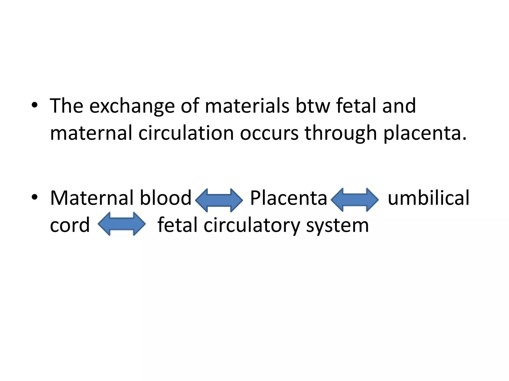
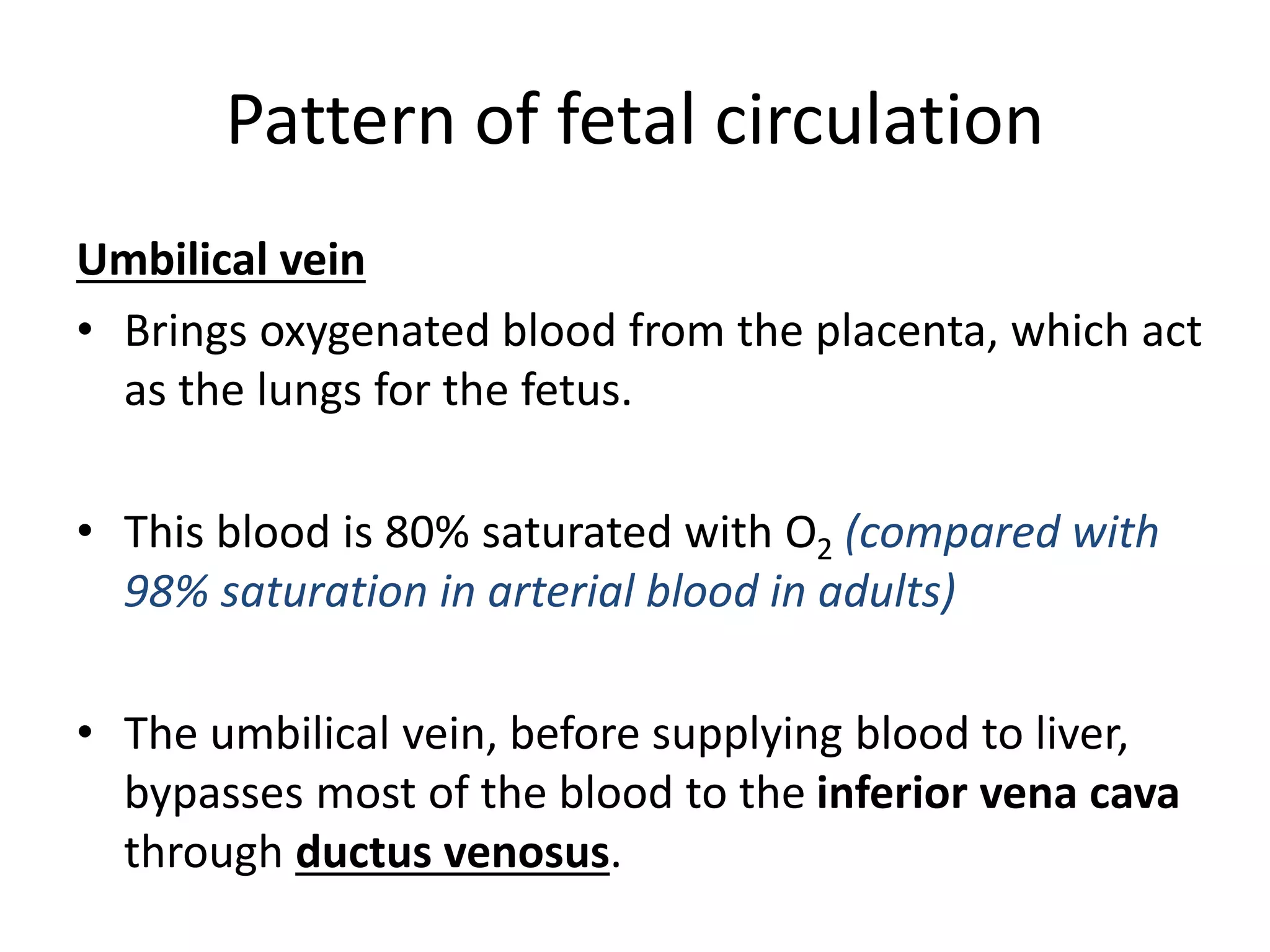
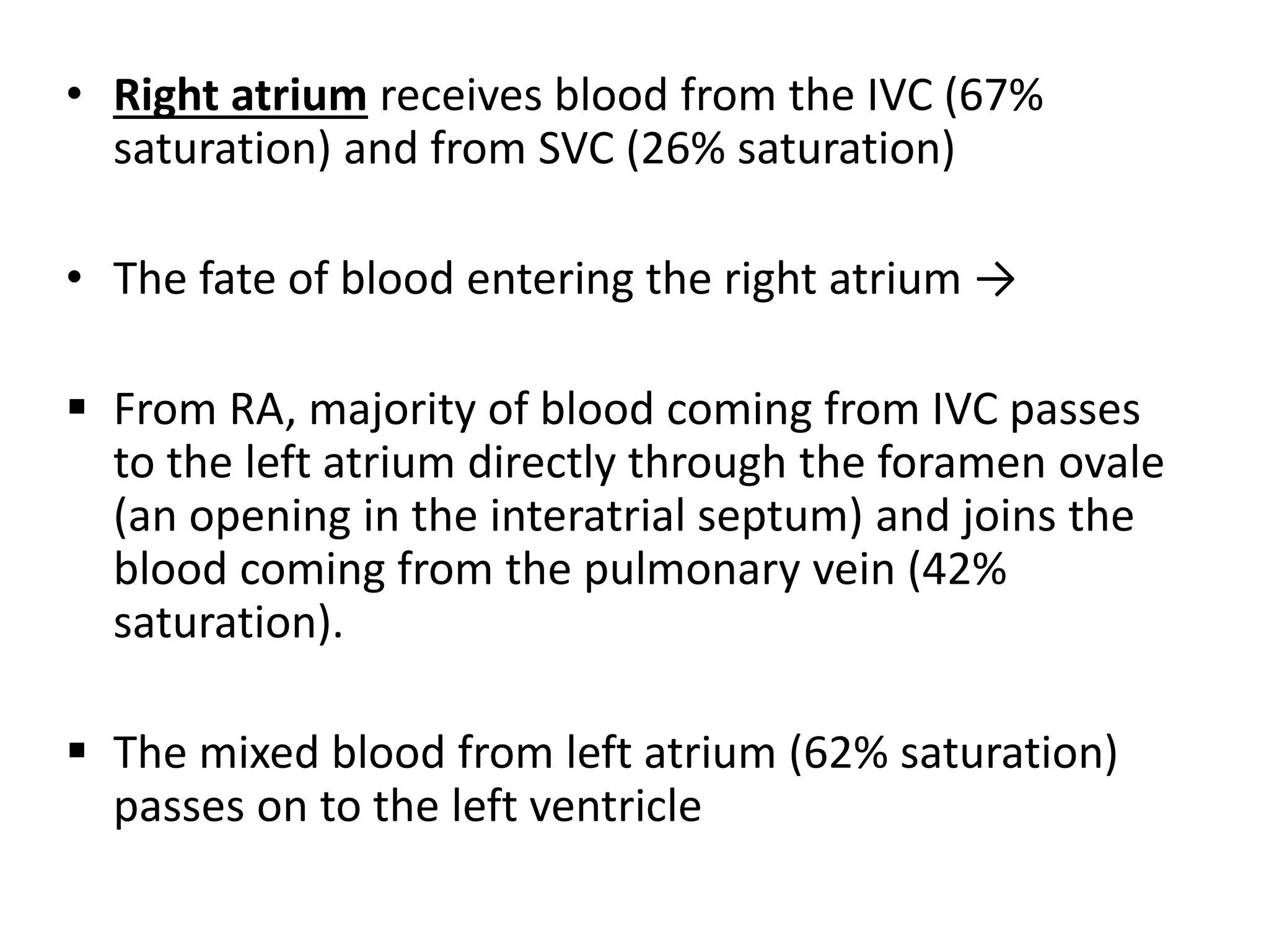
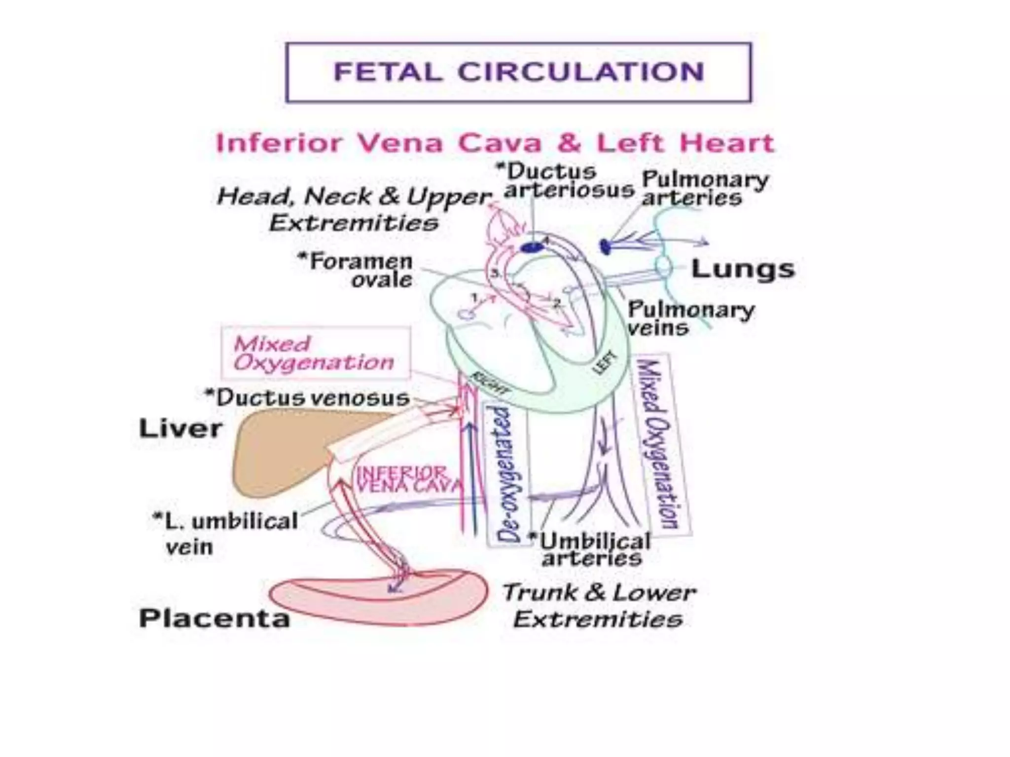
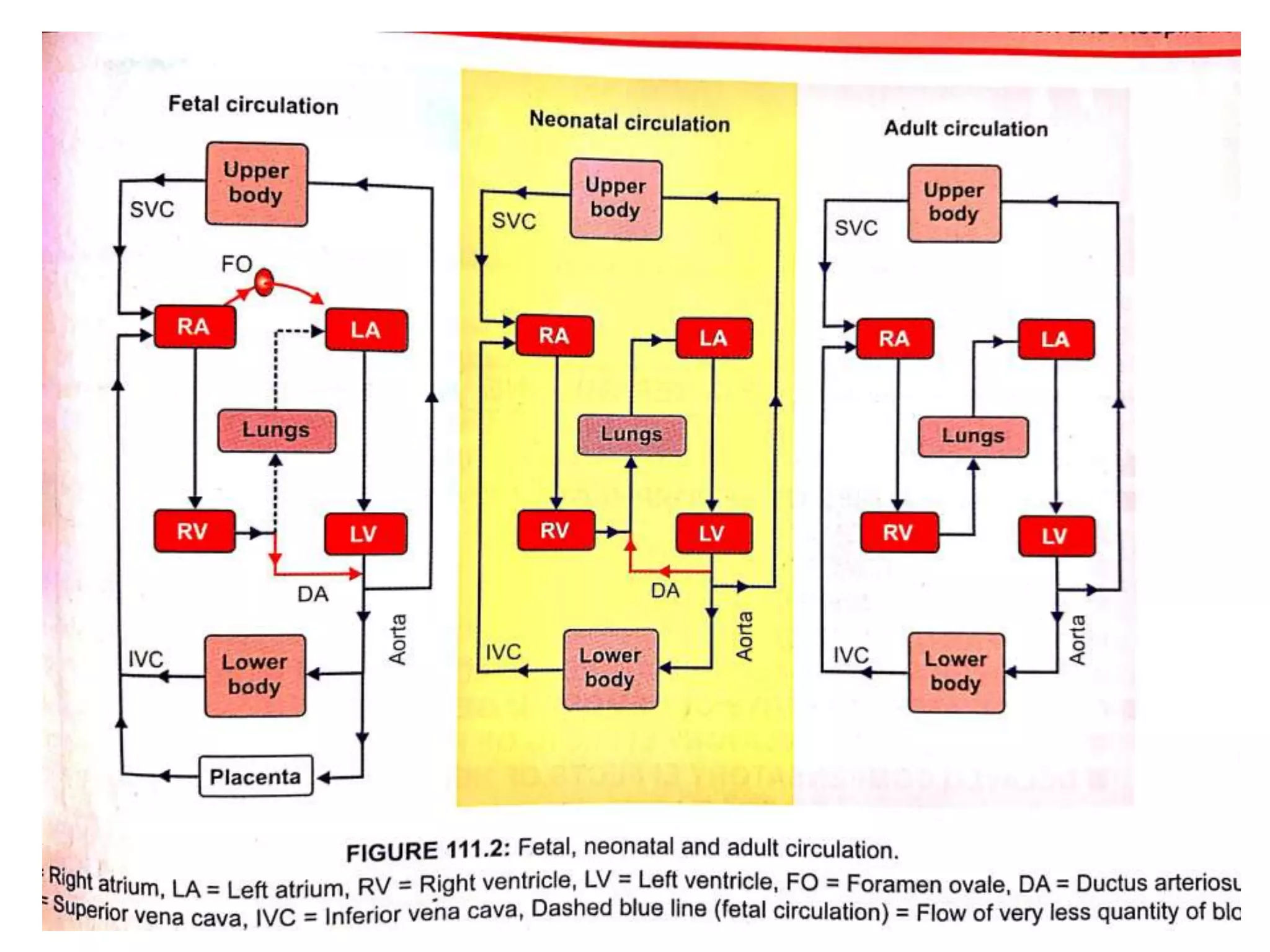
3. • The circulatory system of a fetus is called fetal
circulation.
• It exists only in fetus and contains special structures
that allow the developing fetus to exchange
materials with its mother.
• It differs from the postnatal (after birth) circulation
because the lungs, kidneys and GIT do not begin to
function until birth.
• The fetus obtains O2 and nutrients from the maternal
blood and eliminates CO2 and other wastes into it.
4. • The exchange of materials btw fetal and
maternal circulation occurs through placenta.
• Maternal blood Placenta umbilical
cord fetal circulatory system
5. Pattern of fetal circulation
Umbilical vein
• Brings oxygenated blood from the placenta, which act
as the lungs for the fetus.
• This blood is 80% saturated with O2 (compared with
98% saturation in arterial blood in adults)
• The umbilical vein, before supplying blood to liver,
bypasses most of the blood to the inferior vena cava
through ductus venosus.
6.
7.
8.
9.
10.
11. • IVC thus receives some blood (80% saturated O2) from
the umbilical vein through ductus venosus, and other
blood from the hepatic veins and systemic veins
draining from the trunk and inferior extremities (26%
saturated with O2)
• The mixed blood from the IVC (with approximate 67%
saturation) then enters the right atrium.
12.
13. • Right atrium receives blood from the IVC (67%
saturation) and from SVC (26% saturation)
• The fate of blood entering the right atrium →
From RA, majority of blood coming from IVC passes
to the left atrium directly through the foramen ovale
(an opening in the interatrial septum) and joins the
blood coming from the pulmonary vein (42%
saturation).
The mixed blood from left atrium (62% saturation)
passes on to the left ventricle
19. • From the right atrium, most of the blood coming
from the superior vena cava (26% saturation) and
small amount of blood from IVC (67%), passes into
right ventricle.
• This mixed blood (52% saturation) is pumped into
the pulmonary artery.
• But, since the fetal lungs are collapsed, their vascular
resistance is very high.
20. • Hence only a small amount of blood passes through
the lungs to reach the left atrium via pulmonary
veins.
• Majority of the pulmonary artery blood enters the
descending aorta directly by a vascular connection
called ductus arteriosus.
21.
22.
23. • Left ventricle pumps the blood (62% sat) into the
ascending aorta, from where the most of the blood goes
into the vessels of head and neck and upper limbs and
only small amount of blood goes to the descending
aorta.
• Descending aorta, thus receives blood mainly from the
pulmonary artery through ductus arteriosus (52% sat)
and only a small amount from the left ventricle (62% sat).
• The descending aorta then supplies the blood (58% sat)
to the whole body (except head, neck and upper limbs)
24.
25. • 2 Umbilical arteries, which are the branches
of anterior division of internal iliac artery
brings the blood (58% sat) back to the
placenta for another exchange of materials.
26.
27.
28.
29. Changes after birth
• After birth, when lungs, kidneys and GIT begin to
function, the following changes occur:
1. When the umbilical cord is tied off, blood no longer
flows through the umbilical arteries, they fill with
connective tissue, and the distal portion of UA
become fibrous cords called medial umbilical
ligament.
(Although the arteries are closed functionally only a few
minutes after birth, complete obliteration of the lumens
may take 2- 3 months)
30. 2. The umbilical vein collapses but remains as
the ligamentum teres , a structure that attaches
the umbilicus to the liver.
3. The ductus venosus collapses but remains as
the ligamentum venosum, a fibrous cord on the
inferior surface of the liver.
4. Placenta is expelled as the ‘afterbirth’
31. 5. The foramen ovale normally closes shortly after birth
to become the fossa ovalis, a depression in the inter
atrial septum.
(When an infant takes its first breath, the lungs expand
and blood flow to the lungs increases. Blood returning
from the lungs to heart through pulmonary veins
increases pressure in the left atrium. This closes the
foramen ovale by pushing the valve that guards it
against the interatrial septum. Permanent closure
occurs in about a year)
32. 6. The ductus arteriosus close by
vasoconstriction almost immediately after birth
and becomes the ligamentum arteriosum.
(Complete anatomical obliteration of the lumen
takes 1-3 months.)