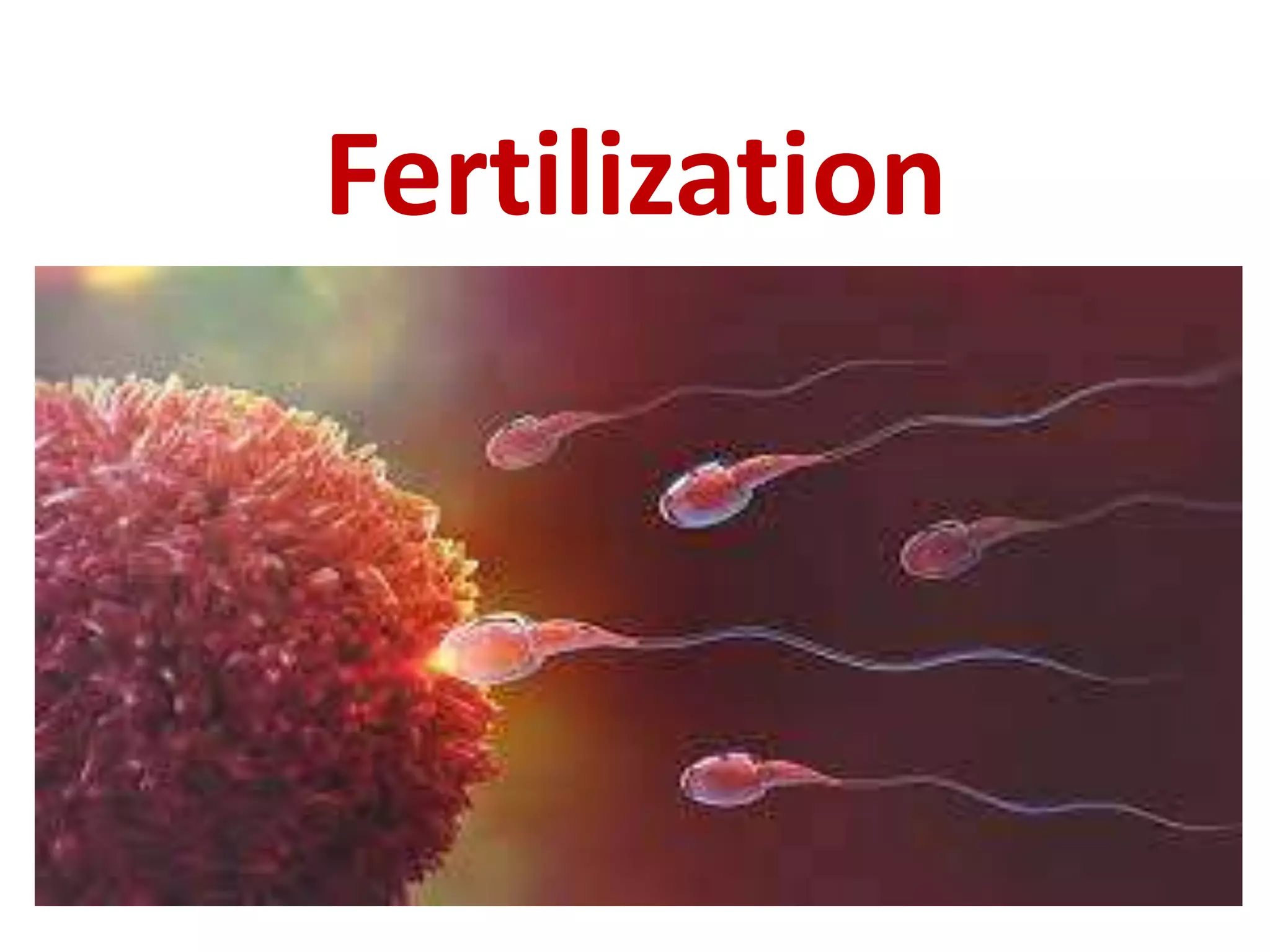
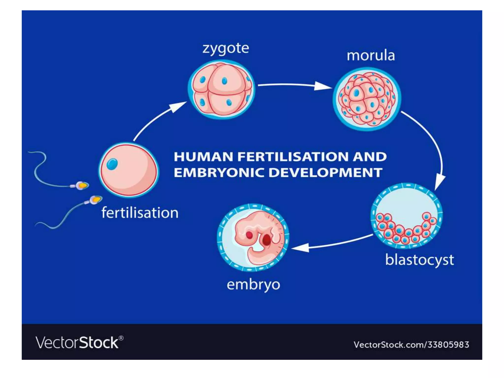
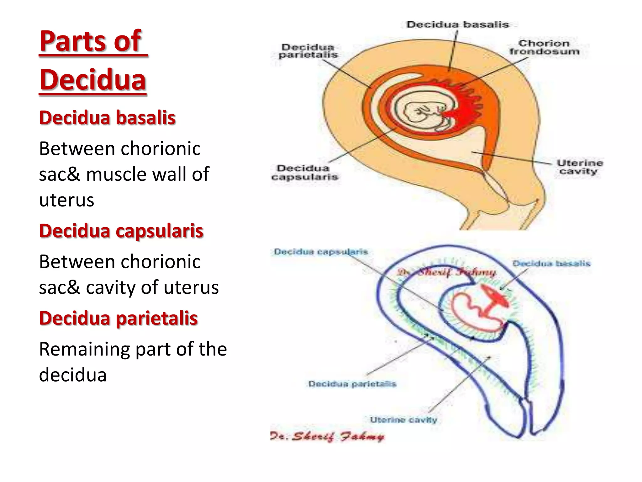
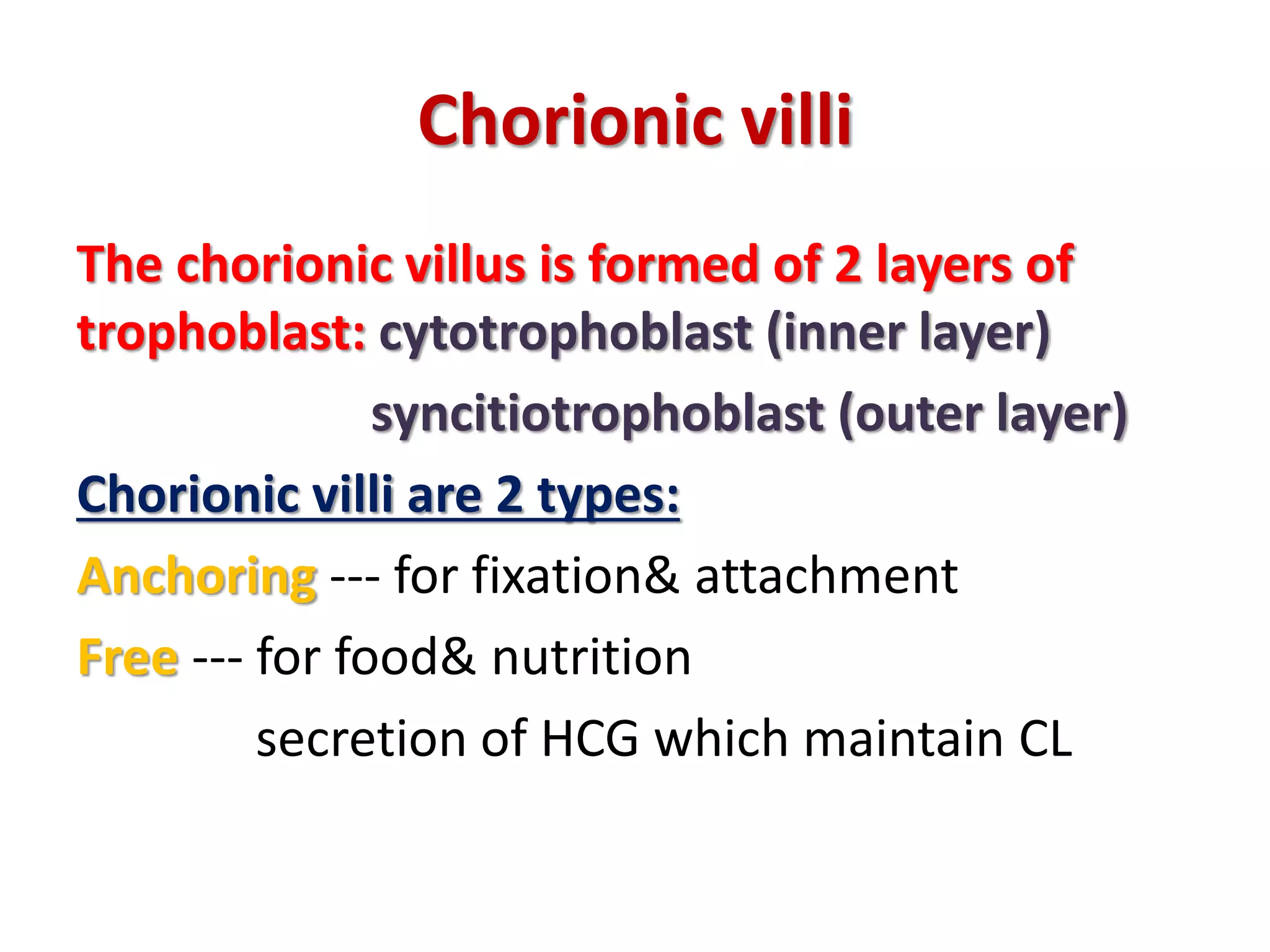
Fertilization, placenta, cord& AF
BY
Dr. Ahmed Nasef
Assistant lecturer of obstetrics&
gynecology – Benha university
Fertilization
Participants
What is fertilization
It is the act of fusion of male and
female gametes (sperm& ovum) to
form zygote
Where does fertilization
happen?
In the ampullary part of the fallopian tube
What happens before fertilization
Ovulation
What happens to sperm to be able for
fertilizing the egg
After insemination sperms pass from vagina into uterus to uterine tubes
Aided by contractions of muscles of uterus& uterine tubes
Only 300-500 sperms reach fertilization site from about 200-300 million sperms
deposited in vagina
These sperms undergo 2 processes to be capable of fertilizing the egg:
1- Capacitation: occur within female genital tract, removal of glycoprotein coat&
seminal plasma proteins
2- Acrosome reaction: secretion of certain substances from acrosomal cap close to 2ry
oocyte which include:
 Hyaluronidase enzyme--- assist in penetration of corona radiata
 Trypsin like substances& acrosin ------- help to penetrate zona pellucida
Phases of fertilization
1- penetration of corona radiata:
Only one sperm is required to fertilize the ovum. Others help the fertilizing sperm in penetration
of corona radiata by hyaluronidase enzyme
2- penetration of zona pellucida:
By trypsin like substances& acrosin
3- zona reaction:
Oocyte secrete lysosomal enzymes which make zona impermeable for other sperms
4- fusion of sperm with ovum to form zygote:
After entry of sperm the 2ry oocyte finishes its 2nd meiotic division
The chromosomes of both sperm and ovum become arranged and called male and female
pronuclei which lose their nuclear membranes and fuse forming the zygote
Results of fertilization
Formation of the zygote
Determination of sex of the embryo
Initiation of cleavage and differentiation
Differentiation
 Z y g o t e rapidly divides by mitosis ------ B l a s t o m e r e (8 cells)
 Then M o r u l a (16 cells)----- reaching uterine cavity after 3 days of
fertilization
 Fluid then accumulate forming B l a s t o c y s t ------ remains free for 3
days in uterine cavity
Then divide into 2 masses of cells:
Inner cell mass---- will form embryo
Outer cell mass (trophoblast)
Implantation
Process of embedding of the fertilized ovum into
the endometrium of uterus
Normal site of implantation:
Upper part of posterior or anterior wall of the
uterus
When: it occurs on 6th to 7th day of fertilization
How: apposition --- adhesion then invasion of
trophoblast (chorion) into Decidua
Implantation
Decidua
Modified secretory endometrium under effect of both
progesterone& estrogen
Functions:
 Site of implantation& nutrition of blastocyst
 Site of formation of placenta
 Protection against invasive power of trophoblast as
invasion stops at a fibrinoid layer called Nitabuch
Parts of
Decidua
Decidua basalis
Between chorionic
sac& muscle wall of
uterus
Decidua capsularis
Between chorionic
sac& cavity of uterus
Decidua parietalis
Remaining part of the
decidua
Chorionic villi
The chorionic villus is formed of 2 layers of
trophoblast: cytotrophoblast (inner layer)
syncitiotrophoblast (outer layer)
Chorionic villi are 2 types:
Anchoring --- for fixation& attachment
Free --- for food& nutrition
secretion of HCG which maintain CL
Chorion parts
Chorion frondosum part of chorion facing
decidua basalis. It will form the fetal part of
placenta
Chorion leave remaining part of chorion facing
decidua capsularis. It will atrophy
In vitro fertilization
What is IVF
Process in which fertilization is performed
outside the body then transfer of the
developed embryo in the uterus
placenta
Formation of the placenta
Its formed from 2 parts:
Fetal part Chorion frondosum
Maternal part Decidua basalis
It has 2 surfaces:
Fetal smooth and covered by amnion
Maternal divided into 15-20 cotyledons
Normal structure
Shape discoid
Weight 500 gm
Site implantation site (upper uterine segment)
Size 18-20 cm in diameter
Thickness 2.5 cm in center gradually thins
towards periphery
Cord insertion eccentric
Functions of placenta
Mechanical attachment
Respiration gas exchange
Nutrition& excretion
Placental barrier making placenta impermeable
to many drugs& organisms
Enzyme production as oxytocinase
Protein production as PAPP
Hormone production sex steroids (E&P)
Abnormalities of placenta
Shape
Bipartite placenta 2 equal lobes connected by membrane
Bilobate placenta 2 equal lobes connected by placental tissue
Placenta fenestrata part of placenta is missed
Placenta succenturiata presence of small accessory cotyledons attached to
placenta by membranes
Placenta circumvallate chorionic plate is < basal plate so fetal margin of
placenta shows white ring of decidua
Placenta membranacea chorion leave doesn’t atrophy --- large thin placenta
Abnormalities of site
In LUS ---- placenta previa
On uterine septum
Abnormalities of size
Small leading to placental insufficiency--- IUGR
Large in cases of syphilis, DM, twins, Rh
incompatibility& placenta membranacea
Abnormal adherence
Due to absent Nitabuch layer, may be:
P. Accreta placenta reaches basal layer of
decidua
P. Increta placenta penetrates muscle but
doesn’t reach serosal surface
P. Percreta placenta perforates uterus
Umbilical cord
Normal structure
Length about 50 cm
Diameter about 2 cm
Contents 2 arteries, 1 vein in a myxomatous
tissue (Wharton's jelly)
Abnormalities of the cord
Length
very long lead to coiling around fetus, true knots,
cord presentation& prolapse
Very short lead to failure of engagement& fetal
descent, malpresentation& fetal distress
Knots
True lead to fetal distress
False focal varicosity in collection of Wharton's
jelly
Abnormal attachment
Central
Marginal (Battledore)
Velamentous insertion vessels are inserted into
membranes not in placenta
Amniotic fluid
What is amniotic fluid?
It is the fluid surrounding the fetus
Source
 Maternal transudation through placenta& cord esp. in 1st
trim.
 Fetal urine esp. in 2nd half of pregnancy
 Transudation from fetal skin& umbilical cord
 Amniotic epithelium
 Lungs (bronchial& buccal secretions)
Composition of AF
About 99% clear watery
About 1-2% crystalloids& colloids (CHO,
proteins, lipids, hormones& electrolytes)
Measurement of AF
Amniotic fluid index (AFI) is a quantitative estimate of
amniotic fluid and an indicator of fetal well-being. It is a
part of the biophysical profile. AFI is the score
(expressed in cm) given to the amount of amniotic fluid
seen on ultrasonography of a pregnant uterus
Single deepest vertical pocket is the deepest longest
vertical pocket between the fetus and uterine wall
Functions of AF
During pregnancy
Protection against trauma
Prevents adhesions between fetal skin& amnion
Prevents infection
Thermoregulation
Allow free movement of the fetus so improve muscle
development
Development of alveoli
During labor
Help cervical dilatation
Prevent direct fetal& placental compression by uterine wall
Wash birth canal after ROM
Abnormalities of AF
Volume
Polyhydramnios
Oligohydramnios
Meconium staining
Inflammation Chorioamnionitis
Fertilization, placenta, umbilical cord& amniotic fluid

Fertilization, placenta, umbilical cord& amniotic fluid

  • 1.
    Fertilization, placenta, cord&AF BY Dr. Ahmed Nasef Assistant lecturer of obstetrics& gynecology – Benha university
  • 3.
  • 4.
  • 5.
    What is fertilization Itis the act of fusion of male and female gametes (sperm& ovum) to form zygote
  • 7.
    Where does fertilization happen? Inthe ampullary part of the fallopian tube
  • 8.
    What happens beforefertilization
  • 10.
  • 11.
    What happens tosperm to be able for fertilizing the egg After insemination sperms pass from vagina into uterus to uterine tubes Aided by contractions of muscles of uterus& uterine tubes Only 300-500 sperms reach fertilization site from about 200-300 million sperms deposited in vagina These sperms undergo 2 processes to be capable of fertilizing the egg: 1- Capacitation: occur within female genital tract, removal of glycoprotein coat& seminal plasma proteins 2- Acrosome reaction: secretion of certain substances from acrosomal cap close to 2ry oocyte which include:  Hyaluronidase enzyme--- assist in penetration of corona radiata  Trypsin like substances& acrosin ------- help to penetrate zona pellucida
  • 14.
    Phases of fertilization 1-penetration of corona radiata: Only one sperm is required to fertilize the ovum. Others help the fertilizing sperm in penetration of corona radiata by hyaluronidase enzyme 2- penetration of zona pellucida: By trypsin like substances& acrosin 3- zona reaction: Oocyte secrete lysosomal enzymes which make zona impermeable for other sperms 4- fusion of sperm with ovum to form zygote: After entry of sperm the 2ry oocyte finishes its 2nd meiotic division The chromosomes of both sperm and ovum become arranged and called male and female pronuclei which lose their nuclear membranes and fuse forming the zygote
  • 16.
    Results of fertilization Formationof the zygote Determination of sex of the embryo Initiation of cleavage and differentiation
  • 17.
    Differentiation  Z yg o t e rapidly divides by mitosis ------ B l a s t o m e r e (8 cells)  Then M o r u l a (16 cells)----- reaching uterine cavity after 3 days of fertilization  Fluid then accumulate forming B l a s t o c y s t ------ remains free for 3 days in uterine cavity Then divide into 2 masses of cells: Inner cell mass---- will form embryo Outer cell mass (trophoblast)
  • 21.
    Implantation Process of embeddingof the fertilized ovum into the endometrium of uterus Normal site of implantation: Upper part of posterior or anterior wall of the uterus When: it occurs on 6th to 7th day of fertilization How: apposition --- adhesion then invasion of trophoblast (chorion) into Decidua
  • 22.
  • 23.
    Decidua Modified secretory endometriumunder effect of both progesterone& estrogen Functions:  Site of implantation& nutrition of blastocyst  Site of formation of placenta  Protection against invasive power of trophoblast as invasion stops at a fibrinoid layer called Nitabuch
  • 24.
    Parts of Decidua Decidua basalis Betweenchorionic sac& muscle wall of uterus Decidua capsularis Between chorionic sac& cavity of uterus Decidua parietalis Remaining part of the decidua
  • 25.
    Chorionic villi The chorionicvillus is formed of 2 layers of trophoblast: cytotrophoblast (inner layer) syncitiotrophoblast (outer layer) Chorionic villi are 2 types: Anchoring --- for fixation& attachment Free --- for food& nutrition secretion of HCG which maintain CL
  • 27.
    Chorion parts Chorion frondosumpart of chorion facing decidua basalis. It will form the fetal part of placenta Chorion leave remaining part of chorion facing decidua capsularis. It will atrophy
  • 29.
  • 30.
    What is IVF Processin which fertilization is performed outside the body then transfer of the developed embryo in the uterus
  • 33.
  • 34.
    Formation of theplacenta Its formed from 2 parts: Fetal part Chorion frondosum Maternal part Decidua basalis It has 2 surfaces: Fetal smooth and covered by amnion Maternal divided into 15-20 cotyledons
  • 35.
    Normal structure Shape discoid Weight500 gm Site implantation site (upper uterine segment) Size 18-20 cm in diameter Thickness 2.5 cm in center gradually thins towards periphery Cord insertion eccentric
  • 37.
    Functions of placenta Mechanicalattachment Respiration gas exchange Nutrition& excretion Placental barrier making placenta impermeable to many drugs& organisms Enzyme production as oxytocinase Protein production as PAPP Hormone production sex steroids (E&P)
  • 38.
    Abnormalities of placenta Shape Bipartiteplacenta 2 equal lobes connected by membrane Bilobate placenta 2 equal lobes connected by placental tissue Placenta fenestrata part of placenta is missed Placenta succenturiata presence of small accessory cotyledons attached to placenta by membranes Placenta circumvallate chorionic plate is < basal plate so fetal margin of placenta shows white ring of decidua Placenta membranacea chorion leave doesn’t atrophy --- large thin placenta
  • 41.
    Abnormalities of site InLUS ---- placenta previa On uterine septum
  • 42.
    Abnormalities of size Smallleading to placental insufficiency--- IUGR Large in cases of syphilis, DM, twins, Rh incompatibility& placenta membranacea
  • 43.
    Abnormal adherence Due toabsent Nitabuch layer, may be: P. Accreta placenta reaches basal layer of decidua P. Increta placenta penetrates muscle but doesn’t reach serosal surface P. Percreta placenta perforates uterus
  • 44.
  • 45.
    Normal structure Length about50 cm Diameter about 2 cm Contents 2 arteries, 1 vein in a myxomatous tissue (Wharton's jelly)
  • 46.
    Abnormalities of thecord Length very long lead to coiling around fetus, true knots, cord presentation& prolapse Very short lead to failure of engagement& fetal descent, malpresentation& fetal distress Knots True lead to fetal distress False focal varicosity in collection of Wharton's jelly
  • 47.
    Abnormal attachment Central Marginal (Battledore) Velamentousinsertion vessels are inserted into membranes not in placenta
  • 49.
  • 50.
    What is amnioticfluid? It is the fluid surrounding the fetus Source  Maternal transudation through placenta& cord esp. in 1st trim.  Fetal urine esp. in 2nd half of pregnancy  Transudation from fetal skin& umbilical cord  Amniotic epithelium  Lungs (bronchial& buccal secretions)
  • 51.
    Composition of AF About99% clear watery About 1-2% crystalloids& colloids (CHO, proteins, lipids, hormones& electrolytes)
  • 52.
    Measurement of AF Amnioticfluid index (AFI) is a quantitative estimate of amniotic fluid and an indicator of fetal well-being. It is a part of the biophysical profile. AFI is the score (expressed in cm) given to the amount of amniotic fluid seen on ultrasonography of a pregnant uterus Single deepest vertical pocket is the deepest longest vertical pocket between the fetus and uterine wall
  • 54.
    Functions of AF Duringpregnancy Protection against trauma Prevents adhesions between fetal skin& amnion Prevents infection Thermoregulation Allow free movement of the fetus so improve muscle development Development of alveoli During labor Help cervical dilatation Prevent direct fetal& placental compression by uterine wall Wash birth canal after ROM
  • 55.